

Abstract
A one-step reductive ligation mediated disulfide formation of S-nitrosothiols was developed. This reaction involves the reaction of the S-nitroso group with phosphine-thioesters to form sulfenamide and thiolate intermediates, which then undergo a fast intermolecular disulfide formation to form stable conjugates. This reaction can be used to design new biosensors of S-nitrosated proteins.
S-Nitrosation is an important post-translational modification that can affect protein activity, localization, or stability. This reversible modification is suggested to occur as a result of biological nitric oxide (NO) formation and has been thought as a mechanism by which NO can transmit signals both within and between cells and tissues.1 However, the detection of protein S-nitrosation is still problematic because the nitrosation products, i.e. S-nitrosothiols (RSNOs), are very labile moieties.2 As a unique functional group, SNO is expected to have distinct reactivity from other biological functional groups. If new reactions which specifically target SNO and convert unstable SNO to stable products under physiological conditions can be developed, such reactions would hold considerable promise in applications for the detection of protein S-nitrosation.
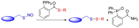
In 2008, our group reported a fast reductive ligation of RSNOs, which can selectively convert SNO to relatively stable sulfenamide product under very mild conditions (Scheme 1, eq. 1). 3 This reaction proceeds through a Staudinger-ligation type mechanism. 4 Recently, in the study of a “traceless” version of the reductive ligation, we discovered an unexpected bis-ligation, which led to the formation of stable disulfide-iminophosphorane products from primary RSNOs (Scheme 1, eq 2).5 In this process, the thiolate intermediate undergoes an intra-molecular substitution with the pseudo-sulfenamide linkage to form the disulfide-iminophosphorane product in excellent yields.
Scheme 1.

Based on the high reactivity of the sulfenamide towards thiolate observed in bis-ligation, we envisioned that phosphine-thioester substrates like 6 should undergo a reductive ligation mediated one-step disulfide formation with RSNOs (Scheme 2): the reaction between RSNO 1 and phosphine 6 should generate azaylide 7 first. Then, an intramolecular acyl transfer should occur to provide the intermediate 7b. Upon hydrolysis in aqueous buffer, 7b should be converted to the sulfenamide product 8 and thiolate 9. Finally, the intermolecular reaction between 8 and 9 could proceed spontaneously to provide a stable disulfide 10 and liberate the phosphine oxide 11. As the thioesters are better leaving groups than esters, we expect that substrates like 6 should facilitate the reductive ligation process. In addition, the formation of simple disulfide products, without the bulky phosphine adducts, would be attractive for the applications in protein systems.
Scheme 2.

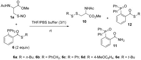
With this idea in mind, a series of phosphine-thioester substrates were prepared and tested with a RSNO model compound-1a (Table 1). As expected, the desired disulfide products were obtained in a mixture solvent THF/PBS buffer (pH 7.4). Other by-products isolated were corresponding phosphine-oxide 12 and amide 11. With primary thiol-based thioester substrates (6a and 6b), the disulfide products were isolated in excellent yields. Thiophenol-based thioester substrates (6c and 6d) also resulted in good yields of disulfide products. However, moderate yield (43%, entry 5) of disulfide was observed when 1a was treated with a tertiary thiol-based substrate 6e. Presumably the t-butylthiolate generated in the reductive ligation was less reactive towards the sulfenamide intermediate, due to the steric hindrance. In all examples, the disulfide formation proved to be a fast reaction which usually completed within 1 hour.
Table 1.
Reaction between RSNO and phosphine-thioesters
 | ||
|---|---|---|
| entry | 6 | yield of 10 [%][a] |
| 1 | 6a | 79 |
| 2 | 6b | 82 |
| 3 | 6c | 63 |
| 4 | 6d | 51 |
| 5 | 6e | 43 |
Yield of isolated product.
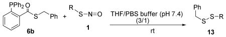
To explore the generality of this disulfide formation, we next tested the reaction of substrate 6b with a series of S-nitrosocysteine derivatives (1a-1h). As shown in Table 2, the desired disulfide products were obtained in good yields in all cases. It should be mentioned that the formation of the sulfenamide intermediate was not observed with these substrates (by TLC). This suggested that the disulfide formation was a faster reaction than the formation of sulfenamide intermediate.
Table 2.
One-step disulfide formation of RSNO
 | ||
|---|---|---|
| entry | RSNO | yield of 13 [%][a] |
| 1 | 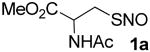 |
82 |
| 2 | 85 | |
| 3 | 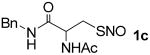 |
68 |
| 4 | 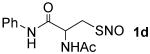 |
66 |
| 5 | 74 | |
| 6 | 73 | |
| 7 | 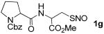 |
72 |
| 8 | 70 | |
Yield of isolated product.
To probe the formation of the sulfenamide intermediates as shown in the mechanism proposal, we carried out the experiment using some different substrates (Scheme 3). When the tertiary RSNO 1i was treated with 6b (eq A), corresponding sulfenamide 14 was isolated in good yield, while the disulfide product was not observed. This result is consistent with previous observations3 that sulfenamides generated from tertiary RSNOs are much more stable than the ones generated from primary RSNOs. In addition, when a homocysteine SNO derivative 1g was treated with 6b and when substrate 1a was treated with a phosphine-thioester (6f) prepared from a secondary thiol, we observed the formation of the sulfenamide intermediates (14a and 14b, eqs B and C). Therefore, the results shown in Scheme 3 clearly demonstrated that sulfenamides were the intermediates in the reaction between RSNOs and phosphine-thioesters.
Scheme 3.

To further prove the presence of sulfenamide intermediate in this reaction, we designed a crossover experiment (Scheme 4). When the reaction between RSNO 1b and phosphine substrate 6b was carried out, a sulfenamide compound 15 was also added into the reaction mixture. We anticipated that the reductive ligation between 1b and 6b should generate sulfenamide 16 and thiolate 17 quickly. Due to the presence of 15 in the reaction, thiolate 17 could react with both 15 and 16. Indeed, we isolated the desired product 13b in 46% yield, as well as the crossover product 13a in 34% yield.
Scheme 4.

If this reaction will be applied for labeling protein SNOs, a concern is that the thiolate intermediate may react with disulfides in proteins to give false positive linkage. To address this question, we tested the reaction between 1a and 6b in the presence of a disulfide compound 18 (Scheme 5). However, only the desired product 13a was obtained from the reaction. No crossover product with 18 was observed.
Scheme 5.

To address the bio-orthogonality of the phosphine-thioester substrates, we tested the reactions between 6b and potential cellular nucleophiles such as thiols and amines. 6b appeared to be quite stable towards lysine derivatives and cysteine derivatives as no reaction was observed (Scheme 6, see supporting information for experimental details).
Scheme 6.

To explore the application of this new reaction in the detection of S-nitrosated proteins, experiments shown in Scheme 7 were carried out. Briefly, COS-7 cells were first treated with S-nitrosocysteine (CysNO). It is known that the treatment of cells with CysNO will result in the S-nitrosation on proteins.6 The cells were then washed, fixed, and permeablized. After blocking free thiols with NEM, the cells were incubated with a biotin-linked phosphine substrate 20. Cellular proteins that were S-nitrosated should react with 20 to form a stable disulfide linkage with biotin. The labeled proteins were detected by NeutrAvidin-HRP and visualized by ECL Plus Western Blotting Detection System. As expected, we observed a number of labeled proteins on SDS-PAGE from CysNO treated cells (lane 2). In the control experiment, non-CysNO treated cells were carried through the same process and no labeled proteins were observed on SDS-PAGE (lane 1). We also found that the signals of CysNO treated cells were reversible by DTT (lane 3). This result confirmed the formation of disulfide linkages between the proteins and biotin. The GAPDH bands shown in lower panel exhibited equal protein loading in these experiments.
Scheme 7.
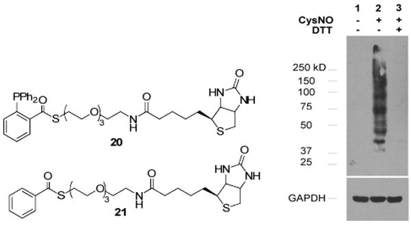
In this biotin-labeling experiment, one concern was that reagent 20 may react with cellular necleophiles (such as amines or thiols) to release biotin-linked thiolate, which might react with the disulfides in proteins to form false positive biotin conjugates. However, experiment results shown in Schemes 5 & 6, and more importantly the fact of lacking any observed biotin-labeled proteins in the control lane (lane 1, Scheme 7) should rule out that possibility. To further clarify this question, a structurally related thioester-compound 21 was prepared and used in CysNO treated cells. No biotin labeling was observed with 21 (even in the presence of PPh3, see Figure S1 in the supporting information). These results clearly demonstrated the efficiency and specificity of the phosphine mediated disulfide formation on SNO moieties.
In summary, a reductive ligation mediated disulfide formation between RSNOs and phosphine-thioester substrates has been developed. This reaction can selectively convert unstable RSNOs to stable disulfides in one step under very mild conditions. We also demonstrated that this reaction can be used to identify SNO proteins in cell extracts. Further explorations of this reaction for the detection of protein S-nitrosation continue in our laboratory.
Supplementary Material
Acknowledgments
This work is funded by the American Heart Association (0930120N) and a CAREER award from NSF (0844931) to M.X. and NIH (R01HL0088559) to A.R.W. We thank Prof. Jack R. Lancaster, Jr. (University of Alabama) for helpful discussion.
Footnotes
Supporting Information Available Synthetic procedures, spectroscopic data, and experimental procedures. This material is available free of charge via the Internet at http://pubs.acs.org.
References
- 1.a) Lancaster JR., Jr Nitric Oxide. 2008;19:68–72. doi: 10.1016/j.niox.2008.04.028. [DOI] [PubMed] [Google Scholar]; b) Zhang Y, Hogg N. Free Radic Biol Med. 2005;38:831–838. doi: 10.1016/j.freeradbiomed.2004.12.016. [DOI] [PubMed] [Google Scholar]; c) Hess DT, Matsumoto A, Kim SO, Marshall HE, Stamler JS. Nat Rev Mol Cell Biol. 2005;6:150–166. doi: 10.1038/nrm1569. [DOI] [PubMed] [Google Scholar]; d) Foster MW, McMahon TJ, Stamler JS. Trends Mol Med. 2003;9:160–168. doi: 10.1016/s1471-4914(03)00028-5. [DOI] [PubMed] [Google Scholar]
- 2.For a recent RSNO detection method, see: Bechtold E, Reisz JA, Klomsiri C, Tsang AW, Wright MW, Poole LB, Furdui CM, King SB. ACS Chem Biol. 2010;5:405–414. doi: 10.1021/cb900302u. For selected reviews on RSNO detection, see: Gow A, Doctor A, Mannick J, Gaston B. J Chromatogr B. 2007;851:140–151. doi: 10.1016/j.jchromb.2007.01.052.Kettenhofen NJ, Broniowska KA, Keszler A, Zhang Y, Hogg N. J Chromatogr B. 2007;851:152–159. doi: 10.1016/j.jchromb.2007.02.035.MacArthur PH, Shiva S, Gladwin MT. J Chromatogr B. 2007;851:93–105. doi: 10.1016/j.jchromb.2006.12.012.Jaffrey SR. Methods Enzymol. 2005;396:105–118. doi: 10.1016/S0076-6879(05)96011-4. For deficiencies of current methods, See: Giustarini D, Milzani A, Dalle-Donne I, Rossi R. J Chromatogr B. 2007;851:124–139. doi: 10.1016/j.jchromb.2006.09.031.Gladwin MT, Wang X, Hogg N. Free Rad Biol Med. 2006;41:557–561. doi: 10.1016/j.freeradbiomed.2006.05.025.
- 3.Wang H, Xian M. Angew Chem Int Ed. 2008;47:6598–6601. doi: 10.1002/anie.200801654. [DOI] [PubMed] [Google Scholar]
- 4.a) Saxon E, Bertozzi CR. Science. 2000;287:2007–2010. doi: 10.1126/science.287.5460.2007. [DOI] [PubMed] [Google Scholar]; b) Lin FL, Hoyt HM, van Halbeek H, Bergman RG, Bertozzi CR. J Am Chem Soc. 2005;127:2686–2695. doi: 10.1021/ja044461m. [DOI] [PubMed] [Google Scholar]; c) Wang H, Zhang J, Xian M. J Am Chem Soc. 2009;131:13238–13239. doi: 10.1021/ja905558w. [DOI] [PubMed] [Google Scholar]
- 5.a) Zhang J, Wang H, Xian M. Org Lett. 2009;11:477–480. doi: 10.1021/ol802663q. [DOI] [PubMed] [Google Scholar]; b) Zhang J, Wang H, Xian M. J Am Chem Soc. 2009;131:3854–3855. doi: 10.1021/ja900370y. [DOI] [PubMed] [Google Scholar]
- 6.a) Forrester MT, Thompson JW, Foster MW, Nogueira L, Moseley MA, Stamler JS. Nat Biotech. 2009;27:557–559. doi: 10.1038/nbt.1545. [DOI] [PMC free article] [PubMed] [Google Scholar]; b) Jaffrey SR, Erdjument-Bromage H, Ferris CD, Tempst P, Snyder SH. Nat Cell Biol. 2001;3:193–197. doi: 10.1038/35055104. [DOI] [PubMed] [Google Scholar]; c) Forrester MT, Foster MW, Benhar M, Stamler JS. Free Rad Biol Med. 2009;46:119–126. doi: 10.1016/j.freeradbiomed.2008.09.034. [DOI] [PMC free article] [PubMed] [Google Scholar]
Associated Data
This section collects any data citations, data availability statements, or supplementary materials included in this article.