

In a recent visit to the United States, the Dalai Lama gave a speech at the Society for Neuroscience's annual meeting in Washington, D.C. Over the past several years, he has helped recruit Tibetan Buddhist monks for, and directly encouraged research on the brain and meditation in the Waisman Laboratory for Brain Imaging and Behavior at the University of Wisconsin-Madison. The findings from studies in this unusual sample as well as related research efforts, suggest that, over the course of meditating for tens of thousands of hours, the long-term practitioners had actually altered the structure and function of their brains. In this article we discuss neuroplasticity, which encompasses such alterations, and the findings from these studies. Further, we comment on the associated signal processing (SP) challenges, current status and how SP can contribute to advance these studies.
What is Neuroplasticity
Neuroplasticity is a term that is used to describe the brain changes that occur in response to experience. There are many different mechanisms of neuroplasticity ranging from the growth of new connections to the creation of new neurons. When the framework of neuroplasticity is applied to meditation, we suggest that the mental training of meditation is fundamentally no different than other forms of skill acquisition that can induce plastic changes in the brain [1,2].
What is Meditation
The term ‘meditation’ refers to a broad variety of practices, ranging from techniques designed to promote relaxation to exercises performed with a more far-reaching goal such as a heightened sense of well-being. It is thus essential to be specific about the type of meditation practice under investigation. In [3] meditation was conceptualized as a family of complex emotional and attentional regulatory strategies developed for various ends, including the cultivation of well-being and emotional balance. Here we focus on Focused Attention (FA) meditation and Open Monitoring (OM) meditation.
The Focused Attention (FA) meditation entails voluntary focusing attention on a chosen object in a sustained fashion. The Open Monitoring (OM) meditation involves non-reactively monitoring the content of experience from moment-to-moment, primarily as a means to recognize the nature of emotional and cognitive patterns.
OM meditation initially involves the use of FA training to calm the mind and reduce distractions, but as FA advances, the cultivation of the monitoring skill per se becomes the main focus of practice. The aim is to reach a state in which no explicit focus on a specific object is retained; instead, one remains only in the monitoring state, attentive moment-by-moment to anything that occurs in experience.
These two common styles of meditation are often combined, whether in a single session or over the course of practitioner's training. These styles are found with some variation in several meditation systems, including the Buddhist Vipassanā and Mahāmudrā and are also implicated in many popular secular interventions that draw on Buddhist practices.
Findings of Brain Changes in Meditation
In what follows we summarize the changes in the brain that occur during each of these styles of meditation practice. Such changes include alterations in patterns of brain function assessed with functional magnetic resonance imaging (fMRI), changes in the cortical evoked response to visual stimuli that reflect the impact of meditation on attention, and alterations in amplitude and synchrony of high frequency oscillations that probably play an important role in connectivity among widespread circuitry in the brain.
Experimental Setup
The experiments described below that measure hemodynamic changes with functional magnetic resonance imaging (fMRI) require a high field strength MRI scanner equipped with the appropriate pulse sequences to acquire data rapidly and with the necessary fiber optic stimulus delivery devices so that visual stimuli can be presented to the subject while he or she lays in the bore of the magnet. For the studies that measure brain electrical activity, a high-density recording system with between 64 and 256 electrodes on the scalp surface is used.
FA Meditation
A recent study [4] used fMRI to interrogate the neural correlates of FA meditation in experts and novices. The study compared FA meditation on an external visual point to a rest condition during which participants do not use meditation and are simply instructed to adopt a neutral baseline state. The meditation condition was associated with activation in multiple brain regions implicated in monitoring (dorsolateral prefrontal cortex), engaging attention (visual cortex), and attentional orienting (e.g., the superior frontal sulcus, the supplementary motor area, and the intraparietal sulcus).
Although this meditation-related activation pattern was generally stronger for long-term-practitioners compared to novices, activity in many brain areas involved in FA meditation showed in an inverted u-shaped curve for both classes of subjects. Whereas expert meditators with an average of 19,000 hours of practice showed stronger activation in these areas than the novices, expert meditators with an average of 44,000 practice hours showed less activation. This inverted u-shaped function resembles the learning curve associated with skill acquisition in other domains of expertise, such as language acquisition. The findings support the idea that, after extensive FA meditation training, minimal effort is necessary to sustain attentional focus.
Expert meditators also showed less activation than novices in the amygdala during FA meditation in response to emotional sounds. Activation in this affective region correlated negatively with hours of practice in life, as shown in Figure 1(A). This finding may support the idea that, advanced levels of concentration are associated with a significant decrease in emotionally reactive behaviors that are incompatible with stability of concentration.
Fig 1.
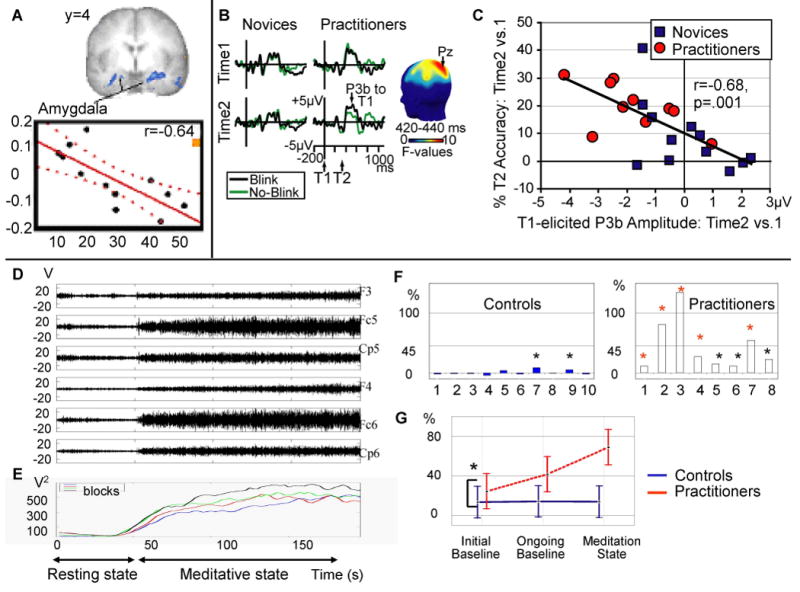
Neuroimaging and neurodynamical correlates of FA and OM meditations.
(a). Relationship between degree of meditation training (in years) and hemodynamic response in the amygdala (in blue) to distractor sounds during FA meditation in long-term Buddhist practitioners. Individual responses in the right amygdala are plotted (adapted from [2]).
(b) the reduction in P3b amplitude (a brain-potential index of resource allocation) to the first of two target stimuli (T1 and T2) presented in a rapid stream of distracter stimuli after three months of intensive Vipassana meditation [3]
(c) shows that generally, the greater the reduction in brain-resource allocation to T1 was over time, the better able an individual became at accurately identifying T2 (adapted from [3]).
(d-e) Example of high-amplitude gamma activity during a form of OM meditation, non-referential compassion meditation, in long-term Buddhist practitioners [4]. (e) Time course of gamma (25–42 Hz) activity power over the electrodes displayed in (d) during four blocks computed in a 20-s sliding window every 2 s and then averaged over electrodes.
(f) Intra-individual analysis on the ratio of gamma to slow oscillations (4–13 Hz) averaged across all electrodes during compassion meditation.
(g) the significant interaction between group (practitioner, control) and state (initial baseline, ongoing baseline, and meditation state) for this ratio.
Collectively these findings support the view that attention is a trainable skill that can be enhanced through the mental practice of FA meditation.
OM Meditation
Another study [5] recently examined the idea that OM meditation decreases elaborative stimulus processing in a longitudinal study using scalp-recorded brain potentials and performance in an attentional blink task. The consequence of decreased elaborative stimulus processing is that the subject is able to better attend moment-to-moment to the stream of stimuli to which they are exposed and less likely to “get stuck” on any one stimulus.
The attentional blink phenomenon illustrates that the information processing capacity of the brain is limited. More specifically, when two targets T1 and T2, embedded in a rapid stream of events, are presented in close temporal proximity, the second target is often not seen. This deficit is believed to result from competition between the two targets for limited attentional resources i.e., when many resources are devoted to T1 processing, too few may be available for subsequent T2 processing.
The study in [5] found that three months of intensive training in Vipassana meditation (a common style of OM meditation) reduced brain-resource allocation to the first target, as reflected in a smaller T1-elicited P3b, a brain-potential index of resource allocation. This is illustrated in Figure 1(B), which shows the reduction in P3B amplitude (a brain-potential index of resource allocation). In this figure, the scalp-recorded brain potentials from electrode Pz, time-locked to T1 onset as a function of T2 accuracy (detected (no-blink) vs. not detected (blink)), time (before or after three months), and group (practitioners vs. novices) are shown. The scalp map shows electrode sites where this three-way interaction was significant between 420 and 440ms.
The reduction in brain-resource allocation to T1 was associated with a smaller attentional blink to T2, as shown in Figure 1(C). As participants were not engaged in formal meditation during task performance, these results provide support for the idea that one long-term effect of OM meditation may be reduction in the propensity to “get stuck” on a target as reflected in (a) less elaborate stimulus processing and (b) the development of efficient mechanisms to engage and then disengage from target stimuli in response to task demands.
Previous studies [6] of high-amplitude pattern of gamma synchrony in expert meditators during an emotional version of OM meditation support the idea that the state of OM may be best understood in terms of a succession of dynamic global states. Compared to a group of novices, the adept practitioners self-induced higher-amplitude sustained electroencephalography (EEG) gamma-band oscillations and long-distance phase synchrony, in particular over lateral fronto-parietal electrodes, while meditating. Importantly, this pattern of gamma oscillations was also significantly more pronounced in the baseline state of the long-term practitioners compared with controls, suggesting a transformation in the default mode of the practitioners as shown in Figure 1(G). Although the precise mechanisms are not clear, such synchronizations of oscillatory neural discharges may play a crucial role in the constitution of transient networks that integrate distributed neural processes into highly ordered cognitive and affective functions.
An example of high-amplitude gamma activity during a form of OM meditation, non-referential compassion meditation, in long-term Buddhist practitioners [6] is shown in Figure 1(D) and (E).
The intra-individual analysis on the ratio of gamma to slow oscillations (4–13 Hz) averaged across all electrodes during compassion meditation in illustrated in Figure 1(F). The abscissa represents the subject numbers, the ordinate represents the difference in the mean ratio between the initial state and meditative state, and the black and red stars indicate that this increase is greater than two and three times, respectively, the baseline standard deviation.
The significant interaction between group (practitioner, control) and state (initial baseline, ongoing baseline, and meditation state) for this ratio is shown in Figure 1(G). The relative gamma increase during meditation was higher in the post-meditation session. In the initial baseline, the relative gamma was already higher for the practitioners than the controls and correlated with the length of the long-term practitioners' meditation training through life (adapted from [6]).
SP Challenges
While SP has a unique opportunity to contribute to this novel effort to chart the manner in which the brain may be transformed through the mental practice of meditation, there are several associated challenges. Among the many challenges include the characterization of different signatures of brain function that distinguish among different meditation practices, the parsing of variance in brain activity that may be due to changes in peripheral physiology such as respiration, and the simultaneous measurement of electrical and hemodynamic signals to harness the best temporal and spatial resolution possible.
Impact on Brain-Computer Interfaces
One of the interesting implications of the research on meditation and brain function is that meditation might help to reduce “neural noise” and so enhance signal-to-noise ratios in certain types of tasks. In contexts where brain-computer interfaces are being developed that are based upon electrical recordings of brain function, training in meditation may facilitate more rapid learning. This idea warrants systematic evaluation in the future.
Future Work
Ongoing and future work focuses on a few distinct directions. One of the crucial areas needing attention is the characterization of changes in connectivity among the various brain circuits that are engaged by these practices. The developmental of new methods to probe different aspects of connectivity (both structural and functional) will be extremely valuable in furthering this line of inquiry. The goal of such work is to better understand how different circuits are integrated during meditation to produce the behavioral and mental changes that are said to occur as a result of such practices, including the promotion of increased well-being.
Contributor Information
Richard J. Davidson, Email: rjdavids@wisc.edu.
Antoine Lutz, Email: alutz@wisc.edu.
References
- 1.Berger A, Kofman O, Livneh U, Henik A. Multidisciplinary perspectives on attention and the development of self-regulation. Progress in Neurobiology. 2007;82:256–286. doi: 10.1016/j.pneurobio.2007.06.004. [DOI] [PubMed] [Google Scholar]
- 2.Poldrack RA. Neural systems for perceptual skill learning. Behavioral and Cognitive Neuroscience Reviews. 2002;1:76–83. doi: 10.1177/1534582302001001005. [DOI] [PubMed] [Google Scholar]
- 3.Lutz A, Dunne JP, Davidson RJ. Meditation and the neuroscience of consciousness: An Introduction. In: Zelazo PD, Thompson E, editors. The Cambridge Handbook of Consciousness. Cambridge University Press; 2006. [Google Scholar]
- 4.Brefczynski-Lewis JA, Lutz A, Schaefer HS, Levinson DB, Davidson RJ. Neural correlates of attentional expertise in long-term meditation practitioners. Proceedings of the National Academy of Sciences. 2007;104:11483–11488. doi: 10.1073/pnas.0606552104. [DOI] [PMC free article] [PubMed] [Google Scholar]
- 5.Slagter HA, Lutz A, Greischar LL, Francis AD, Nieuwenhuis S, Davis JM, Davidson RJ. Mental training affects use of limited brain resources. PLoS Biology. 2007;5(6):e138. doi: 10.1371/journal.pbio.0050138. [DOI] [PMC free article] [PubMed] [Google Scholar]
- 6.Lutz A, Greischar L, Rawlings NB, Ricard M, Davidson RJ. Long-term meditators self-induce high-amplitude synchrony during mental practice. Proceedings of the National Academy of Sciences. 2004;101:16369–16373. doi: 10.1073/pnas.0407401101. [DOI] [PMC free article] [PubMed] [Google Scholar]