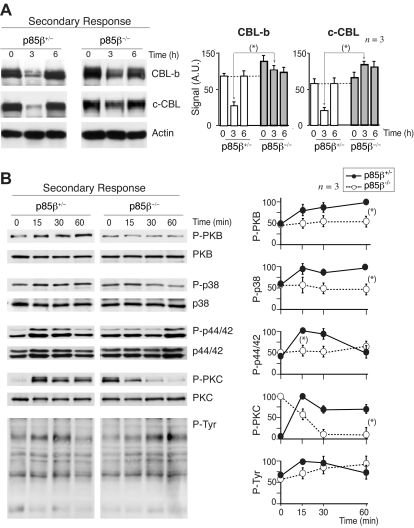
Figure 7.
CBL down-regulation and pPKB defects upon secondary stimulation of p85β−/− T cells. (A,B) We measured CBL expression levels in antigen-experienced T cells; p85β−/− and p85β+/− F5TCRTg mice were injected with the NP366-374 peptide; 11 days later both mice groups were reinjected with the NP366-374 peptide, and CBL-b or c-CBL was analyzed in WB at different times after injection (A). Alternatively, 11 days after first priming, mice were killed and purified T cells were stimulated in vitro with peptide-pulsed APCs for the times indicated. Cell extracts were examined in WB using antibodies as indicated (B). Figures are representative of at least 3 experiments with similar results. Graphs in panel A show the mean plus or minus SD (n = 3) of CBL-b or c-CBL signal. Graphs in panel B show the mean percentage plus or minus SD (n = 3) of the signal for each of the proteins, compared with the maximal signal in control cells (100%) and normalized in comparison with loading controls (n = 3). P values are shown for the times of maximal signal (*P < .05).

