Abstract
Green tea (Camellia sinensis) is rich in catechins, of which (−)-epigallocatechin-3-gallate (EGCG) is the most abundant. Studies in animal models of carcinogenesis have shown that green tea and EGCG can inhibit tumorigenesis during the initiation, promotion and progression stages. Many potential mechanisms have been proposed including both antioxidant and pro-oxidant effects, but questions remain regarding the relevance of these mechanisms to cancer prevention. In the present review we will discuss the redox chemistry of the tea catechins and the current literature on the antioxidant and pro-oxidative effects of the green tea polyphenols as they relate to cancer prevention. We report that although the catechins are chemical antioxidants which can quench free radical species and chelate transition metals, there is evidence that some of the effects of these compounds may be related to induction of oxidative stress. Such pro-oxidant effects appear to be responsible for the induction of apoptosis in tumor cells. These pro-oxidant effects may also induce endogenous antioxidant systems in normal tissues that offer protection against carcinogenic insult. This review is meant point out understudied areas and stimulate research on the topic with the hope that insights into the mechanisms of cancer preventive activity of tea polyphenols will result.
Keywords: green tea, catechins, cancer prevention, pro-oxidant, antioxidant
Introduction
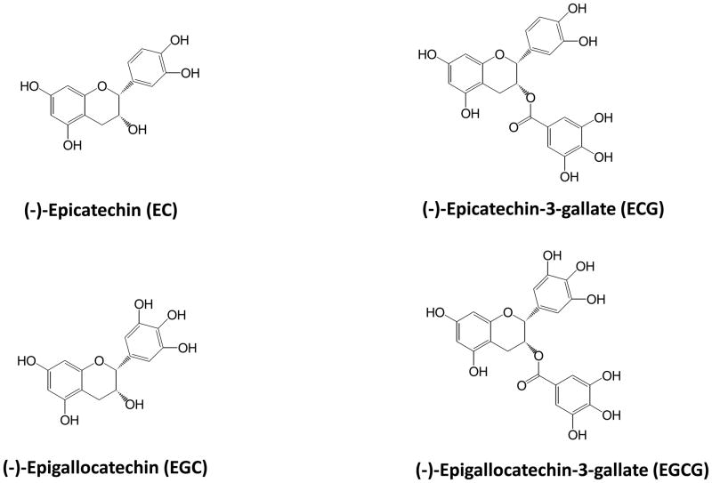
Tea (Camellia sinensis, Theaceae) is second only to water in terms of worldwide popularity [1]. The 3 major forms of tea: green tea, black tea, and oolong tea, differ in terms of their production methods and chemical composition [2]. Green tea, which represents 20% of world consumption, is characterized by the presence of large amounts of flavan-3-ols also known as catechins (Fig. 1). A typical cup of brewed green tea is made using 2 g of tea leaves in 200 mL of hot water and contains approximately 600–900 mg water extractabe solids. Of these solids, approximately 30–40% by weight are the tea catechins. (−)-Epigallocatechin-3-gallate (EGCG) is the most abundant catechin and may represent up to 50% of the catechins by weight. Green tea is prepared by pan-frying or steaming the tea leaves in order to inactive polyphenol oxidase activity in the leaves. By contrast, in the processing of black tea, fresh leaves are crushed and allowed to undergo a polyphenol oxidase mediated oxidation known as “fermentation”. This results in the oxidation of the catechins to form catechin dimers, known as theaflavins, as well as polymeric thearubigins. These compounds are responsible of the characteristic color and taste of black tea.
Figure 1.
Structures of the major tea polyphenols.
Extensively laboratory and epidemiological studies have suggested that green tea and green tea polyphenols, especially EGCG, have preventive effects against chronic diseases including heart disease, diabetes, neurodegenerative disease, and cancer (reviewed in [3–6]). Numerous mechanisms have been proposed to account for the cancer preventive effects of green tea and EGCG in laboratory animal models. These mechanisms include the inhibition of growth factor signaling, inhibition of key cellular enzymes, inhibition of gene transcription, and induction of tumor suppressor genes (reviewed in [7–10]). The antioxidant activity of green tea polyphenols and, more recently, the pro-oxidant effects of these compounds, have also been suggested as potential mechanisms for cancer prevention [11–13]. In the present review, we will discuss the potential role for antioxidant vs. pro-oxidant effects of green tea polyphenols in cancer prevention. We will pay careful attention to the underlying chemical mechanisms involved, the relative strength of the various lines of biological evidence for these effects, and the potential for direct pro-oxidant effects of tea polyphenols resulting in indirect antioxidant effects. Our goal in writing this review is to stimulate research into the role of the redox effects of tea polyphenols as a mechanism for cancer prevention. A better understanding of the chemistry of these compounds, the effects of biological matrices on this chemistry, and the complexity of the biological response to exposure to tea polyphenols will be essential for understanding their ultimate usefulness in preventive chronic diseases including cancer.
Redox Chemistry of Tea Polyphenols
Direct antioxidant effects
The antioxidant activity of (−)-epicatechin (EC), (−)-epigallocatechin (EGC), (−)-epicatechin-3-gallate (ECG), and EGCG has been demonstrated in a number of in vitro and chemical-based assays. The chemistry underlying this activity results mainly from hydrogen atom transfer (HAT) or single electron transfer reactions (SET), or both involving hydroxyl groups. These groups are constituents of the B-rings of EC and EGC, and both B- and D-rings of ECG and EGCG (Fig. 1).
As chain-breaking antioxidants, tea catechins are thought to interrupt deleterious oxidation reactions by HAT mechanisms, the most important being lipid peroxidation:
| (1) |
| (2) |
| (3) |
Lipid peroxidation is a radical chain reaction in which hydrogen atoms are abstracted (Rxn. 1) from unsaturated fatty acids (L1H), yielding alkyl radicals (L1•) that react (Rxn. 2) at near-diffusion limited rates with molecular oxygen to give lipid hydroperoxyl radicals (L1OO•). In the absence of chain-breaking antioxidants, these peroxyl radicals abstract hydrogen atoms (Rxn. 3) from unoxidized lipid substrate (L2), resulting in new lipid alkyl radicals (L2•), thus propagating the chain reaction. Lipid hydroperoxides (L1OOH) are produced concomitantly in Rxn. 3, which are further reduced by transition metal-catalyzed, or Fenton-type, reactions to unstable alkoxyl radicals and, eventually, secondary oxidation products (e.g., malonaldehyde). Fortunately, the reaction between lipid peroxyl radicals and unoxidized lipids (Rxn. 3) is relatively slow (ca. 101 M−1 s−1), affording phenolic antioxidants (PhOH) the opportunity to intercept peroxyl radicals and interrupting chain propagation:
| (4) |
This rate (Rxn. 4) is dependent on the bond dissociation enthalpy of the catechins, as the weaker the O-H bond of the hydroxyl group, the faster the reaction with the peroxyl radical [14]. Furthermore, the resulting semiquinone radical (PhO•) must be relatively stable so that it reacts only slowly with unoxidized lipids. This stability is achieved by resonance stabilization of the PhO• species (Fig. 2) [15].
Figure 2.
Resonance stabilization of unpaired eectrons by the gallate ring found in tea catechins.
Tea catechins are also known to quench free radicals by SET reactions, wherein phenolic radical cations are first formed followed by deprotonation:
| (5) |
| (6) |
| (7) |
SET reactions involving phenolic antioxidants and lipid peroxyl radicals give rise to the same products as HAT reactions (i.e., LOOH and PhO•), but are solvent dependent because their rates depend on the ionization potential (IP) of the hydroxyl groups [16]. Whereas the chain-breaking activity of tea catechins are known to involve both HAT and SET mechanisms, it appears HAT mechanisms dominate [14] and that this activity is restricted to the B-ring, even in the case of catechins with galloyl moieties comprising their D-rings (ECG, EGCG) [14, 17].
Flavanols have been reported to scavenge a host of oxygen, nitrogen, and chlorine radical species, which include superoxide, peroxyl radicals, hypochlorous acid, and peroxynitrous acid [18]. The order of radical scavenging activity of the major tea catechins has been calculated on the basis of δBDE using computational methods, and is predicted to follow the order: EC ≤ ECG < EGC ≤ EGCG [14]. This order is consistent with empirical studies showing the rate of reaction with peroxyl radicals followed the order EC < ECG ≈ EGC < EGCG [19].
Scavenging of hydroxyl radicals by flavanols, including tea catechins, has also been reported, yet this activity is suspect given the extremely reactive nature of this species; hydroxyl radicals have been shown to react with organic matter in proportion to its concentration [20], thus given the relatively low bioavailability of the tea catechins, it is hard to envisage that such a reaction plays a major role in vivo.
Trace Metal Catalysis of Polyphenol Oxidation
The rate of catechin oxidation increases as a function of increasing pH [21–23]. The base-catalyzed oxidation of phenolic compounds is often referred to as “autooxidative” because it is thought that oxygen reacts faster with phenolate anions. While this reaction is thermodynamically favorable, it is kinetically unfavorable, as it would result in new orbitals containing electrons with the same quantum number which is forbidden by the Pauli Exclusion Principle [24–27]. However, transition metas (e.g., iron and copper) are capable of initiating phenolic oxidation and are essential catalysts in this process [24]. This reaction also yields a reactive oxygen species, namely superoxide (O2−•) or its protonated form, the hydroperoxyl radical (HO2•), under acidic conditions (Rxn. 9), that is further reduced to hydrogen peroxide (Rxn. 11):
| (8) |
| (9) |
| (10) |
| (11) |
Whereas many have observed rapid phenolic oxidation in aqueous solution without added iron or copper, it is known that such metals are present as contaminants in chemical reagents, buffer, cell culture media, solvents, etc. [25]. The importance of iron catalysis in catechol oxidation has been demonstrated by removal of the metal with desferrioxamine [26, 28]. Catechol “autoxidation” was also stopped at pH 8.0 by the addition of diethylenetriaminepentaacetic acid, catalase, and superoxide dismutase (SOD) [27, 29].
The metal-catalyzed oxidation of catechins has consequences beyond that of reactive oxygen species generation. Semiquinone radicals and, eventually, quinones are generated in the process, which are highly electrophilic species that can react with free thiol-bearing compounds to form stable conjugates [30, 31]. Furthermore, catechol and galloyl oxidation is coupled to metal reduction (e.g., Fe3+ → Fe2+; Rxn. 5). The reduced forms of transition metals are known to catalyze lipid hydroperoxide and hydrogen peroxide decomposition to lipid alkoxyl (LO•) and hydroxyl radicals (HO•), respectively. These results suggest a role for transition metals in some of the pro-oxidant effects of green tea polyphenols observed in vitro and in vivo (Discussed below). Although the levels of transition metals are tightly regulated in vivo, the catalytic amounts necessary for the reactions describe above would indicate that such effects may still occur.
Chelation of Metal Catalysts
The association of metals and phenolic compounds does not always lead to deleterious consequences, a phenomenon that appears linked to pH, among other factors. Catechoate- and gallate-ferric complexes appear to be less stable under acidic conditions, liberating ferrous (Fe2+) ions and semiquinone radicals as they fall apart. On the other hand, phenolic-metal complexes appear to be more stable at neutral pH [32]. In some cases, the catalytic activity of phenol-bound transition metals is decreased in such complexes, despite the fact that complexation typically results in metal reduction [33]. Under conditions when the bound metal retains its catalytic ability, the associated phenolic compound may serve to intercept radical species as they are formed on the complexed metal [33]. These so-called “site-specific” reactions, where radical damage is directed at the site of formation, may explain some of the purported hydroxyl radical scavenging activity of tea catechins. Evidence of site-specific metal catalyzed reactions have been reported in biological membranes [34–37].
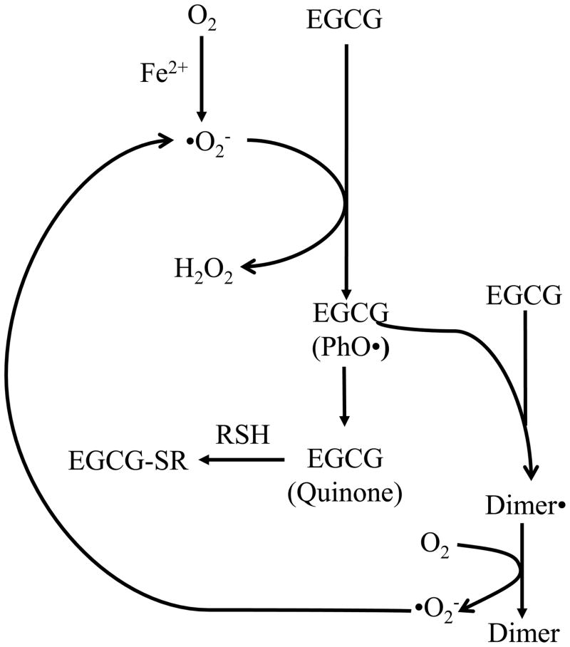
The many elegant chemistry experiments that have been conducted on the redox effects of tea polyphenols provide some potential mechanisms by which to understand the antioxidant and pro-oxidant effects of these compounds in biological systems (Fig. 3). These data must, however, be integrated with the complex nature of cell-lines, animal models, and human physiology in order to determine the relevance of these redox effects to human disease prevention.
Figure 3.
Oxidative reaction between EGCG, superoxide, and ferric iron resulting in the production of oxidative stress, EGCG dimers, and EGCG-cysteine conjugates (EGCG-SR). PhO = semiquinone radical.
Role of Antioxidant Effects in the Cancer Preventive Activity of Green Tea Polyphenols
Direct antioxidant effects
As described above, tea polyphenols are strong radical scavengers and metal chelators in model chemical systems, and these effects correlate with the presence of the dihydroxy and trihydroxy groups (reviewed in [3, 4]). An increasing number of studies have also demonstrated these antioxidative effects in vivo.
For example treatment of 24 month old rats with 100 mg/kg, i.g. EGCG decreased the hepatic levels of lipid peroxides (50% decrease) and protein carbonyls (39% decrease) [38]. EGCG treatment also increased the hepatic levels of both small moecule antioxidants and antioxidant enzymes compared to control rats. A second study by the same group found similar results using a much lower dose of EGCG (2 mg/kg, i.g.) over a relatively long period of time (30 d) [39]. These effects were not observed in young rats, suggesting that EGCG offered no improvement in antioxidant status in the absence of pre-existing oxidative stress. This may explain why other studies have failed to observe an effect of tea polyphenol treatment [4, 40].
Cooking oil fumes represent an important environmental toxicant that has been associated with lung diseases including cancer. Exposure of Wistar rats to cooking oil fumes for 30 min increased ROS levels in the blood and increased levels of ROS and 4-hydroxynonenal (4-HNE) levels in the alveolar lavage fluid [41]. Pre-treatment with green tea catechins for two weeks significantly reduced the levels of 4-HNE and reactive oxygen species (ROS) in the lavage fluid.
Treatment of C3(1) SV40 T,t antigen transgenic multiple mammary adenocarcinoma (TAg) mice mice with 0.05% green tea catechins or black tea theaflavins as the sole source of drinking fluid for 25 weeks results in an increase in mean survival time (5–7% increase) and decreased tumor load (25%–39% reduction) [42]. These changes in cancer parameters correlated with tea-polyphenol mediated decreases in the levels of 3-(2-deoxy--D-erythro-pentofuranosyl)pyrimido[l,2-]purin-10(3H)one, a malonyldialdehyde-DNA adduct, in the tumors by (63–78% decrease). Likewise, decreased tumor burden was associated with increased apoptosis.
Supplementation of healthy human volunteers with 500 mg/d catechins for 4 weeks decreased plasma oxidized low density lipoprotein by 18% compared to control [43]. Tea catechins have also been shown to blunt plasma levels of Fas ligand, interleukin (IL)-6 soluble receptor, hydrogen peroxide, and C-reactive protein in hemodialysis patients [44]. By contrast, a randomized, double blind crossover study of endurance-trained men for 3 wk found no significant effect of green tea extract (160 mg/d catechins) on markers of oxidative stress or inflammation [45]. This lack of effect may be due to the small study size (n = 10) or the high level of physical training of the subjects. Perhaps a greater effect would be observable in more sedentary study population. Such hypotheses should be further explored in larger studies.
Both Schwartz et al., [46] and Hakim et al. [47] have shown that green tea treatment has antioxidant effects in smokers. In the former study, green tea treatment (400–500 mg, 5 times per day for 4 wks) reduced the levels of benzo[a]pyrene adducted deoxyguanosine (B[a]P-dG) by 50% compared to baseline. Tea treatment reduced the number of 8-OHdG positive cells in smokers to a similar extent. In the study by Hakim et al., supplementation of heavy smokers (> 10 cigarettes per day) with 4 cups of decaffeinated green tea (73.5 mg catechins per cup) per day for 4 months reduced urinary 8-OHdG levels by 31% compared to control.
Induction of endogenous antioxidants
Studies have shown that tea treatment can induce Phase II metabolism and antioxidant enzymes in both animal models and humans. Treatment of piglets with 0.2% green tea extract for 3 weeks increased the ex vivo formation of aflatoxin (AF)B1 glutathione conjugates by small intestinal microsomes [48]. Similarly, treatment of female Wistar rats with 2% green tea solution for four weeks was shown to increase cytosolic glutathione S transferase (GST) activity in the liver [49]. A later study with pure tea polyphenols, however, found no significant effect on hepatic GST activity in Wistar rats [50]. The reasons for these differences are unclear but suggest that non-polyphenolic compounds in tea may play a role in increasing hepatic GST in this model.
Treatment of C57bl/6J mice with EGCG (200 mg/kg, i.g.) has been reported to increase gene expression of γ-glutamyltransferase, glutamate cysteine ligase, and hemeoxygenase 1 in an Nrf2-antioxidant response element-dependent manner [12]. Similar results have been reported in the tumor cells of colon cancer xenograft-bearing nude mice treated with dietary EGCG [51].
These effects have been re-capitulated in vitro [52]. Treatment of human mammary epithelial cells with EGCG resulted in increased expression of hemeoxygenase-1 and superoxide dismutase. This effect was reduced by small-interfering RNA (siRNA)-mediated disruption of Nrf2, suggesting a role for this pathway in the EGCG-mediated induction of these endogenous antioxidant systems.
Khan et al., have reported that green tea supplementation could reduce the oxidative renal damage induced by cisplatin treatment in Wister rats [53]. Cisplatin increased the levels of lipid peroxides (by 41%), and reduced the levels of SOD (by 23%) and catalase (by 17%) in the cortex of the kidney compared to vehicle-treated controls. Co-treatment with 3% green tea extract prevented the observed increase in lipid peroxides and caused an increase in the levels of SOD and catalase beyond those observed at baseline. Interestingly, treatment with green tea extract alone also increased SOD and catalase levels in renal cortex, but caused a reduction in the levels of total thiols. These results suggest that green tea extract may induce some level of oxidative stress on its own, and that this oxidative stress may lead to the induction of endogenous antioxidant systems. In contrast to the protective effects observed in the renal cortex, green tea extract did not have any significant protective effect in the renal medulla. The underlying reasons for these tissue specific effects remain to be determined, but may be related to differences in tissue blood flow or the distribution of the tea catechins.
In human studies, tea polyphenol treatment has been associated with changes in carcinogen metabolism. For example, a study in China of subjects in high risk areas for liver cancer found that supplementation with green tea polyphenols (500 or 1000 mg/d) resulted in increased urinary excretion of aflatoxin B1 (AFB1)mercapturic acid conjugates (AFB1-NAC) compared to baseline [54]. Supplementation with green tea polyphenols for 3 months also resulted in a dose-dependent decrease in urinary 8-hydroxydeoxyguanosine (8-OHdG) levels compared to placebo. Another study in healthy volunteers found that treatment with Polyphenon E (800 mg/d) for 4 wks increased the activity of GST-π in blood lymphocytes [55]. A result that indicates a potential mechanism for the increased excretion of NAC-conjugated carcinogen metabolites observed in China.
Role of pro-oxidant effects in the cancer preventive activity of green tea polyphenols
Generation of Reactive Oxygen Species by Tea Polyphenols
Under typical cell culture conditions, green tea polyphenols are unstable and undergo auto-oxidative reactions resulting in the production of ROS [56]. EGCG incubated in the absence of cells in McCoy’s 5A cell culture medium at 37°C under 5% CO2 atmosphere resulted in the time-dependent formation of H2O2 [57]. At 50 μM EGCG, the maximum level of H2O2 achieved was 25 μM after 120 min. Incubation of EGCG in the same medium in the presence of HT-29 human colon cancer cells decreased the maximum concentration of H2O2 produced to 10 μM, and caused the levels of H2O2 to peak after 30 min and to decrease back to baseline. This decrease is likely due to cell-derived catalase eliminating the H2O2. EGCG-mediated production of H2O2 is accompanied by a decrease in the concentration of EGCG (half-life < 30 min) and an increase in theasinesin A, an oxidative dimer of EGCG.
Sang et al., determined that the stability of EGCG is dependent on the concentration of EGCG present, the pH of the system, the presence of oxygen, and the temperature of the incubation [58]. For example, the half-life of EGCG in water at 37°C increased from 30 min to 150 min as the concentration of EGCG increased from 20–96 μM. Similarly, if EGCG (20 μM) is incubated in phosphate buffer (pH 7.4) under an air atmosphere at 37°C, the half-life is 30 min, whereas under a N2 atmosphere there is no appreciable loss of EGCG for at least 360 min.
The presence of transition metals has also been shown to play an important role in EGCG-mediated H2O2. Incubation of HL-60 human leukemia cells with tea catechins in the presence of Cu(II) resulted in the formation of 8-oxo-guanosine [59]. Co-incubation with the catalase or the copper chelator, bathocuproine, reduced catechin-mediated oxidative damage.
Induction of Apoptosis and Inhibition of Cell Proliferation
Yang et al., and others have reported that production of ROS plays an important role in the pro-apoptotic effects of EGCG and other tea polyphenols against cancer cell lines [60–62]. Treatment of 21BES Ha-Ras-transformed human bronchial epithelial cells with EGCG and theaflavin-3,3′-digallate (TFdiG) resuted in dose-dependent growth inhibition with IC50 values of 24 and 22 μM [60]. Both EGCG and TFdiG induced apoptosis in 21BES cells. EGCG-mediated apoptosis was reduced by approximately 50% by inclusion of exogenous catalase, whereas inclusion of catalase had no effect on the pro-apoptotic effects of TFdiG. These results suggested that TFdiG can induce apoptosis by an ROS independent mechanism in this model system.
Similar results have been reported in the RAW 264.7 mouse macrophage cell and with HL-60 human promyelocytic leukemia cells [63]. Treatment of HL-60 cells with 50 μM EGCG resulted in the formation of ROS as measured by dichlorofluoroscein (DCF) fluorescence, and a concomitant increase in the number of apoptotic cells. Inclusion of either exogenous catalase or SOD reduced both the levels of ROS and the number of apoptotic cells.
Treatment of H1299 cells with EGCG have been shown to inhibit cell growth and induce intracellular levels of ROS as measured by oxidation of intracellular DCF [64]. EGCG treatment also increased phosphorylation of histone 2A.X (γH2A.X), a marker of oxidative DNA damage. These effects were reduced, but not eliminated by the inclusion of exogenous SOD and catalase suggesting that EGCG may undergo intracellular oxidation, and suggesting the possibility that EGCG-mediated pro-oxidant activity has relevance in vivo.
Effects on cell signaling
Several studies have shown that the effect of EGCG and other tea polyphenols on key cell signaling pathways may be dependent on polyphenol-mediated reactive oxygen species. Vittal et al., have reported that treatment of 21BES cells with EGCG in the presence of absence of catalase results in differential gene expression patterns [62]. Of note, treatment of cells with 25 μM EGCG results in increased expression of genes involved in transforming growth factor (TGF)β signaling. These included TGFβ1 and β2, SMAD2, 3 and 6, and SMAD anchor for receptor activation. These effects were ablated by inclusion of exogenous catalase. By contrast, EGCG-mediated down-regulation of genes involved in the bone morphogenetic (BMP) pathway (BMP receptor 5, SMAD7, and cullin) were unaffected by the addition of catalase. These results suggest that the effects of EGCG on TGFβ signaling are dependent on EGCG-mediated reactive oxygen species, whereas the effects on BMP signaling are not.
It has been reported by several laboratories that EGCG and TFdiG can inhibit epidermal growth factor (EGF) mediated signaling. Initially, it was hypothesized that EGCG competed for receptor binding with EGF, or alternatively inhibited the kinase activity of the EGF receptor (EGFR) [65, 66]. The work of Liang et al., which showed that EGCG competed with EGF for binding to EGF, reported that inhibition the receptor was dependent on the pre-incubation of the receptor with EGCG prior to addition of EGF. This would suggest that the inhibition is in fact not competitive since true competitive inhibition should require no pre-incubation.
Studies by Hou et al., suggest that the effects of EGCG on EGFR-signaling are mediated by EGCG-induced oxidative stress [11]. Pre-incubation of KYSE150 human esophageal cancer cells and A431 human epidermoid carcinoma cells with EGCG inhibited the EGF-mediated phosphorylation of EGFR and also decreased the total amount of EGFR protein present. Interestingly, total EGFR protein was decreased by EGCG pre-treatment even in the absence of EGF stimulation. Under the cell culture conditions used in these experiments, EGCG was unstable, underwent polymerization, and produced ROS. Inclusion of superoxide dismutase (SOD), stabilized EGCG and reduced production of reactive oxygen species. SOD also ablated the inhibitory effects of EGCG on EGFR. These results indicate that EGCG-mediated inhibition of EGF-signaling is at least partially dependent on EGCG-mediated oxidative stress.
Muzolf-Panek et al., have reported that EGCG-mediated ROS production may underlie it ability to induce endogenous antioxidants, at least in vitro [67]. Treatment of Hepa1c1c7 human hepatoma cells with EGCG and (−)-gallocatechin-3-gallate (GCG) resulted in dose-dependent increases in NADPH:quinone reductase-1 mediated transcriptional activity. Transcription of this gene is mediated through the electrophile-responsive element (EpRE). Co-treatment of cells with N-acetylcysteine (NAC) blunted the catechin-mediated increase in EpRE-mediated transcription, whereas co-treatment with buthionine sulfoximine, which reduced intracellular GSH, enhanced the increased effects. These results suggest that the pro-oxidant effects of EGCG and GCG are important for their ability to regulate EpRE-mediated transcription. LC-MS analysis of the cell culture medium revealed the presence of EGCG-2′-glutathione further supporting this conclusion.
The relevance of these mechanisms remains to be determined in vivo. The differences in oxygen tension in cell culture (~155 mmHg) compared to many tissues in vivo (< 40 mmHg) raise questions about whether such oxidation effects can occur [68]. Inside the cells of the body, there is the possibility of superoxide leakage, which could cause the oxidation of EGCG and initiate pro-oxidant effects, but this remains to be tested. Additionally, the concentrations of tea catechins used in these studies exceed those available in vivo following tea consumption [69]. In order to establish the relevance of the pro-oxidant effects of tea polyphenols in vivo, careful mechanistic animal model and human intervention studies are needed.
Pro-oxidant effects in animal models
Hou et al., have previously observed that treatment of NCR nu/nu mice bearing H1299 xenograft tumors with a single daily dose of 40 mg/kg, i.p. EGCG or 0.2% EGCG in the diet resulted in significantly increased expression of γH2A.X in the tumors of the mice compared to control-treated mice [64]. These results indicate that at non-toxic doses of EGCG, which are comparable to those to which humans are exposed [70, 71], oxidative stress is induced in xenograft tumors. These results must be reproduced in other animal models of cancer to determine the general applicability of this mechanism.
Studies using higher doses of EGCG also show that pro-oxidant effects may play a role in the potential toxic effects of EGCG that have been reported in vivo. For example, Galati et al., have reported that treatment of freshly isolated mouse hepatocytes with 200 μM EGCG resulted in time and dose-dependent cytotoxicity that correlated with the production of ROS as measured by oxidation of dichlorofluorescin [72]. Inclusion of GSH, catalase, or ascorbic acid reduced levels of ROS and EGCG-mediated cytoxicity.
Following treatment of CF-1 mice with 400 mg/kg, i.p. EGCG, EGCG-2′-cysteine and EGCG-2″-cysteine could be detected in the urine [30]. These metabolites are hypothesized to arise from the reaction of EGCG quinone intermediates with the thiol moiety of cysteine, and suggests that at high doses EGCG may have pro-oxidant effects in vivo (Fig. 3). Our laboratory has recently reported that treatment of mice with high oral doses of EGCG result in the dose-dependent hepatotoxicity [73]. These hepatotoxic effects were correlated with increased hepatic lipid peroxidation and expression of hepatic metallothionein I/II and hepatic levels of γH2A.X. These biomarkers all suggest the pro-oxidant effects of EGCG may underlie the hepatotoxicity of EGCG in mice.
Potential Toxicity of High Doses of Tea Polyphenols in Human Subjects
Tea polyphenols are generally regarded as safe based on their long history of dietary usage. Moreover, there have been a number of human intervention studies using relatively high (600–1800 mg/d) doses of tea polyphenols which have reported no adverse reactions [69, 74, 75]. There are, however, an increasing number of case reports of hepatoxicity in humans associated with intake of green tea dietary supplements (reviewed in [76]). The dose range of green tea supplements in most case reports ranges from 700–2100 mg/d and clinical presentation involves elevated serum transaminase and bilirubin levels, abdominal pain, and occasionally jaundice. In some cases, liver biopsies showed portal and periportal inflammation and necrosis [77, 78]. Although most case-reports involved the use of non-traditional dosage forms (e.g. pills or capsules), there has been a report of green tea beverage causing hepatotoxicity [79]. A 45-yr old man developed jaundice and elevated serum ALT following consumption of 6 cups/d green tea infusion for 4 months. The underlying reasons for this differences in sensitivity to potential green tea toxicity are unclear, but may be related to intra-individual differences in the metabolism and bioavailability of green tea polyphenols.
Conclusions
Green tea polyphenols have been shown to have strong antioxidant activity in vitro by virtue of their ability to quench free radical species and chelate transition metals. Studies in animal models and in human subjects have been less conclusive regarding the direct antioxidant effects of the tea polyphenols. As discussed in previous sections, the direct effects on markers of oxidative stress have tended to be rather weak compared to effects on other pathways involved in carcinogenesis. It is possible that the direct antioxidant effects of tea polyphenols may play an important role under certain circumstances. For example, in conditions of high oxidative stress I(e.g. ulcerative colitis, hepatitis, etc.), tea polyphenols may be able to directly react with and scavenge free radicals, thus blocking tissue damage. Alternatively, it may be that the bioavailability and tissue distribution of tea polyphenols in limited to such an extent that the antioxidative effects observed in vitro very unlikely or impossible in vivo. Further studies correlating in vivo bioavailability with in vitro effective concentrations, and using models of chronic, but localized, oxidative stress should aid in answering these questions.
By contrast, there is considerable and increasing evidence that green tea and EGCG can enhance the expression of endogenous antioxidant systems in both animal models and human subjects. These effects include modulation of plasma and tissue levels of SOD, catalase, and enzymes related to glutathione metabolism [53–55]. Given the relatively greater capacity of antioxidant enzymes to deal with ROS compared to the lower capacity of presumably low concentrations of tea polyphenols, stimulation of these pathways may ultimately be of more importance to chemoprevention. Further studies on the robustness and longevity of the tea polyphenol-mediated induction of endogenous systems will be very important. A key question is, “How long does the induction of endogenous antioxidant effects last after the tea polyphenols are cleared from the body?” If the answer to that question is that the effects are long-lasting, then these induced antioxidant systems could be of considerable importance.
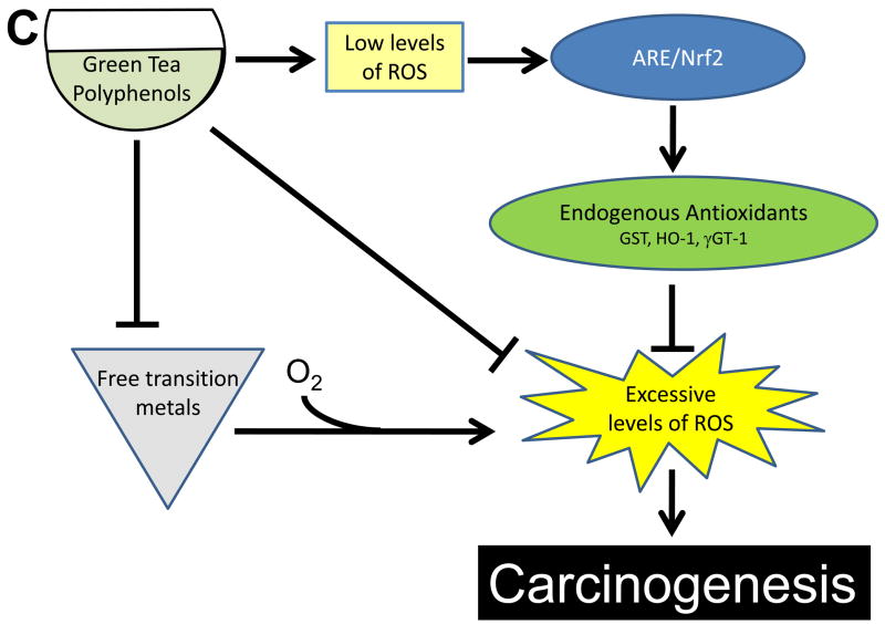
Although tea polyphenols have generally been regarded as antioxidants, the emerging evidence for the pro-oxidant effects of these compounds is interesting and raises many potential questions. Although a limited amount of data has shown that these pro-oxidant effects can occur in vivo, there have been no careful dose-response studies and there is no strong data relating these pro-oxidant effects and cancer prevention in vivo [64, 73]. It remains unclear how universal these effects might be. Pro-oxidant effects of EGCG have been reported in large xenograft tumors, but it has not been established if such effects occur in normal or hyperplastic tissue at low doses. Further at what point do such pro-oxidant effects transition from a potential beneficial effect to the hepatotoxic effects observed at higher doses of EGCG? Studies answering these questions are needed 1.) to determine appropriate doses for human intervention studies, 2.) to identify potentially responsive subjects, and 3.) to determine individual with increased sensitivity to EGCG-induced toxicity. One particularly interesting possibility is that tea polyphenols, at dietary levels, induce low levels of oxidative stress which “prime” endogenous antioxidant systems to deal with larger oxidative insults arising from ultraviolet light and other carcinogens (Fig. 4). At this point, this idea remains hypothetical and requires careful dose-response studies comparing induction of oxidative stress, induction of endogenous antioxidant systems, and prevention of carcinogenesis by tea polyphenols
Figure 4.
Proposed model for the mechanism by which the antioxidant and/or pro-oxidant effects tea polyphenols inhibit the carcinogenic process. ROS = reactive oxygen species, ARE = antioxidant response element, GST = glutathione S transferase, γGT = gamma glutamyltransferase.
It is likely that the cancer preventive effects of green tea polyphenols arise by a relatively small number of mechanisms that affect different aspects of the carcinogenic process. Tea polyphenol-induced antioxidative or pro-oxidative effects may be one such mechanism, and the importance of such effects may depend on the stage of carcinogenesis. For example increased endogenous antioxidant capacity may be more important prior to carcinogen exposure, whereas pro-oxidant cell killing effects may be more important in clearing transformed cells from the body and limiting tumor growth. Careful mechanistic studies in animal models representing different stages of carcinogenesis, and an integrated approach to analyzing the data from these studies will be essential to understanding when and to what extent the antioxidant or pro-oxidant effects of tea polyphenols are important to cancer prevention.
In summary, the cancer preventive effects of tea polyphenols have been demonstrated in a number of laboratory model systems. Similarly, the chemical antioxidative effects of these compounds have been well-studied. It remains for chemists, biochemists, and cancer prevention researchers to incorporate these data into a coherent picture of the role of tea polyphenol-mediated redox effects in cancer prevention.
Acknowledgments
This work was supported in part by the National Center for Complementary and Alternative Medicine Grant AT004678 (to JDL).
Footnotes
Abbreviations: 4-HNE, 4-hydroxynonenal; 8-OHdG, 8-hydroxydeoxyguanosine; AFB1, aflatoxin B1; AFB1-NAC, AFB1 mercapturic acid conjugate; B[a]P, benozo[a]pyrene; B[a]P-dG; B[a]P-adducted deoxyguanosine; EC, (−)-epicatechin; ECG, (−)-epicatechin-3-gallate; EGCG, (−)-epigallocatechin-3-gallate; EGC, (−)-epigallocatechin; EpRE, EGF, epidermal growth factor; electrophile responsive element; γH2A.X, phosphorylated histone 2A.X; GCG, (−)-gallocatechin-3-gallate; GSH, reduced glutathione; GST, glutathione S transferase; HAT, hydrogen atom transfer; IL, interleukin; IP, ionization potential; NAC, N acetylcysteine; PhO, semiquinone radical; ROS, reactive oxygen species; SET, single electron transfer; SOD, superoxide dismutase; Tag, C3(1)SV40 T,t antigen transgenic multiple mammary adenocarcinoma; TFdiG, theaflavin-3,3′-digallate; TGFβ, transforming growth factor β
Publisher's Disclaimer: This is a PDF file of an unedited manuscript that has been accepted for publication. As a service to our customers we are providing this early version of the manuscript. The manuscript will undergo copyediting, typesetting, and review of the resulting proof before it is published in its final citable form. Please note that during the production process errors may be discovered which could affect the content, and all legal disclaimers that apply to the journal pertain.
References
- 1.Yang CS, Wang X, Lu G, Picinich SC. Cancer prevention by tea: animal studies, molecular mechanisms and human relevance. Nat Rev Cancer. 2009;9:429–439. doi: 10.1038/nrc2641. [DOI] [PMC free article] [PubMed] [Google Scholar]
- 2.Balentine DA, Wiseman SA, Bouwens LC. The chemistry of tea flavonoids. Crit Rev Food Sci Nutr. 1997;37:693–704. doi: 10.1080/10408399709527797. [DOI] [PubMed] [Google Scholar]
- 3.Higdon JV, Frei B. Tea catechins and polyphenols: health effects, metabolism, and antioxidant functions. Crit Rev Food Sci Nutr. 2003;43:89–143. doi: 10.1080/10408690390826464. [DOI] [PubMed] [Google Scholar]
- 4.Yang CS, Maliakal P, Meng X. Inhibition of carcinogenesis by tea. Annu Rev Pharmacol Toxicol. 2002;42:25–54. doi: 10.1146/annurev.pharmtox.42.082101.154309. [DOI] [PubMed] [Google Scholar]
- 5.Siddiqui IA, Afaq F, Adhami VM, Ahmad N, Mukhtar H. Antioxidants of the beverage tea in promotion of human health. Antioxid Redox Signal. 2004;6:571–582. doi: 10.1089/152308604773934323. [DOI] [PubMed] [Google Scholar]
- 6.Grove KA, Lambert JD. Laboratory, Epidemiological, and Human Intervention Studies Show That Tea (Camellia sinensis) May Be Useful in the Prevention of Obesity. J Nutr. 2010 doi: 10.3945/jn.109.115972. [DOI] [PMC free article] [PubMed] [Google Scholar]
- 7.Yang CS, Sang S, Lambert JD, Hou Z, Ju J, Lu G. Possible mechanisms of the cancer-preventive activities of green tea. Mol Nutr Food Res. 2006;50:170–175. doi: 10.1002/mnfr.200500105. [DOI] [PubMed] [Google Scholar]
- 8.Khan N, Afaq F, Saleem M, Ahmad N, Mukhtar H. Targeting multiple signaling pathways by green tea polyphenol (−)-epigallocatechin-3-gallate. Cancer Res. 2006;66:2500–2505. doi: 10.1158/0008-5472.CAN-05-3636. [DOI] [PubMed] [Google Scholar]
- 9.Chen D, Daniel KG, Kuhn DJ, Kazi A, Bhuiyan M, Li L, Wang Z, Wan SB, Lam WH, Chan TH, Dou QP. Green tea and tea polyphenols in cancer prevention. Front Biosci. 2004;9:2618–2631. doi: 10.2741/1421. [DOI] [PubMed] [Google Scholar]
- 10.Tachibana H. Molecular basis for cancer chemoprevention by green tea polyphenol EGCG. Forum Nutr. 2009;61:156–169. doi: 10.1159/000212748. [DOI] [PubMed] [Google Scholar]
- 11.Hou Z, Sang S, You H, Lee MJ, Hong J, Chin KV, Yang CS. Mechanism of Action of (−)-Epigallocatechin-3-Gallate: Auto-oxidation-Dependent Inactivation of Epidermal Growth Factor Receptor and Direct Effects on Growth Inhibition in Human Esophageal Cancer KYSE 150 Cells. Cancer Res. 2005;65:8049–8056. doi: 10.1158/0008-5472.CAN-05-0480. [DOI] [PubMed] [Google Scholar]
- 12.Shen G, Xu C, Hu R, Jain MR, Nair S, Lin W, Yang CS, Chan JY, Kong AN. Comparison of (−)-epigallocatechin-3-gallate elicited liver and small intestine gene expression profiles between C57BL/6J mice and C57BL/6J/Nrf2 (−/−) mice. Pharm Res. 2005;22:1805–1820. doi: 10.1007/s11095-005-7546-8. [DOI] [PubMed] [Google Scholar]
- 13.Butt MS, Sultan MT. Green tea: nature’s defense against malignancies. Crit Rev Food Sci Nutr. 2009;49:463–473. doi: 10.1080/10408390802145310. [DOI] [PubMed] [Google Scholar]
- 14.Wright JS, Johnson ER, DiLabio GA. Predicting the activity of phenolic antioxidants: Theoretical method, analysis of substituent effects, and application to major families of antioxidants. J Am Chem Soc. 2001;123:1173–1183. doi: 10.1021/ja002455u. [DOI] [PubMed] [Google Scholar]
- 15.Moran JF, Klucas RV, Grayer RJ, Abian J, Becana M. Complexes of iron with phenolic compounds from soybean nodules and other legume tissues: Prooxidant and antioxidant properties. Free Rad Biol Med. 1997;22:861–870. doi: 10.1016/s0891-5849(96)00426-1. [DOI] [PubMed] [Google Scholar]
- 16.Prior RL, Wu XL, Schaich K. Standardized methods for the determination of antioxidant capacity and phenolics in foods and dietary supplements. Amer Chemical Soc. 2005:4290–4302. doi: 10.1021/jf0502698. [DOI] [PubMed] [Google Scholar]
- 17.Zhu NQ, Sang SM, Huang TC, Bai NS, Yang CS, Ho CT. Antioxidant chemistry of green tea catechins: Oxidation products of (−)-epigallocatechin gallate and (−)-epigallocatechin with peroxidase. J Food Lipids. 2000;7:275–282. [Google Scholar]
- 18.Halliwell B. Are polyphenols antioxidants or pro-oxidants? What do we learn from cell culture and in vivo studies? Arch Biochem Biophys. 2008;476:107–112. doi: 10.1016/j.abb.2008.01.028. [DOI] [PubMed] [Google Scholar]
- 19.Jovanovic SV, Hara Y, Steenken S, Simic MG. Antioxidant potential of gallocatechins- a pulse-radiolysis and laser photolysis study. J Am Chem Soc. 1995;117:9881–9888. [Google Scholar]
- 20.Symons MCR, Gutteridge JMC. Free radicals and iron: chemistry, biology, and medicine. Oxford University Press; Oxford; New York: 1998. [Google Scholar]
- 21.Cilliers JJL, Singleton VL. Nonenzymic Autoxidative Reactions of Caffeic Acid in Wine. Am J Enol Viticulture. 1990;41:84–86. [Google Scholar]
- 22.Singleton VL, Cilliers JJL. Phenolic Browning-a Perspective from Grape and Wine Research. Enzymatic Browning and Its Prevention. 1995;600:23–48. [Google Scholar]
- 23.Wildenradt HL, Singleton VL. Production of aldehydes as a result of oxidation of polyphenolic compounds and its relation to wine aging. Am J Enol Viticulture. 1974;25:119–126. [Google Scholar]
- 24.Danilewicz JC. Review of reaction mechanisms of oxygen and proposed intermediate reduction products in wine: Central role of iron and copper. Am J Enol Viticulture. 2003;54:73–85. [Google Scholar]
- 25.Miller DM, Buettner GR, Aust SD. Transition-metals as catalysts of autoxidation reactions. Free Rad Biol Med. 1990;8:95–108. doi: 10.1016/0891-5849(90)90148-c. [DOI] [PubMed] [Google Scholar]
- 26.Bandy B, Davison AJ. Interactions between metals, ligands, and oxygen in the autoxidation of 6-hydroxydopamine–mechanisms by which metal chelation enhances inhibition by superoxide-dismutase. Arch Biochem Biophys. 1987;259:305–315. doi: 10.1016/0003-9861(87)90497-8. [DOI] [PubMed] [Google Scholar]
- 27.Gee P, Davison AJ. 6-hydroxydopamine does not reduce molecular-oxygen directly, but requires a coreductant. Arch Biochem Biophys. 1984;231:164–168. doi: 10.1016/0003-9861(84)90373-4. [DOI] [PubMed] [Google Scholar]
- 28.Bandy B, Walter PB, Moon J, Davison AJ. Reaction of oxygen with 6-hydroxydopamine catalyzed by Cu, Fe, Mn, and V complexes: Identification of a thermodynamic window for effective metal catalysis. Arch Biochem Biophys. 2001;389:22–30. doi: 10.1006/abbi.2001.2285. [DOI] [PubMed] [Google Scholar]
- 29.Gee P, Davison AJ. Autoxidation of 6-hydroxydopamine involves a ternary reductant-metal-oxygen compex. Ircs Medical Science-Biochemistry. 1984;12:127–127. [Google Scholar]
- 30.Sang S, Lambert JD, Hong J, Tian S, Lee MJ, Stark RE, Ho CT, Yang CS. Synthesis and structure identification of thiol conjugates of (−)-epigallocatechin gallate and their urinary levels in mice. Chem Res Toxicol. 2005;18:1762–1769. doi: 10.1021/tx050151l. [DOI] [PubMed] [Google Scholar]
- 31.Hagerman AE, Dean RT, Davies MJ. Radical chemistry of epigallocatechin gallate and its relevance to protein damage. Arch Biochem Biophys. 2003;414:115–120. doi: 10.1016/s0003-9861(03)00158-9. [DOI] [PubMed] [Google Scholar]
- 32.Afanasev IB, Dorozhko AI, Brodskii AV, Kostyuk VA, Potapovitch AI. CHELATING AND FREE-Radical scavenging mechanisms of inhibitory-action of rutin and quercetin in lipid-peroxidation. Biochem Pharmacol. 1989;38:1763–1769. doi: 10.1016/0006-2952(89)90410-3. [DOI] [PubMed] [Google Scholar]
- 33.Mira L, Fernandez MT, Santos M, Rocha R, Florencio MH, Jennings KR. Interactions of flavonoids with iron and copper ions: A mechanism for their antioxidant activity. Free Rad Res. 2002;36:1199–1208. doi: 10.1080/1071576021000016463. [DOI] [PubMed] [Google Scholar]
- 34.Minotti G, Aust SD. The Role of Iron in the Initiation of Lipid-Peroxidation. Chem Phys Lipids. 1987;44:191–208. doi: 10.1016/0009-3084(87)90050-8. [DOI] [PubMed] [Google Scholar]
- 35.Fukuzawa K, Tadokoro T, Kishikawa K, Mukai K, Gebicki JM. Site-specific induction of lipid-peroxidation by iron in charged micelles. Arch Biochem Biophys. 1988;260:146–152. doi: 10.1016/0003-9861(88)90435-3. [DOI] [PubMed] [Google Scholar]
- 36.Girotti AW, Thomas JP. Superoxide and Hydrogen-Peroxide Dependent Lipid-Peroxidation in Intact and Triton-Dispersed Erythrocyte-Membranes. Biochem Biophys Res Comm. 1984;118:474–480. doi: 10.1016/0006-291x(84)91327-5. [DOI] [PubMed] [Google Scholar]
- 37.Girotti AW, Thomas JP. Damaging Effects of Oxygen Radicals on Resealed Erythrocyte-Ghosts. J Biol Chem. 1984;259:1744–1752. [PubMed] [Google Scholar]
- 38.Senthil Kumaran V, Arulmathi K, Srividhya R, Kalaiselvi P. Repletion of antioxidant status by EGCG and retardation of oxidative damage induced macromolecular anomalies in aged rats. Exp Gerontol. 2008;43:176–183. doi: 10.1016/j.exger.2007.10.017. [DOI] [PubMed] [Google Scholar]
- 39.Srividhya R, Jyothilakshmi V, Arulmathi K, Senthilkumaran V, Kalaiselvi P. Attenuation of senescence-induced oxidative exacerbations in aged rat brain by (−)-epigallocatechin-3-gallate. Int J Dev Neurosci. 2008;26:217–223. doi: 10.1016/j.ijdevneu.2007.12.003. [DOI] [PubMed] [Google Scholar]
- 40.Rietveld A, Wiseman S. Antioxidant effects of tea: evidence from human clinical trials. J Nutr. 2003;133:3285S–3292S. doi: 10.1093/jn/133.10.3285S. [DOI] [PubMed] [Google Scholar]
- 41.Yang CH, Lin CY, Yang JH, Liou SY, Li PC, Chien CT. Supplementary catechins attenuate cooking-oil-fumes-induced oxidative stress in rat lung. Chin J Physiol. 2009;52:151–159. doi: 10.4077/cjp.2009.amh022. [DOI] [PubMed] [Google Scholar]
- 42.Kaur S, Greaves P, Cooke DN, Edwards R, Steward WP, Gescher AJ, Marczylo TH. Breast cancer prevention by green tea catechins and black tea theaflavins in the C3(1) SV40 T,t antigen transgenic mouse model is accompanied by increased apoptosis and a decrease in oxidative DNA adducts. J Agric Food Chem. 2007;55:3378–3385. doi: 10.1021/jf0633342. [DOI] [PubMed] [Google Scholar]
- 43.Inami S, Takano M, Yamamoto M, Murakami D, Tajika K, Yodogawa K, Yokoyama S, Ohno N, Ohba T, Sano J, Ibuki C, Seino Y, Mizuno K. Tea catechin consumption reduces circulating oxidized low-density lipoprotein. Int Heart J. 2007;48:725–732. doi: 10.1536/ihj.48.725. [DOI] [PubMed] [Google Scholar]
- 44.Hsu SP, Wu MS, Yang CC, Huang KC, Liou SY, Hsu SM, Chien CT. Chronic green tea extract supplementation reduces hemodialysis-enhanced production of hydrogen peroxide and hypochlorous acid, atherosclerotic factors, and proinflammatory cytokines. Am J Clin Nutr. 2007;86:1539–1547. doi: 10.1093/ajcn/86.5.1539. [DOI] [PubMed] [Google Scholar]
- 45.Eichenberger P, Colombani PC, Metter S. Effects of 3-week consumption of green tea extracts on whole-body metabolism during cycling exercise in endurance-trained men. Int J Vitam Nutr Res. 2009;79:24–33. doi: 10.1024/0300-9831.79.1.24. [DOI] [PubMed] [Google Scholar]
- 46.Schwartz JL, Baker V, Larios E, Chung FL. Molecular and cellular effects of green tea on oral cells of smokers: A pilot study. Mol Nutr Food Res. 2005;49:43–51. doi: 10.1002/mnfr.200400031. [DOI] [PubMed] [Google Scholar]
- 47.Hakim IA, Harris RB, Brown S, Chow HH, Wiseman S, Agarwal S, Talbot W. Effect of increased tea consumption on oxidative DNA damage among smokers: a randomized controlled study. J Nutr. 2003;133:3303S–3309S. doi: 10.1093/jn/133.10.3303S. [DOI] [PubMed] [Google Scholar]
- 48.Tulayakul P, Dong KS, Li JY, Manabe N, Kumagai S. The effect of feeding piglets with the diet containing green tea extracts or coumarin on in vitro metabolism of aflatoxin B1 by their tissues. Toxicon. 2007;50:339–348. doi: 10.1016/j.toxicon.2007.04.005. [DOI] [PubMed] [Google Scholar]
- 49.Maliakal PP, Coville PF, Wanwimolruk S. Tea consumption modulates hepatic drug metabolizing enzymes in Wistar rats. J Pharm Pharmacol. 2001;53:569–577. doi: 10.1211/0022357011775695. [DOI] [PubMed] [Google Scholar]
- 50.Liu TT, Liang NS, Li Y, Yang F, Lu Y, Meng ZQ, Zhang LS. Effects of long-term tea polyphenols consumption on hepatic microsomal drug-metabolizing enzymes and liver function in Wistar rats. World J Gastroenterol. 2003;9:2742–2744. doi: 10.3748/wjg.v9.i12.2742. [DOI] [PMC free article] [PubMed] [Google Scholar]
- 51.Yuan JH, Li YQ, Yang XY. Inhibition of epigallocatechin gallate on orthotopic colon cancer by upregulating the Nrf2-UGT1A signal pathway in nude mice. Pharmacol. 2007;80:269–278. doi: 10.1159/000106447. [DOI] [PubMed] [Google Scholar]
- 52.Na HK, Surh YJ. Modulation of Nrf2-mediated antioxidant and detoxifying enzyme induction by the green tea polyphenol EGCG. Food Chem Toxicol. 2008;46:1271–1278. doi: 10.1016/j.fct.2007.10.006. [DOI] [PubMed] [Google Scholar]
- 53.Khan SA, Priyamvada S, Khan W, Khan S, Farooq N, Yusufi AN. Studies on the protective effect of green tea against cisplatin induced nephrotoxicity. Pharmacol Res. 2009;60:382–391. doi: 10.1016/j.phrs.2009.07.007. [DOI] [PubMed] [Google Scholar]
- 54.Tang L, Tang M, Xu L, Luo H, Huang T, Yu J, Zhang L, Gao W, Cox SB, Wang JS. Modulation of aflatoxin biomarkers in human blood and urine by green tea polyphenols intervention. Carcinogenesis. 2008;29:411–417. doi: 10.1093/carcin/bgn008. [DOI] [PubMed] [Google Scholar]
- 55.Chow HH, Hakim IA, Vining DR, Crowell JA, Tome ME, Ranger-Moore J, Cordova CA, Mikhael DM, Briehl MM, Alberts DS. Modulation of human glutathione s-transferases by polyphenon e intervention. Cancer Epidemiol Biomarkers Prev. 2007;16:1662–1666. doi: 10.1158/1055-9965.EPI-06-0830. [DOI] [PubMed] [Google Scholar]
- 56.Sang S, Lee MJ, Hou Z, Ho CT, Yang CS. Stability of Tea Polyphenol (−)-Epigallocatechin-3-gallate and Formation of Dimers and Epimers under Common Experimental Conditions. J Agric Food Chem. 2005;53:9478–9484. doi: 10.1021/jf0519055. [DOI] [PubMed] [Google Scholar]
- 57.Hong J, Lu H, Meng X, Ryu JH, Hara Y, Yang CS. Stability, cellular uptake, biotransformation, and efflux of tea polyphenol (−)-epigallocatechin-3-gallate in HT-29 human colon adenocarcinoma cells. Cancer Res. 2002;62:7241–7246. [PubMed] [Google Scholar]
- 58.Sang SM, Lee MJ, Hou Z, Ho CT, Yang CS. Stability of tea polyphenol (−)-epigallocatechin-3-gallate and formation of dimers and epimers under common experimental conditions. J Ag Food Chem. 2005;53:9478–9484. doi: 10.1021/jf0519055. [DOI] [PubMed] [Google Scholar]
- 59.Oikawa S, Furukawaa A, Asada H, Hirakawa K, Kawanishi S. Catechins induce oxidative damage to cellular and isolated DNA through the generation of reactive oxygen species. Free Radic Res. 2003;37:881–890. doi: 10.1080/1071576031000150751. [DOI] [PubMed] [Google Scholar]
- 60.Yang GY, Liao J, Li C, Chung J, Yurkow EJ, Ho CT, Yang CS. Effect of black and green tea polyphenols on c-jun phosphorylation and H(2)O(2) production in transformed and non-transformed human bronchial cell lines: possible mechanisms of cell growth inhibition and apoptosis induction. Carcinogenesis. 2000;21:2035–2039. doi: 10.1093/carcin/21.11.2035. [DOI] [PubMed] [Google Scholar]
- 61.Nakagawa H, Hasumi K, Woo JT, Nagai K, Wachi M. Generation of hydrogen peroxide primarily contributes to the induction of Fe(II)-dependent apoptosis in Jurkat cells by (−)-epigallocatechin gallate. Carcinogenesis. 2004;25:1567–1574. doi: 10.1093/carcin/bgh168. [DOI] [PubMed] [Google Scholar]
- 62.Vittal R, Selvanayagam ZE, Sun Y, Hong J, Liu F, Chin KV, Yang CS. Gene expression changes induced by green tea polyphenol (−)-epigallocatechin-3-gallate in human bronchial epithelial 21BES cells analyzed by DNA microarray. Mol Cancer Ther. 2004;3:1091–1099. [PubMed] [Google Scholar]
- 63.Elbling L, Weiss RM, Teufelhofer O, Uhl M, Knasmueller S, Schulte-Hermann R, Berger W, Micksche M. Green tea extract and (−)-epigallocatechin-3-gallate, the major tea catechin, exert oxidant but lack antioxidant activities. Faseb J. 2005;19:807–809. doi: 10.1096/fj.04-2915fje. [DOI] [PubMed] [Google Scholar]
- 64.Hou Z, Xiao H, Lambert J, You H, Yang CS. Green tea polyphenol, (−)-epigallocatechin-3-gallate, induces oxidative stress and DNA damage in cancer cell lines, xenograft tumors, and mouse liver. ProcAmer Assoc Cancer Res. 2006 [Google Scholar]
- 65.Liang YC, Lin-shiau SY, Chen CF, Lin JK. Suppression of extracellular signals and cell proliferation through EGF receptor binding by (−)-epigallocatechin gallate in human A431 epidermoid carcinoma cells. J Cell Biochem. 1997;67:55–65. doi: 10.1002/(sici)1097-4644(19971001)67:1<55::aid-jcb6>3.0.co;2-v. [DOI] [PubMed] [Google Scholar]
- 66.Masuda M, Suzui M, Weinstein IB. Effects of epigallocatechin-3-gallate on growth, epidermal growth factor receptor signaling pathways, gene expression, and chemosensitivity in human head and neck squamous cell carcinoma cell lines. Clin Cancer Res. 2001;7:4220–4229. [PubMed] [Google Scholar]
- 67.Muzolf-Panek M, Gliszczynska-Swiglo A, de Haan L, Aarts JM, Szymusiak H, Vervoort JM, Tyrakowska B, Rietjens IM. Role of catechin quinones in the induction of EpRE-mediated gene expression. Chem Res Toxicol. 2008;21:2352–2360. doi: 10.1021/tx8001498. [DOI] [PubMed] [Google Scholar]
- 68.Sherwood L. Human Physiology: From Cells to Systems. Wadsworth Publishing Co; Belmont, CA: 1997. [Google Scholar]
- 69.Chow HH, Cai Y, Hakim IA, Crowell JA, Shahi F, Brooks CA, Dorr RT, Hara Y, Alberts DS. Pharmacokinetics and safety of green tea polyphenols after multiple-dose administration of epigallocatechin gallate and polyphenon E in healthy individuals. Clin Cancer Res. 2003;9:3312–3319. [PubMed] [Google Scholar]
- 70.Lee MJ, Maliakal P, Chen L, Meng X, Bondoc FY, Prabhu S, Lambert G, Mohr S, Yang CS. Pharmacokinetics of tea catechins after ingestion of green tea and (−)-epigallocatechin-3-gallate by humans: formation of different metabolites and individual variability. Cancer Epidemiol Biomarkers Prev. 2002;11:1025–1032. [PubMed] [Google Scholar]
- 71.Lambert JD, Lee MJ, Diamond L, Ju J, Hong J, Bose M, Newmark HL, Yang CS. Dose-dependent levels of epigallocatechin-3-gallate in human colon cancer cells and mouse plasma and tissues. Drug Metab Dispos. 2006;34:8–11. doi: 10.1124/dmd.104.003434. [DOI] [PubMed] [Google Scholar]
- 72.Galati G, Lin A, Sultan AM, O’Brien JP. Cellular and in vivo hepatotoxicity caused by green tea phenolic acids and catechins. Free Radic Biol Med. 2006;40:570–580. doi: 10.1016/j.freeradbiomed.2005.09.014. [DOI] [PubMed] [Google Scholar]
- 73.Lambert JD, Kennett MJ, Sang S, Reuhl KR, Ju J, Yang CS. Hepatotoxicity of high oral dose (−)-epigallocatechin-3-gallate in mice. Food Chem Toxicol. 2010;48:409–416. doi: 10.1016/j.fct.2009.10.030. [DOI] [PMC free article] [PubMed] [Google Scholar]
- 74.Bettuzzi S, Brausi M, Rizzi F, Castagnetti G, Peracchia G, Corti A. Chemoprevention of human prostate cancer by oral administration of green tea catechins in volunteers with high-grade prostate intraepithelial neoplasia: a preliminary report from a one-year proof-of-principle study. Cancer Res. 2006;66:1234–1240. doi: 10.1158/0008-5472.CAN-05-1145. [DOI] [PubMed] [Google Scholar]
- 75.Chow HH, Cai Y, Alberts DS, Hakim I, Dorr R, Shahi F, Crowell JA, Yang CS, Hara Y. Phase I pharmacokinetic study of tea polyphenols following single-dose administration of epigallocatechin gallate and polyphenon E. Cancer Epidemiol Biomarkers Prev. 2001;10:53–58. [PubMed] [Google Scholar]
- 76.Mazzanti G, Menniti-Ippolito F, Moro PA, Cassetti F, Raschetti R, Santuccio C, Mastrangelo S. Hepatotoxicity from green tea: a review of the literature and two unpublished cases. Eur J Clin Pharmacol. 2009;65:331–341. doi: 10.1007/s00228-008-0610-7. [DOI] [PubMed] [Google Scholar]
- 77.Stevens T, Qadri A, Zein NN. Two patients with acute liver injury associated with use of the herbal weight-loss supplement hydroxycut. Ann Intern Med. 2005;142:477–478. doi: 10.7326/0003-4819-142-6-200503150-00026. [DOI] [PubMed] [Google Scholar]
- 78.Bonkovsky HL. Hepatotoxicity associated with supplements containing Chinese green tea (Camellia sinensis) Ann Intern Med. 2006;144:68–71. doi: 10.7326/0003-4819-144-1-200601030-00020. [DOI] [PubMed] [Google Scholar]
- 79.Jimenez-Saenz M, Martinez-Sanchez Mdel C. Acute hepatitis associated with the use of green tea infusions. J Hepatol. 2006;44:616–617. doi: 10.1016/j.jhep.2005.11.041. [DOI] [PubMed] [Google Scholar]


