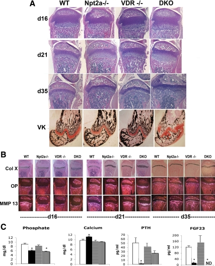
Figure 3.
Histological analyses of the growth plate of VDR and Npt2a null mice. A, Growth plate morphology was evaluated using H&E-stained sections of the tibia obtained at d 16, 21, and 35. Von Kossa staining of the distal femur was performed at 35 d of age to permit evaluation of mineralized matrix. VK, Von Kossa staining. B, In situ hybridization analyses were performed to evaluate markers of hypertrophic chondrocyte differentiation at 16, 21, and 35 d of age. Mice were weaned onto a 2% calcium, 1.25% phosphorus, and 20% lactose diet at 18 d of age. Col X, Collagen X; OP, osteopontin; MMP, matrix metalloproteinase. C, Serum biochemistries at 35 d of age. White bars, WT; black bars, Npt2a−/−; horizontally striped bars, VDR−/−; vertically striped bars, DKO. Data are representative of those obtained from three to six mice of each age and genotype. VDR−/−, VDR null; ND, not detectable.

