

Abstract
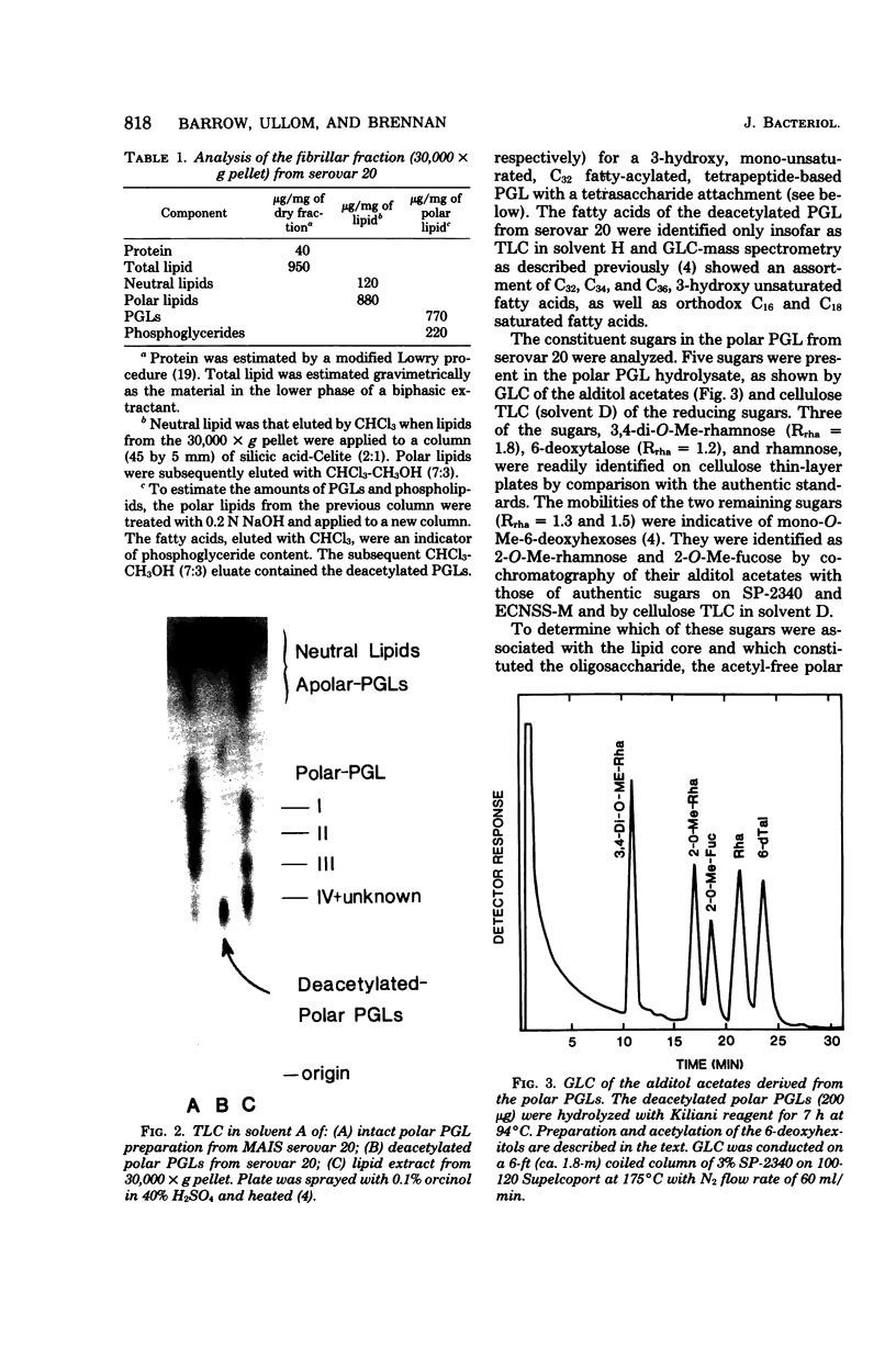
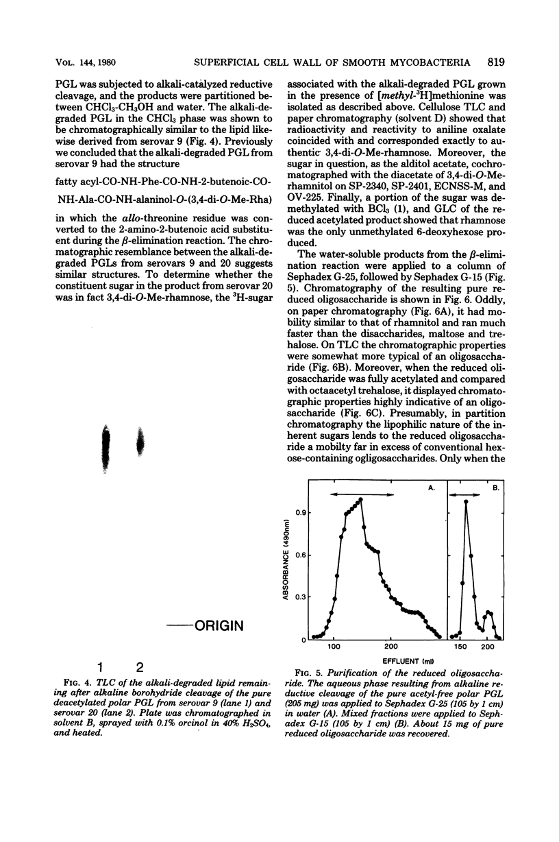
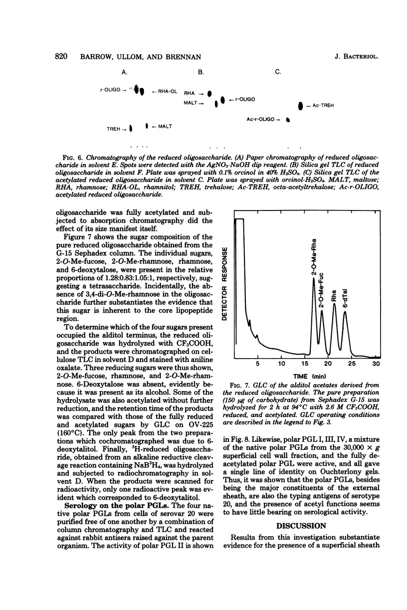
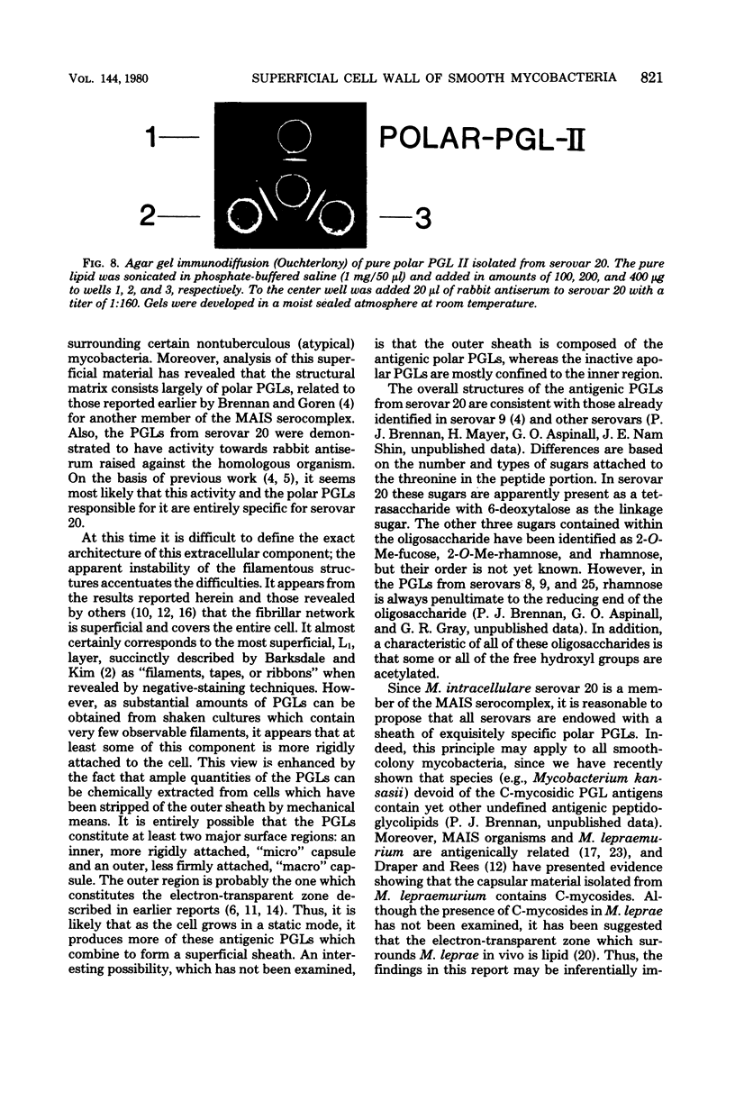
The most superficial cell wall layer present in smooth-colony-forming mycobacteria was isolated from serovar 20 of the Mycobacterium avium-Mycobacterium intracellulare-Mycobacterium scrofulaceum (MAIS) serocomplex and examined chemically and by electron microscopy. Most (70 to 80%) of the fibrillar material consisted of an array of serologically active, acetylated C-myosidic peptidoglycoplipids with the basic structure (formula, see text) but in which the location of acetyl groups and the arrangement of monosaccharides have not been defined. Apparently, all serovars within the MAIS complex are characterized by structurally related superficies in which the monoglycosyl-lipopeptide portion is invariable but the oligosaccharide attachment is peculiar to each serovar. These unique inert structures may be an important factor in shielding the pathogen within phagolysosomes from lysosomal enzymes.
Full text
PDF
Images in this article


Selected References
These references are in PubMed. This may not be the complete list of references from this article.
- BINFORD C. H. Studies on a Mycobacterium obtained from the golden hamster (Cricetus auratus) after inoculation with lepromatous tissue. Lab Invest. 1962 Nov;11:942–955. [PubMed] [Google Scholar]
- Barksdale L., Kim K. S. Mycobacterium. Bacteriol Rev. 1977 Mar;41(1):217–372. doi: 10.1128/br.41.1.217-372.1977. [DOI] [PMC free article] [PubMed] [Google Scholar]
- Brennan P. J., Goren M. B. Structural studies on the type-specific antigens and lipids of the mycobacterium avium. Mycobacterium intracellulare. Mycobacterium scrofulaceum serocomplex. Mycobacterium intracellulare serotype 9. J Biol Chem. 1979 May 25;254(10):4205–4211. [PubMed] [Google Scholar]
- Brennan P. J., Souhrada M., Ullom B., McClatchy J. K., Goren M. B. Identification of atypical mycobacteria by thin-layer chromatography of their surface antigens. J Clin Microbiol. 1978 Oct;8(4):374–379. doi: 10.1128/jcm.8.4.374-379.1978. [DOI] [PMC free article] [PubMed] [Google Scholar]
- Brown C. A., Draper P. An electron-microscope study of rat fibroblasts infected with Mycobacterium lepraemurium. J Pathol. 1970 Sep;102(1):21–26. doi: 10.1002/path.1711020105. [DOI] [PubMed] [Google Scholar]
- CHAPMAN G. B., HANKS J. H., WALLACE J. H. An electron microscope study of the disposition and fine structure of Mycobacterium lepraemurium in mouse spleen. J Bacteriol. 1959 Feb;77(2):205–211. doi: 10.1128/jb.77.2.205-211.1959. [DOI] [PMC free article] [PubMed] [Google Scholar]
- Change Y. T., Andersen R. N., Vaituzis Z. Growth of Mycobacterium lepraemurium in cultures of mouse peritoneal macrophages. J Bacteriol. 1967 Mar;93(3):1119–1131. doi: 10.1128/jb.93.3.1119-1131.1967. [DOI] [PMC free article] [PubMed] [Google Scholar]
- Draper P., Rees R. J. Electron-transparent zone of mycobacteria may be a defence mechanism. Nature. 1970 Nov 28;228(5274):860–861. doi: 10.1038/228860a0. [DOI] [PubMed] [Google Scholar]
- Draper P., Rees R. J. The nature of the electron-transparent zone that surrounds Mycobacterium lepraemurium inside host cells. J Gen Microbiol. 1973 Jul;77(1):79–87. doi: 10.1099/00221287-77-1-79. [DOI] [PubMed] [Google Scholar]
- Draper P. The mycoside capsule of Mycobacterium Avium 357. J Gen Microbiol. 1974 Aug;83(2):431–433. doi: 10.1099/00221287-83-2-431. [DOI] [PubMed] [Google Scholar]
- Edwards R. P. Electron-microscope illustrations of division in Mycobacterium leprae. J Med Microbiol. 1970 Aug;3(3):493–499. doi: 10.1099/00222615-3-3-493. [DOI] [PubMed] [Google Scholar]
- Kim K. S., Salton M. R., Barksdale L. Ultrastructure of superficial mycosidic integuments of Mycobacterium sp. J Bacteriol. 1976 Feb;125(2):739–743. doi: 10.1128/jb.125.2.739-743.1976. [DOI] [PMC free article] [PubMed] [Google Scholar]
- Kwapinski J. B., Kwapinski E. H. Immunological reactions of Mycobacterium leprae and Mycobacterium lepraemurium grown in cayman. Can J Microbiol. 1973 Jun;19(6):764–766. doi: 10.1139/m73-124. [DOI] [PubMed] [Google Scholar]
- LOWRY O. H., ROSEBROUGH N. J., FARR A. L., RANDALL R. J. Protein measurement with the Folin phenol reagent. J Biol Chem. 1951 Nov;193(1):265–275. [PubMed] [Google Scholar]
- Laneelle G., Asselineau J. Structure d'un glycoside de peptidolipide isolé d'une mycobactérie. Eur J Biochem. 1968 Sep 24;5(4):487–491. doi: 10.1111/j.1432-1033.1968.tb00396.x. [DOI] [PubMed] [Google Scholar]
- NISHIURA M. The electron microscopic basis of the pathology of leprosy. Int J Lepr. 1960 Oct-Dec;28:357–400. [PubMed] [Google Scholar]
- Schaefer W. B. Serologic identification and classification of the atypical mycobacteria by their agglutination. Am Rev Respir Dis. 1965 Dec;92(6):85–93. doi: 10.1164/arrd.1965.92.6P2.85. [DOI] [PubMed] [Google Scholar]
- Stanford J. L. An immunodiffusion analysis of Mycobacterium lepraemurium Marchoux and Sorel. J Med Microbiol. 1973 Nov;6(4):435–439. doi: 10.1099/00222615-6-4-435. [DOI] [PubMed] [Google Scholar]
- Stellner K., Saito H., Hakomori S. I. Determination of aminosugar linkages in glycolipids by methylation. Aminosugar linkages of ceramide pentasaccharides of rabbit erythrocytes and of Forssman antigen. Arch Biochem Biophys. 1973 Apr;155(2):464–472. doi: 10.1016/0003-9861(73)90138-0. [DOI] [PubMed] [Google Scholar]
- Voiland A., Bruneteau M., Michel G. Etude du mycoside C 2 de Mycobacterium avium. Détermination de la structure. Eur J Biochem. 1971 Jul 29;21(2):285–291. doi: 10.1111/j.1432-1033.1971.tb01468.x. [DOI] [PubMed] [Google Scholar]
- YAMAMOTO T., NISHIURA M., HARADA N., IMAEDA T. Electron microscopy of Mycobacterium leprae murium in ultra-thin sections of murine leprosy lesions. Int J Lepr. 1958 Apr-Jun;26(2):111–114. [PubMed] [Google Scholar]
- Yoshizumi M. O., Asbury A. K. Intra-axonal bacilli in lepromatous leprosy. A light and electron microscopic study. Acta Neuropathol. 1974 Feb 7;27(1):1–10. doi: 10.1007/BF00687235. [DOI] [PubMed] [Google Scholar]