Abstract
Background
Complications are frequent with osteoarticular allografts, and their long-term survivorship in the distal femur is unclear. Thus, the benefits of osteoarticular allografting remain controversial.
Questions/purposes
We therefore determined the frequency of complications in osteoarticular allografts of the distal femur relative to their potential long-term survival.
Methods
We retrospectively reviewed 26 patients who had osteoarticular allograft reconstruction of the distal femur after resection of a malignant or aggressive benign tumor of bone. The minimum followup was 15 months (average, 156 months; range, 15–283 months) for all patients and 98 months (average, 191 months; range, 98–283 months) for the surviving patients.
Results
At last followup, 16 of the 26 original allografts were still in place. The overall 5-year and 10-year allograft survival rates were 69% and 63%, respectively. The 5-year and 10-year survival rates of the joint surface were 79% and 65%, respectively. Eleven patients retained their original osteoarticular allograft without a resurfacing procedure, and nine had been converted to allograft-prosthetic composites. Five patients were converted to megaprostheses and one had an amputation for local recurrence. At last followup, 25 of 26 patients retained a functional limb.
Conclusions
Osteoarticular allograft reconstructions of the distal femur can provide long term survival and restore function but the risk of complications and their physical and monetary costs for patients are not trivial. Lacking the benefit of improved soft tissue attachments inherent in other anatomic sites, we believe this option is most appropriate for restoring bone stock in young patients with expectations of long-term survival.
Level of Evidence
Level IV, therapeutic study. See the Guidelines for Authors for a complete description of levels of evidence.
Introduction
The distal femur is one of the most common sites for primary destructive neoplasms of bone. Options for restoring a functional lower extremity after wide resection at this site include megaprostheses [2–4, 6, 14–19, 26–30], osteoarticular allografts [2, 5, 10–12, 21–25, 32], and allograft-prosthetic composites (APC) [1, 16, 17, 35], each with unique advantages and disadvantages. Osteoarticular allografts offer the potential benefit of biologic bone union, soft tissue attachment, restoration of bone stock, and preservation of the proximal tibial physis in skeletally immature patients. However, they often require prolonged healing times of 1 year to 4 years [7, 9, 21], are associated with fracture rates of 17% to 45% [20, 28, 31, 34], are of limited supply, carry some potential risk of disease transmission, and have perioperative infection rates of 5% to 10% [20, 22]. Ligamentous reconstruction is technically demanding and some reconstructed joints progress to arthritis over time [18–21]. Particular to the distal femur, there is no soft tissue attachment advantage as in sites such as the proximal tibia and femur. Megaprostheses, most often cemented, provide immediate fixation and rapid return to weightbearing, are less technically demanding to implant, and have no risk of disease transmission; reported survival rates of megaprostheses range from 87% at 3 years [33] to 59% at 5 years [13] to 50% at 25 years [27]. Complications include aseptic loosening (6% to 84% at 5 to 10 years), infection (7% to 15%), component failure rates (5% to 20%), periprosthetic fracture in 5% to 15% of patients, and dissociation of modular components [2, 4, 6, 13, 15–19, 26–30].
Allograft-prosthetic composites share the benefits and liabilities of the other two. They restore bone stock and allow biologic union while providing a stable knee; however, they are technically difficult reconstructions that sacrifice the opposite physis and have some risk of disease transmission, nonunion, and fracture [1, 11, 16, 17, 35]. Mankin et al. [18] reported good or excellent results in 77% of 98 patients treated with APCs; however, this was an overall rating and results were not reported by anatomic location. Several reports dedicated exclusively to the long term outcomes of osteoarticular allografts of the distal femur [20, 22–24] suggest relatively high rates of infection and joint degeneration.
To confirm the findings of these previous papers we determined the long-term viability of distal femoral allografts by evaluating (1) allograft survival; (2) preservation of the joint surface; and (3) the frequency and nature of complications in a modern cohort of patients treated at a single institution.
Patients and Methods
We retrospectively reviewed prospectively collected data from our institution’s orthopaedic oncology database and identified 26 patients with 26 osteoarticular allograft reconstructions of the distal femur performed between January 1985 and January 1999 (Table 1). We chose this time period because MRI images were available for all patients and modern chemotherapy regimens were being used. The 18 female and eight male patients had an average age at the time of surgery of 23 years (range, 10–58 years). Seventeen patients were skeletally mature at the time of reconstruction, and nine were skeletally immature. The most frequent histologic diagnosis was osteosarcoma (19 patients); other diagnoses were giant cell tumor (three patients), malignant fibrous histiocytoma (two patients), chondrosarcoma (one patient), and angiosarcoma (one patient). According to the Musculoskeletal Tumor Society [MSTS] staging system described by Enneking et al. [8], 19 neoplasms were classified as Stage IIB, three as IB, and one as IA; three tumors were Stage III with metastatic disease noted at diagnosis. Eighteen patients were treated with preoperative chemotherapy and two with postoperative chemotherapy. No patient received radiation therapy. The minimal followup was 15 months (average, 156 months; range, 15–283 months). Six patients, five with osteosarcoma and one with malignant fibrous histiocytoma, died from their disease at an average of 32 months (range, 15–66 months) after surgery, and one patient died at 174 months from reasons not related to the primary disease. When the seven patients who died of their disease were excluded, minimal followup increased to 98 months (average, 191 months; range, 98–283 months). No patients were lost to followup, and none were recalled specifically for this report. We received prior Institutional Review Board approval.
Table 1.
Osteoarticular allografts in 26 patients: diagnoses, complications, treatment, and outcomes
| Patient information | Complications | |||||||||
|---|---|---|---|---|---|---|---|---|---|---|
| Patient number | Diagnosis | Chemotherapy | Superficial infection | Deep infection | Fracture | Resorption | DJD | Nonunion | Instability | Local recurrence |
| 1 | Osteosarcoma, NOS | Yes | No | No | No | No | No | No | No | No |
| 2 | Osteosarcoma, NOS | Yes | No | No | No | No | No | No | No | No |
| 3 | Giant cell tumor | No | No | No | No | No | No | No | No | No |
| 4 | Osteosarcoma, telangiectatic | Yes | No | No | No | No | No | No | No | No |
| 5 | Osteosarcoma, NOS | Yes | No | No | No | No | No | No | No | No |
| 6 | Osteosarcoma, osteoblastic | Yes | No | No | No | No | No | No | No | No |
| 7 | Osteosarcoma, osteoblastic | Yes | No | No | No | No | No | No | No | No |
| 8 | Giant cell tumor | No | No | No | Yes | No | Yes | No | No | No |
| 9 | Giant cell tumor | No | No | No | No | No | Yes | No | No | No |
| 10 | Osteosarcoma, periosteal | Yes | No | No | No | No | Yes | No | No | No |
| 11 | Osteosarcoma, NOS | Yes | No | No | No | No | Yes | No | No | No |
| 12 | Osteosarcoma, NOS | Yes | No | No | Yes | No | Yes | No | No | No |
| 13 | Chondrosarcoma, NOS | No | No | No | No | No | Yes | No | No | No |
| 14 | Osteosarcoma, NOS | Yes | No | No | No | No | Yes | No | Yes | No |
| 15 | Osteosarcoma | Yes | No | No | No | No | No | Yes | No | No |
| 16 | Angiosarcoma | No | No | No | No | Yes | Yes | Yes | Yes | No |
| 17 | MFH | Yes | No | No | No | No | No | Yes | No | No |
| 18 | Osteosarcoma, NOS | Yes | No | No | No | No | No | No | No | No |
| 19 | MFH | Yes | No | Yes | Yes | No | No | Yes | No | No |
| 20 | Osteosarcoma, osteoblastic | Yes | No | Yes | No | No | No | No | No | No |
| 21 | Osteosarcoma, periosteal | No | Yes | No | No | No | No | No | No | No |
| 22 | Osteosarcoma, osteoblastic | Yes | Yes | No | No | No | No | No | No | No |
| 23 | Osteosarcoma, osteoblastic | Yes | Yes | Yes | No | No | No | Yes | No | No |
| 24 | Osteosarcoma, NOS | Yes | No | Yes | Yes | No | No | No | No | No |
| 25 | Osteosarcoma, NOS | Yes | No | No | Yes | No | No | No | No | No |
| 26 | Osteosarcoma, NOS | Yes | No | No | No | No | No | No | No | Yes |
| Patient information | Treatment | Outcome | |||||||
|---|---|---|---|---|---|---|---|---|---|
| Patient number | Diagnosis | Chemotherapy | Additional surgeries | Convert to APC | Convert to Mega | Amp | Allograft retained | Graft survival (months) | Followup (months) |
| 1 | Osteosarcoma, NOS | Yes | 0 | No | No | No | Yes | 189 | 189 |
| 2 | Osteosarcoma, NOS | Yes | 0 | No | No | No | Yes | 165 | 165 |
| 3 | Giant cell tumor | No | 0 | No | No | No | Yes | 153 | 153 |
| 4 | Osteosarcoma, telangiectatic | Yes | 0 | No | No | No | Yes | 106 | 106 |
| 5 | Osteosarcoma, NOS | Yes | 0 | No | No | No | Yes | 15 | 15 |
| 6 | Osteosarcoma, osteoblastic | Yes | 0 | No | No | No | Yes | 98 | 98 |
| 7 | Osteosarcoma, osteoblastic | Yes | 0 | No | No | No | Yes | 158 | 158 |
| 8 | Giant cell tumor | No | 2 | Yes | No | No | No | 44 | 236 |
| 9 | Giant cell tumor | No | 1 | Yes | No | No | Yes | 283 | 283 |
| 10 | Osteosarcoma, periosteal | Yes | 1 | Yes | No | No | Yes | 182 | 182 |
| 11 | Osteosarcoma, NOS | Yes | 1 | Yes | No | No | Yes | 194 | 194 |
| 12 | Osteosarcoma, NOS | Yes | 2 | Yes | No | No | No | 55 | 236 |
| 13 | Chondrosarcoma, NOS | No | 2 | Yes | Yes | No | No | 186 | 242 |
| 14 | Osteosarcoma, NOS | Yes | 1 | Yes | No | No | Yes | 238 | 238 |
| 15 | Osteosarcoma | Yes | 1 | No | No | No | Yes | 20 | 20 |
| 16 | Angiosarcoma | No | 2 | No | Yes | No | No | 212 | 258 |
| 17 | MFH | Yes | 2 | No | No | No | Yes | 46 | 46 |
| 18 | Osteosarcoma, NOS | Yes | 1 | Yes | No | No | No | 54 | 187 |
| 19 | MFH | Yes | 2 | No | Yes | No | No | 30 | 174 |
| 20 | Osteosarcoma, osteoblastic | Yes | 1 | No | No | No | Yes | 22 | 22 |
| 21 | Osteosarcoma, periosteal | No | 2 | No | Yes | No | No | 49 | 115 |
| 22 | Osteosarcoma, osteoblastic | Yes | 1 | No | No | No | Yes | 66 | 66 |
| 23 | Osteosarcoma, osteoblastic | Yes | 3 | No | Yes | No | No | 23 | 156 |
| 24 | Osteosarcoma, NOS | Yes | 2 | Yes | No | No | No | 113 | 222 |
| 25 | Osteosarcoma, NOS | Yes | 2 | No | No | No | Yes | 237 | 237 |
| 26 | Osteosarcoma, NOS | Yes | 1 | No | No | Yes | No | 13 | 22 |
DJD = degenerative joint disease; APC = allograft-prosthesis composite; NOS = not otherwise specified; MFH = malignant fibrous histiocytoma.
The surgical approach has been previously described [30] and was determined by the location of the soft tissue extension of the tumor. The biopsy track was included in the incision and excised down to the lesion with a normal cuff of soft tissue with the goal of obtaining a wide margin. An intraarticular resection was performed on all patients in this study. The extensor mechanism was preserved, and exposure was enhanced by everting the patella or sliding the patella laterally with the knee flexed. Neurovascular structures were protected and dissected from the posterior portion of the tumor. Levels of bone resection were determined preoperatively from the MRI images obtained as part of the staging studies. The soft tissue structures of the knee, including the anterior and posterior cruciate ligaments, medial and lateral collateral ligaments, and joint capsule, were sharply dissected near their tibial insertions striving for a balance between adequate oncologic margins and maintaining length for reconstruction. A pathologist was consulted intraoperatively to confirm that a wide margin existed before proceeding with reconstruction. The overall average length of bone resection was 19 ± 3.4 cm (range, 13.5–28 cm). A size-matched fresh-frozen allograft distal femur was obtained before surgery. The allograft was chosen based on measurements derived from preoperative AP and lateral radiographs of the host distal femur. These measurements included the width of the distal femur on the AP view and the thickness of the distal femur on the lateral view. Secondary considerations were the width of the femoral shaft and size of the intramedullary canal. If bone destruction was severe, radiographs of the contralateral femur were used as a template. The fresh-frozen allograft was allowed to thaw in the operating room at room temperature in a solution of antibiotic-impregnated saline. The soft tissue attachments were not removed. Osteotomies were made in the allograft to remove a length of bone that matched the amount of bone removed during tumor resection. Most allografts were secured with an intramedullary nail. The allograft and host bone were reamed, in situ, over a guidewire to accommodate an appropriate-sized locked nail. When preparing the host proximal femur, the guidewire was passed in a retrograde fashion through the greater trochanter and out the soft tissues through a separate incision. The allograft and host bone junctions were modified to achieve maximal cortical contact, but step cuts were not performed. An intramedullary nail was advanced antegrade across the junctions, and proximal and distal interlocking screws were placed in a standard fashion. In one patient, a unicortical four-hole bridging plate was added for stability. The ligamentous and capsular structures that remained on the allograft were repaired primarily to their counterpart structures on the proximal tibia. The sutures were tied with the knee in 45° of flexion. The knee was then moved through a ROM to confirm full extension, joint congruity, and stability. Local muscle transpositions were used in six patients to provide adequate soft tissue coverage of the allograft: biceps femoris (one patient), gastrocnemius (two patients), gracilis (one patient), and sartorius (two patients). In four patients, a laterally based plate rather than an intramedullary nail was used to secure the osteoarticular allograft to the host bone.
The extremity was immobilized postoperatively in a splint and later a brace. Mobilization with nonweightbearing of the affected extremity was begun in the early postoperative period. Postoperative administration of intravenous antibiotics was based on multiple factors, including time of drain retention, medical comorbidities, potential wound healing problems, and the individual attending surgeon’s preference. Adjuvant chemotherapy was initiated 3 weeks postoperatively according to the specific protocol being followed. Partial weightbearing and physical therapy to promote motion of the knee were begun at 8 weeks after surgery.
Patients were seen in the outpatient clinic at 2 weeks, 6 weeks, 3 months, 6 months, 9 months, and 12 months after surgery. Patients with malignant tumors were seen every 3 months until 3 years, every 6 months until 5 years, and yearly thereafter. Patients with benign tumors were followed annually after 2 years. Physical examination and radiographic evaluation to monitor healing of the graft-host junction and any progressive degenerative changes were performed at these clinic visits. At the last followup, functional evaluations of the 15 surviving patients who retained their original allografts were obtained using the MSTS evaluation [8] and the Toronto Extremity Salvage Score (TESS) [4], each of which has a maximal score of 100. The MSTS functional score is determined from participants’ ratings of six areas: pain, function, emotional acceptance, supports used, walking, and gait. Additionally, patients are asked to choose a statement that indicates their overall feeling about the surgical management of their tumor. The TESS functional evaluation is a self-administered survey containing 30 questions that are answered by the patient to indicate his or her level of difficulty associated with accomplishing a variety of tasks.
The primary outcome measurement was survival of the allograft, which was defined as the retention of the originally implanted osteoarticular allograft. However, if enough of the allograft was maintained to allow conversion of the osteoarticular allograft to an APC when the allograft failed, we considered the original surgery a success. We judged the original operation a failure if any portion of the allograft was removed by amputation or conversion to a megaprosthesis; failure of the joint surface was deemed to have occurred if the joint surface was resurfaced or removed through another surgical procedure. For patients who died during the study, allograft followup ended on the date of death.
Infections that necessitated further surgery were categorized as superficial or deep based on clinical judgment at the time of surgery. If the infection was deep to the fascia or involved the allograft directly, it was designated as a deep infection. Some infections initially designated as superficial infections were later found at subsequent surgeries to have progressed to deep infections.
Results are presented as mean ± SD and range or, in the case of categorical variables, as observed frequencies or ratios. Kaplan-Meier survival curves [14] were used to estimate the censored survival rates of both the allograft and the joint surface along with their 95% confidence intervals.
Results
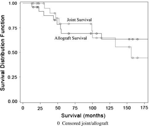
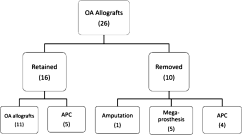
The allograft 5- and 10-year survival rates for allograft survival were estimated to be 69% (95% confidence interval, 50%–88%) and 63% (95% confidence interval, 43%–84%), respectively (Fig. 1). Overall, 16 of the 26 patients retained the osteoarticular allograft, whereas the allograft was sacrificed in the remaining 10 (Table 2; Fig. 2). At the time of last review, seven patients had died. Five patients died with their original osteoarticular allograft in place at an average of 34 months (range, 15–66 months) and were asymptomatic at that time. The sixth patient had a successful two-stage conversion because of an infection at 30 months but died 174 months after the initial implantation because of other complications. The final patient died at 22 months after undergoing a hemipelvectomy at 13 months.
Fig. 1.
Kaplan-Meier survival curves show allograft and joint survival rates. The allograft 5- and 10-year survival rates for allograft survival were estimated to be 69% (95% confidence interval, 50%–88%) and 63% (95% confidence interval, 43%–84%), respectively.
Table 2.
Multiple complications necessitated subsequent surgical interventions
| Patient number | Conversion time (months) | Allograft preserved | Treatment | Complication after conversion | Total graft survival (months) |
|---|---|---|---|---|---|
| Progressive joint degeneration | |||||
| 8 | 31 | No | Conversion to APC | Allograft fracture (44)* | 44 |
| 9 | 156 | Yes | Conversion to APC | Patellar clunk | 283 |
| 10 | 47 | Yes | Conversion to APC | None | 182 |
| 11 | 99 | Yes | Conversion to APC | None | 194 |
| 12 | 39 | No | Conversion to APC | Allograft fracture (55)* | 55 |
| 13 | 133 | No | Conversion to APC | Instability† | 186 |
| 14 | 100 | Yes | Conversion to APC | None | 238 |
| 16 | 212 | Partial | Conversion to Mega‡ | None | 212 |
| Patient number | Initial soft tissue coverage | Infection | Time to presentation | Allograft preserved | Treatment | Final outcome |
|---|---|---|---|---|---|---|
| Infection | ||||||
| 19 | None | Deep | 30 months | No | 2-stage exchange | Limb preservation |
| 20 | Local flap§ | Deep | 20 days | Yes | N/A** | Limb preservation** |
| 21 | None | Superficial | 2 months | Yes | Local flap, STSG | Limb preservation |
| 22 | Local flap|| | Superficial | 2 weeks | Yes | Wound revision | Limb preservation |
| 23 | Local flap¶ | Superficial → deep | 20 days | No | 2-stage exchange | Limb preservation |
| 24 | None | Deep | 114 months | No | 2-stage exchange | Limb preservation |
| Patient number | Time to intervention | Treatment | Final outcome |
|---|---|---|---|
| Nonunion | |||
| 15 | 13 months | Exchange nail | Union |
| 16 | 24 months | Vascularized fibula | Union |
| 17 | 19 months | ICBG; exchange nail (second surgery) | Union |
| 19 | 9 months | ICBG | Developed infxn†† |
| 23 | 11 months | Exchange nail | Developed infxn†† |
| Patient number | Type of implant | Treatment | Allograft preservation |
|---|---|---|---|
| Fracture | |||
| 8 | Nail | Revised APC (new allograft) | No |
| 12 | Plate | Revised APC (new allograft) | No |
| 19 | Nail | Revised to new OA allograft | No |
| 24 | Plate | ORIF fracture | Yes |
| 25 | Nail | Revised APC (new allograft) | No |
| Patient number | Time from initial surgery (months) | Allograft preserved | Treatment | Final outcome |
|---|---|---|---|---|
| Resorption | ||||
| 16 | 24 | Yes‡‡ | Vascularized fibula§§ | Resorption halted |
*Conversion to APC (new allograft); †conversion to mega-prosthesis; ‡megaprosthesis; §sartorius; ||gracilis; ¶gastrocnemius; **patient died before explantation; ††developed infection; ‡‡later developed degenerative joint disease; §§see Fig. 4; APC = allograft-prosthesis composite; STSG = split-thickness skin graft; OA = osteoarthritis; ORIF = open reduction and internal fixation.
Fig. 2.
This flow chart describes retention (16) or removal (10) and subsequent surgical procedures of 26 osteoarticular allografts used for reconstruction of the distal femur.
The joint surface 5- and 10-year survival rates were calculated to be 79% (95% confidence interval, 61%–97%) and 65% (95% confidence interval, 41%–88%), respectively (Fig. 1). Seven patients did not require additional surgical intervention and retained the original osteoarticular allograft at an average of 126 ± 54 months (range, 15–189 months).
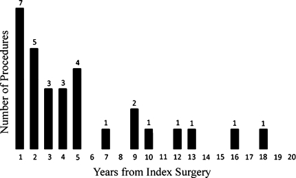
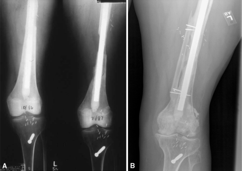
Nineteen of the original 26 patients had complications that necessitated 30 additional surgeries (Table 3). Twenty-two of the 30 procedures were performed in the first 5 years after the index surgery, and the remaining eight were performed throughout the next 13 years (Fig. 3). One patient had resorption of the allograft at 2 years after resection of an angiosarcoma. Resorption was successfully arrested with transfer of a vascularized fibular graft, and no additional surgery was required until the patient developed symptomatic degenerative changes of the joint 17 years later (Fig. 4). One of the 26 patients developed a local tumor recurrence with regional metastases to the groin after distal femoral resection of a Stage IIB osteosarcoma. The advanced disease process required external hemipelvectomy at 13 months, and the patient died 22 months after the initial surgery.
Table 3.
Complications and survival of distal femoral osteoarticular allografts
| Study | Pts (grafts) | Followup | DJD | Fracture | Nonunion | Infection | Graft survival | Joint survival |
|---|---|---|---|---|---|---|---|---|
| Mnaymneh et al. (1994) [20] | 83 (83) | 53 months (24–168) | None | 14% | 12% | 6% | Information not provided | Information not provided |
| Muscolo et al. (2005) [22] | 62 (58) | 82 months (1–368) | 35% | 5% | 6% | 10% | 78% at 5 and 10 years | 71% at 5 and 10 years |
| Toy et al. (2010) [Current study] | 26 (26) | 156 months (15–283) | 31% | 19% | 19% | 15% | 69% at 5 years 63% at 10 years | 79% at 5 years 65% at 10 years |
Fig. 3.
This bar graph illustrates the number of secondary procedures (30) required in 19 patients according to time from index surgery; 22 were performed in the first 5 years after the index surgery, and the remaining eight throughout the next 13 years.
Fig. 4A–B.
(A) The radiograph shows resorption of the allograft 2 years after resection of an angiosarcoma. (B) Insertion of a vascularized fibular graft arrested resorption, and no additional surgery was required until the patient developed symptomatic degenerative changes of the joint 17 years later.
Discussion
Reconstructive options after distal femur resection include: intercalary allograft arthrodesis, prosthetic TKA, rotationplasty, or osteoarticular allograft placement. This review was undertaken to determine graft survival, joint preservation, nature and frequency of complications in osteoarticular allograft reconstructions of the distal femur.
There are limitations in the data presented in this study. First, the small number of patients did not allow sufficient power to explore differences between subgroups of patients or trends between clinical variables and outcomes. The limited study size is related to the rarity of the disease itself and the infrequent use of osteoarticular allograft reconstruction. Second, the heterogeneous nature of these tumors, the patients themselves, the required bone and soft tissue resection, and the varied complications made standardized treatment difficult. Surgeon preference also introduced heterogeneity in treatment because multiple surgeons were involved in this period of treatment.
The overall 5-year and 10-year allograft survival rates were 69% and 63%, respectively; joint surface survival rates were 79% and 65%, respectively. At latest followup, 16 of the original 26 allografts were still in place and 25 of the 26 patients had successful limb salvage. Our survival rates were not quite as high as those reported by Muscolo et al. [22]—78% graft survival, 71% joint preservation, and 97% limb preservation at 5 and 10 years in 75 distal femoral allografts— but are similar to other reports in which poor results, defined as amputation or graft removal, occurred in approximately one-third of patients [10, 17, 20]. The impressive results reported by Musculo et al. [22] are likely a result of their well-known dedication to allograft surgery, extensive bone bank, and the matching of allografts to patients using CT measurements; resources not routinely available to US surgeons.
As in other series [22, 32], progressive degenerative change was the most common complication requiring further surgery (Table 2); eight of our 26 patients required further surgery at a mean of 86 ± 45 months (range, 31–156 months) after the initial allograft procedure. Joint deterioration is considered an inevitable late complication of osteoarticular allograft implantation, but salvage usually is possible with a joint-resurfacing procedure or megaprosthesis. In a large series of osteoarticular allografts of the distal femur, 8% of allografts that were still in place at latest followup had required a resurfacing procedure for joint deterioration or instability [22].
Infection is a potentially devastating complication of osteoarticular allograft reconstruction [15, 22–24]. Early aggressive intervention such as débridement and wound coverage is indicated for wounds showing signs of compromise such as necrosis or persistent drainage. One of the three superficial wound infections in our series of patients progressed to a deep infection that ultimately required removal of the allograft and eventually a two-stage conversion to a megaprosthesis. Of the additional three patients treated for deep infection, only one retained the allograft. This patient may have eventually required removal of the allograft but died from causes unrelated to his tumor or its treatment. In a review of 83 patients treated with allografts of the distal femur, deep infection occurred in five (6%) and was more prevalent in patients treated with chemotherapy. Amputation was required in two of the five patients. Allograft salvage was successful in the remaining three patients, but function was rated as fair [20]. Limb preservation may be possible in a patient with a deep infection, but there is additional morbidity. Meticulous attention to aseptic technique, use of antibiotics, and aggressive soft tissue coverage can minimize the risk of deep infection. Our approach to the treatment of deep infection involves removal of the allograft and associated hardware and placement of an antibiotic-impregnated cement spacer. After appropriate culture-specific intravenous antibiotics and clinical evidence suggesting the infection is eliminated, an attempt is made to reconstruct the limb. The choice of the construct is patient-specific and may be a new osteoarticular allograft, APC, or megaprosthesis.
Nonunion at the graft-host interface is another frequent complication (Table 2). Healing occurs primarily from external callus in cortical-cortical junctions and primarily by internal callus in cancellous-cancellous junctions and is slow to develop between the allograft and host bone [9]. Furthermore, time to callous maturation and union is dependent on the quality of contact and stability of the junction [7, 9, 19]. Chemotherapy has been suggested to negatively affect union of the allograft-host junction [20]; however, Muscolo et al. [22] found no relationship between allograft survival and the use of chemotherapy, age, gender, or amount of femoral resection. With the small number of patients available in our study, nonunion was not affected by age, exposure to chemotherapy, amount of bone resected, or method of initial fixation.
Although osteoarticular allografts should be considered in the reconstruction of skeletal defects about the distal femur, with reported graft survival rates of up to almost 80% at 5 and 10 years after implantation [22], over 70% of patients in our long-term followup study had complications that required further surgical intervention. Despite the high complication rate, others have recommended this procedure [20, 22, 23] for tumors of the distal femur. Because of the frequency of these complications, our institution has not used osteoarticular allografts for reconstruction of the distal femur in the past 8 years, but rather has chosen a megaprosthesis or APC for reconstruction. Additionally, soft tissue attachments to the distal femur are not as critical to maintaining function when compared with other sites such as the proximal tibia or proximal humerus. Osteoarticular allografts may be best suited to younger patients who have a good oncologic prognosis, because considerable time is needed to allow allograft-host union. Patients who cannot tolerate the postoperative rehabilitation or have an anticipated shortened lifespan may be better treated with another type of reconstruction such as a megaprosthesis. If revision is required after implanting an osteoarticular allograft, conversion to an APC or megaprosthesis can be performed when the physical demands of the patient have decreased, which will extend the lifetime of the nonbiologic reconstruction. Despite the frequent complications associated with the osteoarticular allografts, all patients but one retained a functional limb at last followup.
Footnotes
Each author certifies that he or she has no commercial associations (eg, consultancies, stock ownership, equity interest, patent/licensing arrangements, etc) that might pose a conflict of interest in connection with the submitted article.
Each author certifies that his or her institution approved the protocol for this study and that all investigations were conducted in conformity with ethical principles of research.
This work was performed at the University of Florida Orthopaedic and Sports Medicine Institute, Gainesville, FL.
References
- 1.Biau DJ, Larousserie F, Thévenin, Piperno-Neumann S, Anract P. Results of 32 allograft-prosthesis composite reconstructions of the proximal femur. Clin Orthop Relat Res. 2010;468:834–845. doi: 10.1007/s11999-009-1132-z. [DOI] [PMC free article] [PubMed] [Google Scholar]
- 2.Bradish CF, Kemp HB, Scales JT, Wilson JN. Distal femur replacement by custom-made prosthesis: clinical follow-up and survivorship analysis. J Bone Joint Surg Br. 1987;69:276–284. doi: 10.1302/0301-620X.69B2.3818760. [DOI] [PubMed] [Google Scholar]
- 3.Capanna R, Morris HG, Campanacci D, Del Ben M, Campanacci M. Modular uncemented prosthetic reconstruction after resection of tumours of the distal femur. J Bone Joint Surg Br. 1994;76:178–186. [PubMed] [Google Scholar]
- 4.Davis AM, Punniyamoorthy S, Griffin AM, Wunder JS, Bell RS. Symptoms and their relationship to disability following treatment for lower extremity tumours. Sarcoma. 1999;3:73–77. doi: 10.1080/13577149977677. [DOI] [PMC free article] [PubMed] [Google Scholar]
- 5.Dick HM, Malinin TI, Mnaymneh WA. Massive allograft implantation following radical resection of high-grade tumors requiring adjuvant chemotherapy treatment. Clin Orthop Relat Res. 1985;197:88–95. [PubMed] [Google Scholar]
- 6.Eckardt JJ, Matthews JG, Eilber FR. Endoprosthetic reconstruction after bone tumor resections of the proximal tibia. Orthop Clin North Am. 1991;22:149–160. [PubMed] [Google Scholar]
- 7.Enneking WF, Campanacci DA. Retrieved human allografts: a clinicopathological study. J Bone Joint Surg Am. 2001;83:971–986. [PubMed] [Google Scholar]
- 8.Enneking WF, Dunham W, Gebhardt MC, Malawar M, Pritchard DJ. A system for the functional evaluation of reconstructive procedures after surgical treatment of tumors of the musculoskeletal system. Clin Orthop Relat Res. 1993;286:241–246. [PubMed] [Google Scholar]
- 9.Enneking WF, Mindell ER. Observations on massive retrieved human allografts. J Bone Joint Surg Am. 1991;73:1123–1142. [PubMed] [Google Scholar]
- 10.Gebhardt MC, Flugstad DI, Springfield DS, Mankin HJ. The use of bone allografts for limb salvage in high grade extremity osteosarcoma. Clin Orthop Relat Res. 1991;270:181–196. [PubMed] [Google Scholar]
- 11.Gitelis S, Heligman D, Quill G, Piasecki P. The use of large allografts for tumor reconstruction and salvage of the failed total hip arthroplasty. Clin Orthop Relat Res. 1988;231:62–70. [PubMed] [Google Scholar]
- 12.Hornicek FJ, Jr, Mnaymneh W, Lackman RD, Exner GU, Malinin TI. Limb salvage with osteoarticular allografts after resection of proximal tibial bone tumors. Clin Orthop Relat Res. 1998;352:179–186. doi: 10.1097/00003086-199807000-00021. [DOI] [PubMed] [Google Scholar]
- 13.Horowitz SM, Glasser DB, Lane JM, Healey JH. Prosthetic and extremity survivorship after limb salvage for sarcoma: how long do the reconstructions last? Clin Orthop Relat Res. 1993;293:280–286. [PubMed] [Google Scholar]
- 14.Kaplan EL, Meier P. Nonparametric estimation from incomplete observations. J Amer Statistical Assoc. 1958;53:457–481. doi: 10.2307/2281868. [DOI] [Google Scholar]
- 15.Kawai A, Muschler GF, Lane JM, Otis JC, Healey JH. Prosthetic knee replacement after resection of a malignant tumor of the distal part of the femur. Medium to long-term results. J Bone Joint Surg Am. 1998;80:636–647. doi: 10.1302/0301-620X.80B4.8216. [DOI] [PubMed] [Google Scholar]
- 16.Langlais F, Lambotte JC, Collin P, Thomazeau H. Long-term results of allograft composite total hip prostheses for tumors. Clin Orthop Relat Res. 2003;414:197–211. doi: 10.1097/01.blo.0000079270.91782.23. [DOI] [PubMed] [Google Scholar]
- 17.Mankin HJ, Doppelt SH, Sullivan RT, Tomford WW. Osteoarticular and intercalary allograft transplantation in the management of malignant tumors of bone. Cancer. 1982;50:613–630. doi: 10.1002/1097-0142(19820815)50:4<613::AID-CNCR2820500402>3.0.CO;2-L. [DOI] [PubMed] [Google Scholar]
- 18.Mankin HJ, Gebhardt MC, Jennings LC, Springfield DS, Tomford WW. Long-term results of allograft replacement in the management of bone tumors. Clin Orthop Relat Res. 1996;324:86–97. doi: 10.1097/00003086-199603000-00011. [DOI] [PubMed] [Google Scholar]
- 19.Mankin HJ, Gebhardt MC, Tomford WW. The use of frozen cadaveric allografts in the management of patients with bone tumors of the extremities. Orthop Clin N Am. 1987;18:275–289. [PubMed] [Google Scholar]
- 20.Mnaymneh W, Malinin TI, Lackman RD, Hornicek FJ, Mnaymneh LG. Massive distal femoral osteoarticular allografts after resection of bone tumors. Clin Orthop Relat Res. 1994;303:103–115. [PubMed] [Google Scholar]
- 21.Muscolo DL, Ayerza MA, Aponte-Tinao LA. Survivorship and radiographic analysis of knee osteoarticular allografts. Clin Orthop Relat Res. 2000;373:73–79. doi: 10.1097/00003086-200004000-00010. [DOI] [PubMed] [Google Scholar]
- 22.Muscolo DL, Ayerza MA, Aponte-Tinao LA, Ranalletta M. Use of distal femoral osteoarticular allografts in limb salvage surgery. J Bone Joint Surg Am. 2005;87:2449–2455. doi: 10.2106/JBJS.D.02170. [DOI] [PubMed] [Google Scholar]
- 23.Muscolo DL, Ayerza MA, Aponte-Tinao LA, Ranalletta M. Use of distal femoral osteoarticular allografts in limb salvage surgery. Surgical technique. J Bone Joint Surg Am. 2006;88:305–321. doi: 10.2106/JBJS.F.00324. [DOI] [PubMed] [Google Scholar]
- 24.Muscolo DL, Petracchi LJ, Ayerza MA, Calabrese ME. Massive femoral allografts followed for 22 to 36 years. J Bone Joint Surg Br. 1992;74:887–892. doi: 10.1302/0301-620X.74B6.1447252. [DOI] [PubMed] [Google Scholar]
- 25.Ortiz-Cruz E, Gebhardt MC, Jennings LC, Springfield DS, Mankin HJ. The results of transplantation of intercalary allografts after resection of tumors. A long term follow-up study. J Bone Joint Surg Am. 1997;79:97–106. doi: 10.2106/00004623-199701000-00010. [DOI] [PubMed] [Google Scholar]
- 26.Roberts P, Chan D, Grimer RJ, Sneath RS, Scales JT. Prosthetic replacement of the distal femur for primary bone tumours. J Bone Joint Surg Br. 1991;73:762–769. doi: 10.1302/0301-620X.73B5.1894662. [DOI] [PubMed] [Google Scholar]
- 27.Schwartz AJ, Kabo M, Eilber FC, Eilber EF, Eckardt JJ. Cemented distal femoral endoprostheses for musculoskeletal tumor: improved survival of modular versus custom implants. Clin Orthop Relat Res. 2009 Dec 22. [Epub ahead of print] [DOI] [PMC free article] [PubMed]
- 28.Thompson RC, Jr, Pickvance EA, Garry D. Fractures in large-segment allografts. J Bone Joint Surg Am. 1993;75:1663–1673. doi: 10.2106/00004623-199311000-00011. [DOI] [PubMed] [Google Scholar]
- 29.Unwin PS, Cannon SR, Grimer RJ, Kemp HB, Sneath RS, Walker PS. Aseptic loosening in cemented custom-made prosthetic replacements for bone tumours of the lower limb. J Bone Joint Surg Br. 1996;78:5–13. [PubMed] [Google Scholar]
- 30.Wirganowicz PZ, Eckardt JJ, Dorey FJ, Eilber FR, Kabo JM. Etiology and results of tumor endoprosthesis revision surgery in 64 patients. Clin Orthop Relat Res. 1999;358:64–74. doi: 10.1097/00003086-199901000-00009. [DOI] [PubMed] [Google Scholar]
- 31.Wunder JS, Leitch K, Griffin AM, Davis AM, Bell RS. Comparison of two methods of reconstruction for primary malignant tumors at the knee: a sequential cohort study. J Surg Oncol. 2001;77:89–99. doi: 10.1002/jso.1076. [DOI] [PubMed] [Google Scholar]
- 32.Zatsepin ST, Burdygin VN. Replacement of the distal femur and proximal tibia with frozen allografts. Clin Orthop Relat Res. 1994;303:95–102. [PubMed] [Google Scholar]
- 33.Zeegen EN, Aponte-Tinao LA, Hornicek FJ, Gebhardt MC, Mankin HJ. Survivorship analysis of 141 modular metallic endoprostheses at early followup. Clin Orthop Relat Res. 2004;420:239–250. doi: 10.1097/00003086-200403000-00034. [DOI] [PubMed] [Google Scholar]
- 34.Zehr RJ, Enneking WF, Heare T, Liang TS. Fractures in large structural allografts. In: Brown KLB, ed. Complications of Limb Salvage. Montreal, Quebec: 6th International Symposium. International Society of Limb Salvage; 1991:3–9.
- 35.Zehr RJ, Enneking WF, Scarborough MT. Allograft-prosthesis composite versus megaprosthesis in proximal femoral reconstruction. Clin Orthop Relat Res. 1996;322:207–223. doi: 10.1097/00003086-199601000-00026. [DOI] [PubMed] [Google Scholar]


