Abstract
Background
Many prognostic markers have been identified in chronic lymphocytic leukemia, but there have been few opportunities to assess their relative importance in a large randomized trial. The aim of this study was to determine which of the available markers independently predicted outcome in patients requiring treatment and to use these to define new risk groups.
Design and Methods
A broad panel of clinical and laboratory markers, measured at randomization in patients entering the LRF CLL4 trial, was assessed with respect to treatment response, progression-free and overall survival, at a median follow-up of 68 months.
Results
Using the factors identified as independent predictors for progression-free survival, patients were subdivided into three risk groups: 6% had poor risk with known TP53 loss of greater than 10%; 72% had an intermediate risk without TP53 loss (≤10%) and with at least one of: unmutated IGHV genes and/or IGHV3-21 usage, 11q deletion, β-2 microglobulin greater than 4 mg/L; 22% had a good risk (with none of the above and mutated IGHV genes). The 5-year progression-free survival rates for these three groups were 0%, 12% and 34%, respectively, and the corresponding 5-year overall survival rates were 9%, 53% and 79% (both P<0.00005 independent of treatment allocation). In the intermediate risk group 250 patients, with data for all three risk factors, were further subdivided into intermediate-low (one risk factor) or intermediate-high (2 or 3 risk factors). The 5-year progression-free survival rates were 18% and 7% (P=0.0001) and the 5-year overall survival rates were 68% and 40% (P<0.00005), respectively.
Conclusions
This study demonstrates the role of biomarkers in prognosis and shows that, in patients requiring treatment, disease stage may no longer be an independent predictor of outcome. If validated independently, the risk groups defined here may inform the design of future trials in chronic lymphocytic leukemia. (Clinicaltrials.gov identifier: NCT00004218; controlled-trials.com identifier ISRCTN58585610).
Keywords: chronic lymphocytic leukemia, prognostic factors, TP53, IGHV mutation status, progression-free survival, overall survival
Introduction
Considerable effort has been made to identify clinical and laboratory factors that predict the natural history and response to therapy of patients with chronic lymphocytic leukemia (CLL). Early studies highlighted the importance of disease stage and factors such as lymphadenopathy, hepatosplenomegaly, anemia, thrombocytopenia, lymphocyte count, the pattern of bone marrow infiltration and serum markers, all of which at least in part reflect tumor burden.1 More recently, attention has focused on both intrinsic and acquired features of the leukemic clone which are independent of tumor burden. These biomarkers include IGHV gene mutational status and usage, CD38 and ZAP-70 expression and genomic abnormalities.2–8 Many of the published data derive from retrospective analyses of patients studied from presentation to first treatment and evaluate the role of prognostic factors as markers of disease progression. Information on the value of biomarkers, other than TP53 loss, in predicting outcome in patients with a clinical indication for treatment is more limited. In particular there are few published data from prospective studies evaluating the role of prognostic markers, measured at trial entry, in patients entered into randomized clinical trials.9
The LRF CLL4 trial randomized 777 previously untreated patients with Binet stage progressive A, B or C disease between January 1999 and October 2004 to receive either chlorambucil, fludarabine or fludarabine and cyclophosphamide.10 This report, with a median follow-up of 68 months, represents the most comprehensive report published to date on prognostic markers in CLL, measured at the time of randomization into a large trial.
Design and Methods
Full details of the design, conduct and outcome of the LRF CLL4 trial have been published.10 All patients provided written informed consent. The trial was approved by the UK South Thames Multicentre Research Ethics Committee [MREC (1) 98/101] and followed UK Medical Research Council guidelines for good clinical practice.
Laboratory markers
The non-clinical prognostic factors were measured on blood samples taken at the time of trial entry. Fluorescence in situ hybridization (FISH), mutational analysis of immunoglobulin variable region genes (IGHV), and evaluation of CD38 and ZAP-70 expression were performed centrally. Serum lactate dehydrogenase and beta-2 microglobulin (β-2M) were assayed at referring hospitals and absolute values were reported.
FISH was performed using commercially available probes according to the manufacturers' instructions; the panel used (Vysis from Abbott Laboratories, Berkshire, UK) comprised TP53 (17p13.1); D12Z3 (centromere12); D13S25 (13q14.3); either 11q23 or, later in the trial, ATM (11q22.3); and 6q21 (courtesy of Dr S Stilgenbauer). At least 200 cells were examined for each probe in all cases. The cut-off points for defining loss were greater than 5% for 11q, 13q14 and 6q21, and greater than 3% for trisomy 12. In view of the clinical importance of identifying patients with a TP53 abnormality, all cases originally found to have between 5% and 30% TP53 loss were reviewed independently by three to five experienced cytogeneticists at the Royal Marsden and Bournemouth hospitals. The selected cut off defining TP53 loss, based on the mean of the results obtained from this review, was 10%, as discussed in more detail below.
IGHV genes were sequenced from either cDNA or gDNA. cDNA was synthesized by reverse transcription according to the manufacturer’s protocol (Promega). cDNA was amplified as previously described.11
Sequences were aligned to current databases (V-BASE and IMGT). IGHV gene stereotypes were classified according to the criteria used by Stamatopoulos et al.12 CD38 expression was measured by flow cytometry using fresh samples stained with CD5-FITC (BD Biosciences), CD38-PE (clone HB-7, BD Biosciences), and CD19-TC (Caltag, Burlingame, CA, USA), as described by Morilla et al.13 The choice of cut-off for both IGHV mutation and CD38 positivity is described in the results section. ZAP-70 expression was measured by flow cytometry using samples frozen in dimethylsulfoxide, as previously described.7 The previously validated7 cut-off of 10% was used to define ZAP-70 positivity, based on correlations with IGHV gene mutational status, ZAP-70 mRNA expression and clinical outcome.
Statistical analysis
Group-wise comparisons of clinical, laboratory and genetic data were performed using the Kruskal-Wallis test or the χ2 test, as appropriate. Overall survival was calculated from randomization to death from any cause. Progression-free survival was calculated from randomization to initial non-response, progression, or death from any cause. Survival curves were constructed by the Kaplan-Meier method and compared using the log-rank test. Multivariate analyses of variables significant in the univariate log-rank tests were performed by means of the Cox proportional hazards model using step-wise forward selection. Treatment allocation was included as a covariate in all multivariate models. Data on β-2M and the biomarkers were not available for all patients and, since some variables were missing for different sets of patients, the numbers available for some multivariate analyses were reduced, so caution is required in their interpretation. Odds-ratio plots show the relative effect of prognostic markers within treatment groups with tests for heterogeneity to indicate any evidence of a different effect between treatments. The follow-up was to 31st October 2008, with a median follow-up for survivors of 68 months (range, 48 months – 117 months). Statistical analyses were conducted with SAS version 9.1 (SAS Institute Inc, Cary, NC, USA) and in-house programs. All P values given are two-sided.
Results
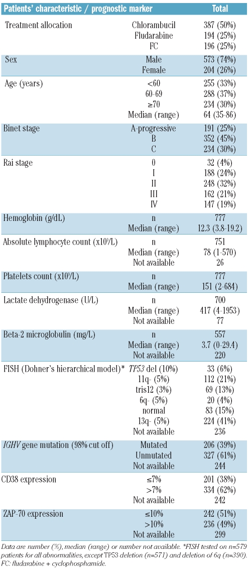
The patients’ characteristics and the number of patients in whom each prognostic marker was measured are shown in Table 1. There was no significant difference in the age, gender, Binet stage, treatment allocation, progression-free survival or overall survival among cases in whom biomarkers were tested compared to the remaining patients. Additionally there was no imbalance in any of the patients’ characteristics by treatment arm (data not shown).
Table 1.
Patients’ characteristics.
Clinical factors
In univariate analysis male gender, Binet stage C or Rai stage 3 or 4 disease, lower hemoglobin, higher lymphocyte count and lower platelet count were all associated with poorer response to treatment. Male gender (P=0.03), lower hemoglobin (P=0.001), higher lactate dehydrogenase concentration (P=0.008) and higher lymphocyte count (P<0.00005) were associated with shorter progression-free survival, while male gender (P=0.02), advanced stage (P=0.0002), increasing age (P<0.00005) and lower hemoglobin (P<0.00005) correlated with shorter overall survival (Online Supplementary Table S1).
Beta-2 microglobulin
β-2M levels were significantly higher in older patients (P<0.0001), in males (P=0.001), in stage C compared to stage B (P=0.0006) and in stage B compared to progressive stage A disease (P<0.0001). Using the cut off of 4 mg/L, there were significant associations between β-2M levels and unmutated IGHV genes and high CD38 expression, and a borderline association with the absence of a 13q deletion. Higher β-2M levels were significantly associated with poorer response to treatment (P=0.0001), shorter progression-free survival (P=0.0001) and shorter overall survival (P<0.00005) (Online Supplementary Table S2).
Fluorescence in situ hybridization analysis
A greater than 10% loss of TP53 was detected in 33 cases (5.8%), of whom 31 had greater than 20% loss. The outcome of the two cases with 10–20% loss was comparable to that of the cases with greater than 20% loss and the outcome of those with 5–10% loss was similar to that of patients with less than 5% loss. Deletion of 13q, 11q, 6q and trisomy 12 was found in 344 (59%), 116 (20%), 33 (8%) and 88 (15%) of cases, respectively. Eighty-three patients (15%) had no FISH abnormality. Four patients had greater than 10% TP53 loss in addition to greater than 5% 11q loss.
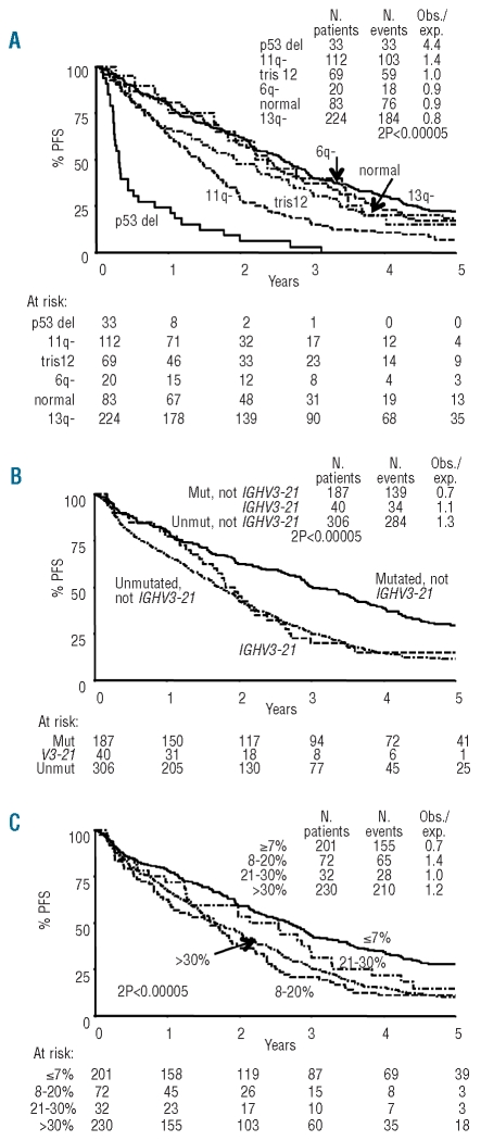
Dohner’s hierarchical model4 was used to examine the influence of genomic abnormalities on response rate, progression-free survival and overall survival (Figure 1A and Online Supplementary Table S2). In those with greater than 10% TP53 loss the overall response rate was low at 29% (compared with 82% in non-TP53 cases, P<0.0001), with only 6% patients achieving a complete or nodular partial response. Outcome overall was very poor indicating that these patients were not salvaged with subsequent therapies. Five patients had TP53 loss in the range of 5–9.99% and their outcome was comparable to that of patients with less than 5% loss. Excluding the patients with TP53 loss, 11q loss was associated with a lower overall response rate (72% versus 84%, P=0.005), shorter progression-free survival (P<0.00005) and shorter overall survival (P=0.0004) compared to those in patients without 11q loss. Deletion of 13q as a sole abnormality was found in 224 patients and was associated with superior progression-free survival (P=0.0003) and overall survival (P<0.00005), but no difference in overall response rate (84% versus 79%, P=0.2), compared to these outcomes in patients with other abnormalities (excluding TP53 loss) or normal FISH (n=284). Trisomy 12 and 6q loss had no influence on response or progression-free survival.
Figure 1.
Factors associated with progression-free survival. (A) Progression-free survival by Dohner’s hierarchical group. (B) Progression-free survival by IGHV mutational status and/or IGHV3-21 use. (C) Progression-free survival by CD38 expression.
IGHV gene analysis
Regardless of whether a 98% or 97% cut-off was used, there were comparable and significant differences in response rate (P=0.01 and P=0.009, respectively) and in progression-free and overall survival between mutated and unmutated cases (P<0.00005 for both cut-offs, for both outcome measures) (Online Supplementary Table S2). A 98% cut-off was chosen for all further analyses given its widespread use in the literature. Forty patients utilized the IGHV3-21 gene of whom 35 had highly homologous CDR3. There were significant differences in progression-free survival (P=0.008) and overall survival (P=0.001) between IGHV3-21 cases and mutated cases without IGHV3-21 gene usage (Figure 1B and Online Supplementary Table S2). There were significant associations between unmutated IGHV genes and male gender, 11q, no loss of 13q, trisomy 12 and increased expression of CD38 and ZAP-70. Whereas some previous studies found that TP53 loss was virtually confined to cases with unmutated IGHV genes, seven (24%) of our cases with greater than 10% TP53 loss had mutated IGHV genes (Online Supplementary Table S3).
CD38 and Zap-70 expression
Since various cut-offs for CD38 have been reported in the literature, we evaluated the prognostic significances of 7%, 20% and 30% cut-offs. There was no correlation between CD38 expression and response rate regardless of the cut-off used. High CD38 expression was associated with shorter progression-free and overall survival (Online Supplementary Table S2). There was no difference in either outcome measure between those with 8–20% and those with 21–30% CD38 expression, or between the 21–30% and greater than 30% groups, suggesting that a 7% cut-off is most appropriate. Patients with 7% or less expression had a significantly better outcome (Figure 1C). ZAP-70 positive patients had a significantly poorer overall response rate (P=0.008) and significantly shorter progression-free survival (P<0.00005), but an only borderline worse overall survival (P=0.07).
Multivariate analysis and risk groups
The independent prognostic value of all clinical and laboratory factors found to be significant in univariate analyses (Online Supplementary Tables S1 and S2) was assessed together, using proportional hazard regression models for progression-free survival and overall survival. In the initial multivariate analysis, deletion of TP53 conferred a markedly inferior prognosis, independently of any other factors. Therefore, in order to determine which factors had independent prognostic value in patients without TP53 deletion, the 33 patients with greater than 10% TP53 deletion were excluded from all further multivariate analyses.
β-2M (P=0.007), 11q deletion (P=0.0006) and IGHV mutational status and/or IGHV3-21 usage (P<0.0001), as well as the treatment allocation (P<0.0001), were identified as independent predictors for progression-free survival (Table 2). ZAP-70 and CD38 expression were only retained as independent predictors for progression-free survival in the absence of information on IGHV mutational status. The most important independent prognostic factors for overall survival were β-2M (P=0.005), IGHV mutational status and/or IGHV3-21 usage (P=0.0001) and age (P<0.0001) (Table 2). There was a suggestion that both CD38 expression and deletion of 11q also had borderline independent effects on overall survival, however, it was not possible to ascertain this with certainty due to missing data and decreasing numbers of patients in the models. None of the clinical variables associated with shorter progression-free survival or overall survival in the univariate analyses (Online Supplementary Table S1) was retained as an independent predictor in the multivariate analysis, other than treatment allocation (progression-free survival) and age (overall survival) (Table 2).
Table 2.
Multivariate analysis of prognostic factors independently associated with progression-free and overall survival (in patients without TP53 loss).
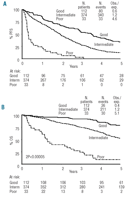
Using the factors identified as independent predictors for progression-free survival, it was possible to sub-divide all CLL4 patients into three risk groups with differing progression-free survival rates as follows: (i) a group of 33 (6%) patients was considered at poor risk based on known TP53 loss of greater than 10%; (ii) a further group of 374 (72%) patients was at intermediate risk defined by TP53 loss of 10% or less and the presence of at least one of the following: unmutated IGHV genes and/or IGHV3-21 usage, 11q deletion or a β-2M level of >4 mg/L; and (iii) 112 (22%) patients with none of the above intermediate risk factors, TP53 loss of 10% or less and mutated IGHV genes, were assigned to a good risk category. The 5-year progression-free survival rate (with 95% confidence interval) (Figure 2A) was 0% for the poor risk group, 12% (9–16%) for the intermediate risk group and 34% (25–43%) for the good risk group. Two hundred and fifty-eight patients from the trial were not assigned to a risk group because of missing genetic information. The 5-year progression-free survival for this group was 19% (14–24%).
Figure 2.
Progression-free survival and overall survival by risk group. (A) Progression-free survival. Poor risk: >10% TP53 loss. Intermediate risk: ≤10% TP53 loss and at least one of: unmutated IGHV genes, IGHV3-21 usage, beta-2 microglobulin >4 mg/L, 11q loss >5%. Good risk: ≤10% TP53 loss, none of the intermediate risk factors and mutated IGHV genes. (B) Overall survival.
These risk groups also predicted for overall survival and response to treatment. For overall survival (Figure 2B) the risk groups separated patients with 5-year overall survival rates of 9% (0–19%), 53% (47–58%) and 79% (71–87%), respectively (P<0.00005). Within each Binet stage the risk groups defined substantially different survival rates (12.5%, 56%, 85% in stage A, 17%, 55%, 77% in stage B, 0%, 44%, 72% in stage C). The 5-year overall survival rate of the patients not assigned to a risk group was 61% (55–67%). In terms of response rates, 89 (87%) of patients in the good risk category achieved a response (complete, nodular partial or partial response) compared with 283 (79%) in the intermediate risk and 9 (29%) in the poor risk (P<0.0001). There was also a highly significant association (P<0.0001) with good response to treatment.
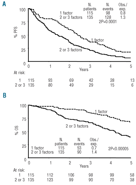
We investigated the effect on outcome of the number of risk factors present in the 250 patients in the intermediate risk group who had data for all three factors which define the intermediate risk group: 13.2% of patients had all three of these poor risk factors, 40.8% had two, and 46.0% had one. Increasing numbers of poor risk features significantly affected both progression-free survival and overall survival. The 5-year progression-free survival rate was 18% (11–25%) for patients with one risk factor, 8% (2–13%) for those with two and 6% (0–14%) for those with three risk factors. The corresponding 5-year overall survival rates were 68% (59–77%), 42% (32–52%) and 30% (13–47%).
In view of the small number of patients with three risk factors and their similar outcome to those with two risk factors, cases with two or three risk factors were combined, comprising 54% of patients in the intermediate group, with a 5-year progression-free survival of 7% (3–11%) and overall survival of 40% (31–48%) (Figure 3A–B). This analysis was not integrated with Figure 2, as it was based on a subset of patients only: those with complete data for all the factors considered.
Figure 3.
Progression-free survival and overall survival in the intermediate risk group by number of adverse risk factors (among 250 patients for whom complete information was available). (A) Progression-free survival. (B) Overall survival.
Effect of prognostic markers within treatment allocation
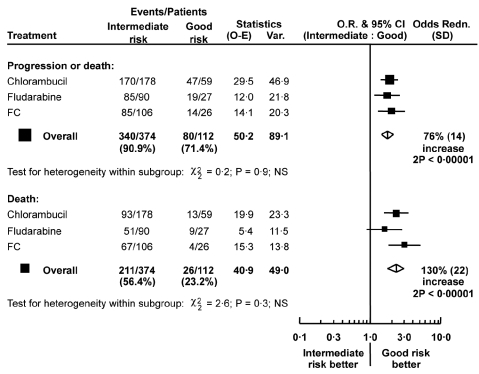
There was no significant difference in the relative effect of 11q deletion, IGHV mutational status, IGHV3-21 use and levels of β-2M between treatment groups for either progression-free survival or overall survival, and consequently risk groups were relevant for all treatments (Figure 4). Overall response rates, as well as progression-free and overall survival were consistently best in the good risk group and worst in the poor risk group, across all three treatment groups (Online Supplementary Table S4).
Figure 4.
Effect of risk group on progression-free survival and overall survival within the randomized treatment arms. The squares and lines show the estimated odds ratio and their 95% confidence interval (95% CI). The size of each square is proportional to the amount of information in the subgroup. Overall estimates are shown by a diamond, with the width representing the 95% CI. O-E: observed minus expected; V: variance; NS=not significant.
Discussion
The LRF CLL4 trial evaluated the prognostic significance of age, gender, risk factors reflecting tumor burden, and biomarkers for prediction of three end-points: treatment response, progression-free survival and overall survival. This was a large study of patients randomized to receive first-line treatment with chlorambucil, fludarabine, or fludarabine with cyclophosphamide, analyzed at a median follow-up of 68 months.10 Multivariate analysis of factors found to be significant in univariate analyses showed that greater than 10% TP53 loss, a β-2M level of greater than 4 mg/L, 11q loss, unmutated IGHV genes and/or use of the IGHV3-21 gene, and treatment allocation were independent predictors of progression-free survival. Greater than 10% TP53 loss, age, β-2M level and IGHV mutational status and/or IGHV3-21 usage were independent predictors of overall survival. It should be noted that disease stage was not an independent predictor of either progression-free survival or overall survival.
As in previous retrospective studies, TP53 loss was the most significant factor predictive of poor response rate and short progression-free and overall survival.9,11,14,15 The choice of a 10% cut-off to define TP53 loss was based on FISH results obtained by experienced cytogeneticists from two centers and correlated well with clinical outcome. However it is possible that other factors such as the presence of TP53 mutation on the remaining allele or the degree of genomic complexity may also influence outcome in patients with comparable levels of TP53 loss. Recent studies have shown that TP53 mutation in the absence of TP53 loss is also associated with poor risk disease.16,17 Patients entered into the UKCLL4 trial were screened for the presence of a TP53 mutation and these results will be reported separately.18
A second important finding from this study is the independent prognostic significance of β-2M levels. Patients with a creatinine clearance of less than 30 mL/min were excluded from this trial so it is unlikely that high β-2M levels reflected poor renal function.19 Previous studies have shown that β-2M is a predictor of progressive disease, progression-free survival and overall survival.20–22 However this is the first study to show that this simple, readily available and inexpensive assay retains prognostic significance even in models that include a comprehensive range of demographic and biological risk factors.
In keeping with previous retrospective studies11,14 and the US Intergroup phase III E2997 trial,9 deletion of 11q using a FISH probe encompassing the ATM gene and a cut-off of 5% correlated with poor response and shorter progression-free survival. The 11q deletion remained a poor risk factor regardless of treatment allocation although those cases receiving fludarabine and cyclophosphamide had a significantly longer progression-free survival than cases in the other two arms (Online Supplementary Figure S1). There is some evidence that the adverse effect of 11q loss might be overcome by the use of chemoimmunotherapy including rituximab, highlighting the fact that the predictive value of prognostic markers may sometimes be dependent on the type of treatment received.23 However, this has not been demonstrated by comparisons of patients with or without 11q deletion in large cohorts of patients with adequate follow-up from first treatment and uncertainty remains.
While the ability of IGHV gene mutational status to predict progressive disease in early stage CLL is well documented, its value as an outcome measure in patients with more advanced disease requiring treatment is less clear.9,24,25 Grever et al.9 found that it had no effect on progression-free survival, while this study demonstrates that IGHV gene mutational status using either a 98% or 97% cut-off is an independent risk factor for both progression-free survival and overall survival in patients requiring treatment by conventional criteria. This disparity may reflect differences in sample size, duration of follow-up and methodology. Recent studies have highlighted the prognostic importance of IGHV gene stereotypes, independent of mutational status. The incidence of the IGHV3-21 stereotype was 7.5%, comparable to that in other Northern European populations and, as expected, this stereotype was associated with a poorer outcome than other mutated cases.5,26 The incidence of other stereotypes, found in 37% of unmutated and 9% of mutated cases (data not shown), was also comparable to the incidence found in other recent studies.12,27
Both high CD38 and ZAP-70 expression were associated with short progression-free survival in univariate analysis. In view of the differing CD38 levels which best predicted outcome in previous studies, we compared cutoffs of 7%, 20% and 30% and found no significant difference in outcome for any value greater than 7%. This is consistent with recent models suggesting that CD38 expression in circulating CLL lymphocytes may be transient and at least partly reflects recent exposure to the microenvironment which supports leukemic proliferation.28–30 Neither ZAP-70 nor CD38 expression was a clear independent poor risk factor in the CLL4 trial when IGHV gene mutational status was included in the multivariate models.
Stratification of CLL patients requiring therapy into risk groups has a number of potential benefits. It can inform patients about their likely outcomes, partially explain differences in outcome data among studies, provide an opportunity to balance randomization by risk group in future clinical trials, and, most importantly, influence the choice of therapy. Accordingly we assigned patients to one of three risk groups using the factors that were predictive of poor progression-free survival in multivariate analysis, with 5-year progression-free survival rates of 0%, 12% and 34% for the poor, intermediate and good risk groups and 5-year overall survival rates of 9%, 53%, and 79%, respectively. A subset analysis of patients in the intermediate risk group for whom data on all the independent risk factors were available further divided this group into those with either one risk factor (low intermediate) or two or three risk factors (high intermediate) with differing progression-free and overall survival rates. The poorer outcome of patients with more than one risk factor is also seen in stage A patients among whom those with more than one adverse risk factor have a shorter treatment-free survival.31–33
A number of important conclusions can be drawn from this study. Firstly, the poor outcome of patients with 17p loss treated with alkylator/purine analog therapy is confirmed, supporting the need to screen patients for a TP53 abnormality prior to initial therapy. Secondly, those patients receiving fludarabine and cyclophosphamide had a better outcome than those in the other treatment arms independently of prognostic factors, supporting the use of more intensive therapy in patients who are fit enough to receive it, regardless of chronological age. Thirdly, IGHV gene mutational status and/or IGHV3-21 usage, β-2M levels and 11q loss identified risk groups with different outcomes, regardless of the treatment allocation. More frequently than not, prognostic factors remain valid, independent of treatment. Nevertheless, we recognize that this requires confirmation in other independent data sets, particularly those in which patients are treated with newer combination therapies, including monoclonal antibodies. In addition, recent trials employing intensive chemotherapy or chemoimmunotherapy regimens have highlighted the prognostic importance of achieving a state of no minimal residual disease. It will be important to determine whether the incidence of negativity for minimal residual disease varies among the risk groups identified in this study or whether minimal residual disease status is independent of biomarkers.
If confirmed, the significant improvement in overall survival for patients in the good risk group who relapse after initial treatment indicates that such patients respond to second-line therapies and, by implication, may not be acquiring poor risk genomic abnormalities. There is current interest in consolidation therapy with alemtuzumab for patients who have a good response to initial treatment but remain positive for minimal residual disease. This improves progression-free survival but may be associated with morbidity and, very occasionally mortality. Further studies will be necessary to determine whether the use of consolidation or maintenance therapy following initial treatment should be based on the presence of the adverse risk factors identified in this trial.
If validated by others in different settings, the risk groups identified in this study could inform the design of future trials and may represent a major shift of emphasis in the prognosis for CLL patients.
Acknowledgments
the authors would like to thank Peter Hillmen, chair of the UK National Cancer Research Institute (NCRI) Chronic Lymphocytic Leukaemia Working Group, for providing editorial assistance to the authors during the preparation of this manuscript; Vasantha Babapulle and Ricardo Morilla, The Institute of Cancer Research, for conducting laboratory studies; the cytogeneticists John Swansbury and Toon Min, The Institute of Cancer Research, and Anne Gardiner and Hazel Robinson, Royal Bournemouth Hospital, for conducting a cytogenetic (FISH) review of a cohort of TP53-deleted cases. We would also like to thank the UK National Cancer Research Institute (NCRI) Chronic Lymphocytic Leukaemia Working Group for participating in this study.
Appendix
The following are members of the UK National Cancer Research Institute (NCRI) Chronic Lymphocytic Leukaemia Working Group: Adrian Bloor, Amit Nathwani, Andrew Duncombe, Andrew Pettitt, Anna Schuh, Ben Kennedy, Chris Pepper, Christopher Fegan, Christopher Pocock, Claire Dearden, Daniel Catovsky, David Oscier, Dena Cohen, Estella Matutes, George Follows, John Gribben, Mike Leach, Monica Else, Nicola Fenwick, Peter Hillmen (chair), Rachel Wade, Scott Marshall, Stephen Devereux, Sue Richards, Terry Hambin, Walter Gregory.
Footnotes
Funding: the LRF CLL4 trial was supported by a core grant from Leukaemia & Lymphoma Research. Laboratory studies were supported by an educational grant from Schering Health Care (UK) and Schering AG (Germany). ME was supported by the Arbib Foundation.
Authorship and Disclosures
The information provided by the authors about contributions from persons listed as authors and in acknowledgments is available with the full text of this paper at www.haematologica.org.
Financial and other disclosures provided by the authors using the ICMJE (www.icmje.org) Uniform Format for Disclosure of Competing Interests are also available at www.haematologica.org.
A complete list of the members of the UK National Cancer Research Institute (NCRI) Chronic Lymphocytic Leukaemia Working Group appears in the “Appendix”.
The online version of this article has a Supplementary Appendix.
References
- 1.Shanafelt TD, Geyer SM, Kay NE. Prognosis at diagnosis: integrating molecular biologic insights into clinical practice for patients with CLL. Blood. 2004;103(4):1202–10. doi: 10.1182/blood-2003-07-2281. [DOI] [PubMed] [Google Scholar]
- 2.Hamblin TJ, Davis Z, Gardiner A, Oscier DG, Stevenson FK. Unmutated Ig V(H) genes are associated with a more aggressive form of chronic lymphocytic leukemia. Blood. 1999;94(6):1848–54. [PubMed] [Google Scholar]
- 3.Damle RN, Wasil T, Fais F, Ghiotto F, Valetto A, Allen SL, et al. IgV gene mutation status and CD38 expression as novel prognostic indicators in chronic lymphocytic leukemia. Blood. 1999;94(6):1840–7. [PubMed] [Google Scholar]
- 4.Döhner H, Stilgenbauer S, Benner A, Leupolt E, Kröber A, Bullinger L, et al. Genomic aberrations and survival in chronic lymphocytic leukemia. N Engl J Med. 2000;343(26):1910–6. doi: 10.1056/NEJM200012283432602. [DOI] [PubMed] [Google Scholar]
- 5.Tobin G, Thunberg U, Johnson A, Thörn I, Söderberg O, Hultdin M, et al. Somatically mutated IgV(H)3–21 genes characterize a new subset of chronic lymphocytic leukemia. Blood. 2002;99(6):2262–4. doi: 10.1182/blood.v99.6.2262. [DOI] [PubMed] [Google Scholar]
- 6.Crespo M, Bosch F, Villamor N, Bellosillo B, Colomer D, Rozman M, et al. ZAP-70 expression as a surrogate for immunoglobulin-variable-region mutations in chronic lymphocytic leukemia. N Engl J Med. 2003;348(18):1764–75. doi: 10.1056/NEJMoa023143. [DOI] [PubMed] [Google Scholar]
- 7.Orchard JA, Ibbotson RE, Davis Z, Wiestner A, Rosenwald A, Thomas PW, et al. ZAP-70 expression and prognosis in chronic lymphocytic leukaemia. Lancet. 2004;363(9403):105–11. doi: 10.1016/S0140-6736(03)15260-9. [DOI] [PubMed] [Google Scholar]
- 8.Rassenti LZ, Huynh L, Toy TL, Chen L, Keating MJ, Gribben JG, et al. ZAP-70 compared with immunoglobulin heavy-chain gene mutation status as a predictor of disease progression in chronic lymphocytic leukemia. N Engl J Med. 2004;351(9):893–901. doi: 10.1056/NEJMoa040857. [DOI] [PubMed] [Google Scholar]
- 9.Grever MR, Lucas DM, Dewald GW, Neuberg DS, Reed JC, Kitada S, et al. Comprehensive assessment of genetic and molecular features predicting outcome in patients with chronic lymphocytic leukemia: results from the US Intergroup Phase III Trial E2997. J Clin Oncol. 2007;25(7):799–804. doi: 10.1200/JCO.2006.08.3089. [DOI] [PubMed] [Google Scholar]
- 10.Catovsky D, Richards S, Matutes E, Oscier D, Dyer MJ, Bezares RF, et al. Assessment of fludarabine plus cyclophosphamide for patients with chronic lymphocytic leukaemia (the LRF CLL4 trial): a randomised controlled trial. Lancet. 2007;370(9583):230–9. doi: 10.1016/S0140-6736(07)61125-8. [DOI] [PubMed] [Google Scholar]
- 11.Oscier DG, Gardiner AC, Mould SJ, Glide S, Davis ZA, Ibbotson RE, et al. Multivariate analysis of prognostic factors in CLL: clinical stage, IGHV gene mutational status, and loss or mutation of the p53 gene are independent prognostic factors. Blood. 2002;100(4):1177–84. [PubMed] [Google Scholar]
- 12.Stamatopoulos K, Belessi C, Moreno C, Boudjograh M, Guida G, Smilevska T, et al. Over 20% of patients with chronic lymphocytic leukemia carry stereotyped receptors: pathogenetic implications and clinical correlations. Blood. 2007;109(1):259–70. doi: 10.1182/blood-2006-03-012948. [DOI] [PubMed] [Google Scholar]
- 13.Morilla A, Gonzalez de Castro D, Del Giudice I, Osuji N, Else M, Morilla R, et al. Combinations of ZAP-70, CD38 and IGHV mutational status as predictors of time to first treatment in CLL. Leuk Lymphoma. 2008;49(11):2108–15. doi: 10.1080/10428190802360810. [DOI] [PubMed] [Google Scholar]
- 14.Kröber A, Seiler T, Benner A, Bullinger L, Brückle E, Lichter P, et al. V(H) mutation status, CD38 expression level, genomic aberrations, and survival in chronic lymphocytic leukemia. Blood. 2002;100(4):1410–6. [PubMed] [Google Scholar]
- 15.Byrd JC, Gribben JG, Peterson BL, Grever MR, Lozanski G, Lucas DM, et al. Select high-risk genetic features predict earlier progression after chemoimmunotherapy with fludarabine and rituximab in chronic lymphocytic leukemia: justification for risk-adapted therapy. J Clin Oncol. 2006;24(3):437–43. doi: 10.1200/JCO.2005.03.1021. [DOI] [PubMed] [Google Scholar]
- 16.Zenz T, Kröber A, Scherer K, Häbe S, Bühler A, Benner A, et al. Mono-allelic TP53 inactivation is associated with poor prognosis in CLL: results from a detailed genetic characterisation with long term follow-up. Blood. 2008;112(8):3322–9. doi: 10.1182/blood-2008-04-154070. [DOI] [PubMed] [Google Scholar]
- 17.Rossi D, erri M, Deambrogi C, Sozzi E, Cresta S, Rasi S, et al. The prognostic value of TP53 mutations in chronic lymphocytic leukaemia is independent of del 17p13: implications for overall survival and chemorefractoriness. Clin Cancer Res. 2009;15(3):995–1004. doi: 10.1158/1078-0432.CCR-08-1630. [DOI] [PubMed] [Google Scholar]
- 18.Gonzalez D, Martinez P, Wade R, Hockley SL, Brito-Babapulle V, Oscier DG, et al. Mutational status of the TP53 gene as a predictor of response and survival in CLL patients with and without 17p deletion [abstract] Blood. 2008;112:784. [Google Scholar]
- 19.Delgado J, Pratt G, Phillips N, Briones J, Fegan C, Nomdedeu J, et al. Beta 2 microglobulin is a better predictor of treatment-free survival in patients with chronic lymphocytic leukaemia if adjusted according to glomerular filtration rate. Br J Haematol. 2009;145(6):801–5. doi: 10.1111/j.1365-2141.2009.07699.x. [DOI] [PubMed] [Google Scholar]
- 20.Keating M, Lerner S, Kantarjian H, Freireich E, O'Brien S. The serum B2-microglobulin level is more powerful than stage in predicting response and survival in chronic lymphocytic leukemia (CLL) [abstract] Blood. 1995;86:606a. [Google Scholar]
- 21.Del Poeta G, Maurillo L, Venditti A, Buccisano F, Epiceno AM, Capelli G, et al. Clinical significance of CD38 expression in chronic lymphocytic leukemia. Blood. 2001;98(9):2633–9. doi: 10.1182/blood.v98.9.2633. [DOI] [PubMed] [Google Scholar]
- 22.Wierda WG, O'Brien S, Wang X, Faderl S, Ferrajoli A, Do KA, et al. Characteristics associated with important clinical endpoints in patients with chronic lymphocytic leukaemia at initial treatment. J Clin Oncol. 2009;27(10):1637–43. doi: 10.1200/JCO.2008.18.1701. [DOI] [PMC free article] [PubMed] [Google Scholar]
- 23.Tsimberidou AM, Tam C, Abruzzo LV, O'Brien S, Wierda WG, Lerner S, et al. Chemoimmunotherapy may overcome the adverse prognostic significance of 11q deletion in previously untreated patients with chronic lymphocytic leukaemia. Cancer. 2009;115(2):373–80. doi: 10.1002/cncr.23993. [DOI] [PMC free article] [PubMed] [Google Scholar]
- 24.Vasconcelos Y, Davi F, Levy V, Oppezzo P, Magnac C, Michel A, et al. Binet’s staging system and VH genes are independent but complementary prognostic indicators in chronic lymphocytic leukaemia. J Clin Oncol. 2003;21(21):3928–32. doi: 10.1200/JCO.2003.02.134. [DOI] [PubMed] [Google Scholar]
- 25.Tobin G, Thunberg U, Laurell A, Karlsson K, Aleskog A, Willander K, et al. Patients with chronic lymphocytic leukaemia with mutated VH genes presenting with Binet stage B or C form a subgroup with a poor outcome. Haematologica. 2005;90(4):465–9. [PubMed] [Google Scholar]
- 26.Thorsélius M, Kröber A, Murray F, Thunberg U, Tobin G, Bühler A, et al. Strikingly homologous immunoglobulin gene rearrangements and poor outcome in IGHV3-21-using chronic lymphocytic leukemia patients independent of geographic origin and mutational status. Blood. 2006;107(7):2889–94. doi: 10.1182/blood-2005-06-2227. [DOI] [PubMed] [Google Scholar]
- 27.Murray F, Darzentas N, Hadzidimitriou A, Tobin G, Boudjogra M, Scielzo C, et al. Stereotyped patterns of somatic hypermutation in subsets of patients with chronic lymphocytic leukemia: implications for the role of antigen selection in leukemogenesis. Blood. 2008;111(3):1524–33. doi: 10.1182/blood-2007-07-099564. [DOI] [PubMed] [Google Scholar]
- 28.Deaglio S, Vaisitti T, Aydin S, Bergui L, D'Arena G, Bonello L, et al. CD38 and ZAP-70 are functionally linked and mark CLL cells with high migratory potential. Blood. 2007;110(12):4012–21. doi: 10.1182/blood-2007-06-094029. [DOI] [PubMed] [Google Scholar]
- 29.Damle RN, Temburni S, Calissano C, Yancopoulos S, Banapour T, Sison C, et al. CD38 expression labels an activated subset within chronic lymphocytic leukemia clones enriched in proliferating B cells. Blood. 2007;110(9):3352–9. doi: 10.1182/blood-2007-04-083832. [DOI] [PMC free article] [PubMed] [Google Scholar]
- 30.Patten PE, Buggins AG, Richards J, Wotherspoon A, Salisbury J, Mufti GJ, et al. CD38 expression in chronic lymphocytic leukaemia is regulated by the tumor microenvironment. Blood. 2008;111(10):5173–81. doi: 10.1182/blood-2007-08-108605. [DOI] [PubMed] [Google Scholar]
- 31.Del Giudice I, Morilla A, Osuji N, Matutes E, Morilla R, Burford A, et al. Zeta-chain associated protein 70 and CD38 combined predict the time to first treatment in patients with chronic lymphocytic leukemia. Cancer. 2005;104(10):2124–32. doi: 10.1002/cncr.21437. [DOI] [PubMed] [Google Scholar]
- 32.Rassenti LZ, Jain S, Keating MJ, Wierda WG, Grever MR, Byrd JC, et al. Relative value of ZAP-70, CD38 and immunoglobulin mutation status in predicting aggressive disease in chronic lymphocytic leukemia. Blood. 2008;112(5):1923–30. doi: 10.1182/blood-2007-05-092882. [DOI] [PMC free article] [PubMed] [Google Scholar]
- 33.Morabito F, Cutrona G, Gentile M, Matis S, Todoerti K, Colombo M, et al. Definition of progression risk based on combinations of cellular and molecular markers in patients with Binet stage A chronic lymphocytic leukaemia. Br J Haematol. 2009;146(1):44–53. doi: 10.1111/j.1365-2141.2009.07703.x. [DOI] [PubMed] [Google Scholar]


