Abstract
Objectives:
To compare the finger pressure applied by dentists during cementation and to examine the effect of gender and time of day on finger pressure.
Methods:
Fifteen dentists (9 males, 6 females) formed a study group and 10 master dies in premolar shape and Turcom Cera all-ceramic crowns were prepared to measure the maximum finger pressure applied by dentists during cementation. The dentists performed a total of 300 cementation processes. One-way analysis of variance and independent t tests were used to evaluate the results.
Results:
A statistically significant difference was found in the amount of pressure applied during cementation (P<.005). However, there was no significant difference for time of day or gender according to one-way analysis of variance.
Conclusions:
Our results show that finger pressure varies by dentist. For this reason, the optimum pressure should be determined exactly. Special equipment or an apparatus could be developed to apply that pressure.
Keywords: Finger pressure, Cementation, Dentists, Retention, All-Ceramic
INTRODUCTION
The final stage of fixing a prosthesis following clinical and laboratory work is the cementation of restorations. The success of a fixed restoration depends on the use of both the correct cement and cementation technique. Various problems, such as loss of restoration and microleakage, can occur when the wrong cement and/or technique is used. Zinc phosphate cement has been widely used in the cementation of fixed restorations; however, more recently, glass ionomer cements have been used. Moreover, the use of resin cements is gradually increasing in popularity.1,2
Various researchers2–4 have investigated bond strength, microleakage, structural deformation during polymerisation, effects on pulp, and the biocompatibility of cements. These findings are then related to objective principles and standard protocols, although the outcome is typically results that cannot be compared between studies. Whereas some researchers cement their samples to dentine and apply certain loads, others cement them using finger pressure. From the point of view of standardization, even if the cementing procedure involved one operator, it is still subjective. The pressure applied during cementation depends on the dentist’s finger power and obviously varies from one dentist to another. Moreover, it may even vary by the mood and tiredness of a single dentist. Clearly, finger pressure as applied by a single operator is variable.2–6
Some recent investigations7,8 have shown that the pressure applied during cementation increases the bonding strength of the cement. Karipidis and Pearson7 reported that the cementation strength of zinc phosphate cement increased in proportion to increased placement force. White et al8 found that cement film thickness decreased in proportion to the increase in placement force.
The purpose of this study was to investigate whether the finger pressure applied by dentists during cementation is dependent on the gender of the dentist or the time of day when the process is performed. The null hypothesis was that the finger pressure applied by dentists does not vary.
MATERIALS AND METHODS
Preparation of master dies and all-ceramic crowns
Ten brass master dies in premolar shape were prepared. In these samples, the crown height was 7 mm and the width was 8 mm in a conical shape, with a 1.5 mm shoulder margin in all directions. To prepare Turcom Cera (Turcom-Ceramic SDN-BHD, Kuala Lumpur, Malaysia) crowns, we made impressions of the master dies with a polysiloxane material using light and heavy bodies (Express Penta H Quick, 3M ESPE, Seefeld, Germany). Ten impressions were made for each master die. The impressions were poured into an improved dental stone (New Fujirock, GC Corporation, Tokyo, Japan) to form stone-work dies. Ten Turcom Cera all-ceramic crowns were prepared on the working models in accordance with the manufacturer’s instructions.
Preparation of special pressure device for cementation
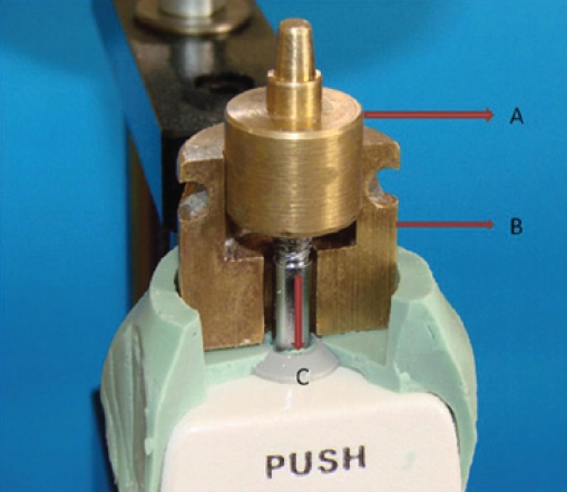
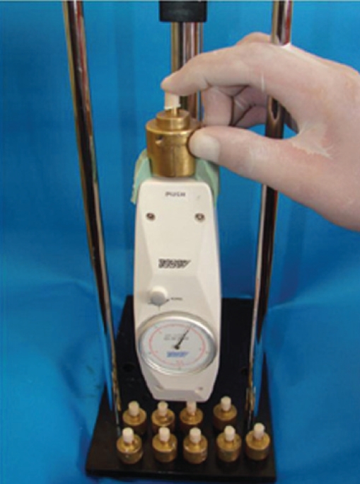
The master die, a split brass mould, and a handy analog force gauge apparatus (Algol Instrument Co, Hsin-Chuang, Taipei, Taiwan) were used to determine the pressure that would be applied in the cementation test. The mould allowed for vertical movement of the master dies and was fixed on the upper section of the test apparatus (Figure 1). Maximum cementation pressure (in newtons) was recorded based on the vertical movement of the master die within the mould.
Figure 1.
The brass mold that allows to move as vertical of master dies (A: Master die, B: Split mold C: Vertical movement).
Selection of dentists and cementation
Dentists (9 male, 6 female) were selected from the Department of Prosthodontics, Faculty of Dentistry, Erciyes University. Dentists were given no information before the study, and their cementation techniques were never interfered with during the process. Glass ionomer cement (Meron, Voco, Cuxhaven, Germany) was selected for cementation. The dentists were asked how many crowns they planned to cement in one session and then were provided with the correct powder/liquid ratio of cement according to the manufacturer’s instructions. The maximum pressure applied during cementations on the master die of all-ceramic crowns was recorded (Figure 2). However, the dentists did not see the amount of pressure they applied. Master dies were cleaned by ultrasonic cleaning after every 10 cementations. Each dentist conducted 10 cementations in the morning and 10 cementations in the afternoon. As a result, 300 cementation processes were conducted.
Figure 2.
Special pressure device to apply standard cementation force during setting of cement.
Statistical analysis
Differences in the finger pressure applied by the 15 dentists during cementation were evaluated using one-way analysis of variance (ANOVA). Cementations performed in the morning were included in this evaluation. An independent specimen t-test was used to evaluate the differences in finger pressure applied in the morning and in the afternoon. Finally, differences in pressure by dentist gender were evaluated using one-way ANOVA.
RESULTS
Mean values and standard deviations of finger pressure are shown in Table 1. The values obtained ranged from 12 to 67 N, revealing a statistically significant difference in finger pressure applied during cementation (Table 2).
Table 1.
Descriptive statistics of SBS values for all groups.
| Dentists | N | Mean±SD |
|---|---|---|
| R.K. | 20 | 24.05±6.81 |
| P.B. | 20 | 26.10±7.29 |
| Z.E. | 20 | 30.35±11.33 |
| F.Y. | 20 | 38.65±10.89 |
| A.S. | 20 | 44.05±7.66 |
| G.D. | 20 | 40.25±9.21 |
| T.B. | 20 | 39.15±8.42 |
| D.K. | 20 | 57.40±6.55 |
| O.C. | 20 | 46.45±8.43 |
| K.P. | 20 | 30.95±7.23 |
| K.K. | 20 | 54.50±6.78 |
| Y.U. | 20 | 45.60±6.45 |
| M.Z. | 20 | 36.30±7.99 |
| H.I.K. | 20 | 46.60±9.53 |
| H.O.G. | 20 | 47.75±9.77 |
Table 2.
Results of independent t test according to time for each dentist (F=female, M=male, *:P<.05; ns:P>.05).
| Dentist | Gender | Morning mean (n =10) | Evening mean (n =10) | Min | Max | P |
|---|---|---|---|---|---|---|
| R.K. | F | 20.50 | 27.60 | 12 | 30 | * |
| P.B. | F | 23.00 | 29.20 | 16 | 30 | ns |
| Z.E. | F | 23.80 | 36.90 | 12 | 35 | * |
| F.Y. | F | 42.40 | 34.90 | 20 | 46 | ns |
| A.S. | F | 43.10 | 45.00 | 32 | 51 | ns |
| G.D. | F | 45.80 | 34.70 | 18 | 43 | * |
| T.B. | F | 46.20 | 32.10 | 27 | 43 | * |
| D.K. | F | 53.00 | 61.80 | 44 | 55 | * |
| O.C. | F | 53.00 | 39.90 | 32 | 50 | * |
| K.P. | M | 27.70 | 34.20 | 15 | 35 | * |
| K.K. | M | 58.20 | 50.80 | 43 | 60 | * |
| Y.U | M | 46.60 | 44.60 | 33 | 54 | ns |
| M.Z. | M | 37.50 | 35.10 | 17 | 47 | ns |
| H.İ.K. | M | 39.50 | 53.70 | 26 | 50 | * |
| H.O.G. | M | 45.10 | 50.40 | 23 | 63 | ns |
According to the independent t-test analyses, there were statistically significant differences between morning and afternoon pressure values for nine dentists, but no difference was found for the other six dentists. Dentists conducted a total of 300 cementations, 150 in the morning and 150 in the afternoon. There was no statistically significant difference for time of day according to a one-way ANOVA (Table 3).
Table 3.
Tukey’s paired comporisons of finger pressures of dentists at the morning (*:P<.05; ns:P>.05).
| R.K. | P.B. | Z.E. | F.Y. | A.S. | G.D. | T.B. | D.K. | O.C. | K.P. | K.K. | Y.U. | M.Z. | H.I.K. | H.O.G. | |
|---|---|---|---|---|---|---|---|---|---|---|---|---|---|---|---|
| R.K. | |||||||||||||||
| P.B. | ns | ||||||||||||||
| Z.E. | ns | ns | |||||||||||||
| F.Y. | * | * | * | ||||||||||||
| A.S. | * | * | * | * | |||||||||||
| G.D. | * | * | * | * | ns | ||||||||||
| T.B. | * | * | * | * | ns | ns | |||||||||
| D.K. | * | * | * | * | ns | ns | ns | ||||||||
| O.C. | * | * | * | * | ns | ns | ns | ns | |||||||
| K.P. | ns | ns | ns | * | * | * | * | * | * | ||||||
| K.K. | * | * | * | * | * | * | * | ns | ns | * | |||||
| Y.U | * | * | * | * | ns | ns | ns | ns | ns | * | * | ||||
| M.Z. | * | * | * | * | ns | ns | ns | * | * | ns | * | ns | |||
| H.İ.K. | * | * | * | * | ns | ns | * | * | * | * | * | * | ns | ||
| H.O.G. | * | * | * | * | ns | ns | ns | * | ns | * | * | ns | ns | ns |
When the cementation pressure values were compared by gender, no statistically significant difference was found. Both the lowest (20.5 N) and highest (61 N) average values were achieved by female dentists (Table 4).
Table 4.
Finger pressure values of dentists according to time and gender
| Time | Gender | n | Mean±SD | P |
|---|---|---|---|---|
| Morning | Female | 90 | 38.98±13.88 | >0.05 |
| Male | 60 | 42.43±12.09 | ||
| Afternoon | Female | 90 | 38.01±12.23 | |
| Male | 60 | 44.80±9.97 |
DISCUSSION
In this study, we used standard brass dies to investigate whether differences in finger pressure applied by dentists during cementation vary according to gender or time of day. The results of this study do not support the hypothesis that the finger pressure applied by dentists does not vary.
In studies of cementation, researchers have cemented samples by applying either static pressure or the finger pressure of a single operator. Some of these studies reported that a finger pressure was applied by a single operator, and no pressure value was provided. Differences in pressure applied during the cementation of fixed prosthetic restorations may affect the uniformity of the cement film thickness. Such differences in cement film thickness can also affect retention, resistance, and marginal adaptation of restoration.9–11
According to Piemjai et al12 increased pressure during cementation may cause the cement to escape, providing for a better marginal adaptation. They suggested that a cementation pressure of 300 N would provide for better marginal adaptation. The maximum pressure provided by the chewing muscles varies from 200 to 600 N. According to this study, this maximum pressure has to be applied by the patient in clinical circumstances. To achieve this, the crown must first be placed on the prepared tooth using finger pressure, and then the patient has to be told to bite with a maximum force in the centric occlusion position. In contrast, the cementation pressure applied by the dentists in our study ranged from 12 to 67 N. Clearly, there is a large difference between the pressure applied by dentists and that which can be applied by patients in cementation. Amoore et al13 compared the cementation pressure of five dentists by fixing a pressure tester on their fingertips. They found that dentists applied 60 N in the first few seconds and 20–30 N thereafter. These results are consistent with the values we obtained in the present study.
We found a statistically significant difference in the dentists’ finger pressure (P<.05). Static load is applied during cementation in most studies. However, differences between the cementation pressure applied by operators and operator techniques have largely been ignored. The effects of differences in cementation pressure during cementation, both in vivo and in vitro, have not been sufficiently considered.
Tuntiparawon14 found that between 25 and 300 N of pressure applied during metal crown cementation significantly affected the marginal adaptation of restoration; however, it had no effect on retention. Goracci et al9 examined the microtensile bonding strength of Maxcem, Rely X, and Panavia F 2.0 resin cements on onlay restorations applied under various pressures. They found that a more powerful placement force was effective in reducing the distribution and frequency of porosity that may develop between the adhesive agent and the interface to be cemented. Moreover, they revealed that closer adaptation between adhesive and substrate optimized the physical interactions, such as van der Waals forces, hydrogen bridges, and charge transfers. This contributes to the micromechanics of retention and chemical bonding in the adhesion process. A recent study15 reported that if 98 N force was applied on the composite overlay during self-polymerization of Panavia F 2.0, an ideal adhesion was obtained at the dentine–cement interface. However, the maximum pressure applied in our study was 67 N. Thus, the ideal cementation pressure likely cannot be applied by the finger alone.9,15
We found that nine dentists applied different cementation pressures in the morning and afternoon. Some of these dentists used greater pressure in the morning, others in the afternoon. We anticipated that the pressure applied in the morning would be greater than that applied in the afternoon. However, this was not the case.
Finally, we found no significant difference in pressure by dentist gender (P>.05). Nevertheless, the average pressure applied by male dentists (42 N) was 4 N greater than that applied by female dentists (38 N).
CONCLUSIONS
Within the limitations of this study, the results of this paper show that the finger pressure applied by dentists varies. Additional studies on finger pressure during cementation are required. In the light of these results, equipment may be developed to apply a controlled pressure during cementation after determination of the optimal pressure. By standardizing this important factor, better cementation restorations can be achieved.
REFERENCES
- 1.Borges GA, Goes MF, Platt JA, Moore K, Menezes FH, Vedovato E. Extrusion shear strength between an alumina-based ceramic and three different cements. J Prosthet Dent. 2007;98:208–215. doi: 10.1016/S0022-3913(07)60057-2. [DOI] [PubMed] [Google Scholar]
- 2.Gorodovsky S, Zidan O. Retentive strength, disintegration, and marginal quality of luting cements. J Prosthet Dent. 1992;68:269–274. doi: 10.1016/0022-3913(92)90328-8. [DOI] [PubMed] [Google Scholar]
- 3.Wang CJ, Millstein PL, Nathanson D. Effects of cement, cement space, marginal design, seating aid materials, and seating force on crown cementation. J Prosthet Dent. 1992;67:786–790. doi: 10.1016/0022-3913(92)90583-v. [DOI] [PubMed] [Google Scholar]
- 4.Koyano E, Iwaku M, Fusayama T. Pressuring techniques and cement thickness for cast restorations. J Prosthet Dent. 1978;40:544–548. doi: 10.1016/0022-3913(78)90090-2. [DOI] [PubMed] [Google Scholar]
- 5.De Freitas OJ, Ishikiriama A, Vieira DF, Mondelli J. Influence of pressure and vibration during cementation. J Prosthet Dent. 1979;41:173–177. doi: 10.1016/0022-3913(79)90303-2. [DOI] [PubMed] [Google Scholar]
- 6.Chieffi N, Chersoni S, Papacchini F, Vano M, Goracci C, Davi Effect of the seating pressure on the adhesive bonding of indirect restorations. Am J Dent. 2006;19:333–336. [PubMed] [Google Scholar]
- 7.Karipidis A, Pearson GJ. The effect of seating pressure and powder/liquid ratio of zinc phosphate cement on the retention of. J Oral Rehabil. 1988;15:333–337. doi: 10.1111/j.1365-2842.1988.tb00165.x. [DOI] [PubMed] [Google Scholar]
- 8.White SN, Yu Z, Kipnis V. Effect of seating force on film thickness of new adhesive luting agents. J Prosthet Dent. 1992;68:476–481. doi: 10.1016/0022-3913(92)90414-6. [DOI] [PubMed] [Google Scholar]
- 9.Goracci C, Cury AH, Cantoro A, Papacchini F, Tay FR, Ferrari M. Microtensile bond strength and interfacial properties of self-etching and self-adhesive resin cement. J Adhes Dent. 2006;8:327–335. [PubMed] [Google Scholar]
- 10.Wang CJ, Millstein PL, Nathanson D. Effects of cement, cement space, marginal design, seating aid materials, and seating force on crown. J Prosthet Dent. 1992;67:786–790. doi: 10.1016/0022-3913(92)90583-v. [DOI] [PubMed] [Google Scholar]
- 11.Yu Z, Strutz JM, Kipnis V, White SN. Effect of dynamic loading methods on cement film thickness in vitro. J Prosthodont. 1995;4:251–255. doi: 10.1111/j.1532-849x.1995.tb00351.x. [DOI] [PubMed] [Google Scholar]
- 12.Piemjai M. Effect of seating force, margin design, and cement on marginal seal and retention of complete metal crowns. Int J Prosthodont. 2001;14:412–416. [PubMed] [Google Scholar]
- 13.Black S, Amoore JN. Measurement of forces applied during the clinical cementation of dental crowns. Physiol Meas. 1993;14:387–392. doi: 10.1088/0967-3334/14/3/018. [DOI] [PubMed] [Google Scholar]
- 14.Tuntiprawon M. Effect of tooth surface roughness on marginal seating and retention of complete metal crowns. J Prosthet Dent. 1999;81:142–147. doi: 10.1016/s0022-3913(99)70241-6. [DOI] [PubMed] [Google Scholar]
- 15.Chieffi N, Chersoni S, Papacchini F, Vano M, Goracci C, Davidson CL, Tay FR, Ferrari M. The effect of application sustained seating pressure on adhesive luting procedure. Dent Mater. 2007;23:159–164. doi: 10.1016/j.dental.2006.01.006. [DOI] [PubMed] [Google Scholar]


