Long considered to be an immune-privileged site because of the presence of the blood-brain-barrier (BBB) and the lack of a lymphatic system, it is now well-established that the brain is fully capable of mounting inflammatory responses in response to invading pathogens, trauma, or ischemic events. Within the microenvironment of the brain, brain-resident macrophages termed microglia serve a critical role in normal central nervous system (CNS) function by mediating innate immune responses. Although microglial cells are the primary mediators of neuroinflammatory responses, astrocytes and oligodendrocytes can also participate in the process. Astrocytes regulate glutamate uptake and thus provide homoeostatic control of the extracellular environment in the CNS; but they too can also become activated by physical damage or ischemia in a process termed reactive gliosis that is characterized by up-regulation of the glial fibrillary acidic protein (GFAP). Activation of oligodendrocytes results in secretion of inflammatory molecules, such as nitric oxide (NO), cytokines, and prostaglandins and most notably in upregulation of several chondroitin sulfate proteoglycans, including NG2, which contributes to the growth-inhibitory environment that prevents regeneration of axons in the injured CNS. Immune-mediated damage to oligodendrocytes as a result of innate and adaptive immune system attack in patients with multiple sclerosis results in extensive demyelination, loss of oligodendrocytes and axonal degeneration; but the extent to which oligodendrocytes participate in chronic neuroinflammatory degenerative disease and the contribution to inflammatory responses is not well understood. In summary, in acute situations and when short-lived, neuroinflammatory mechanisms generally limit injury and promote healing; however, when neuroinflammation becomes chronic it can damage viable host tissue, resulting in compromised neuronal survival and cognitive impairment. For these reasons, inflammation in the CNS has been appropriately described as a two-edged sword (Wyss-Coray and Mucke 2002; McGeer and McGeer 2004).
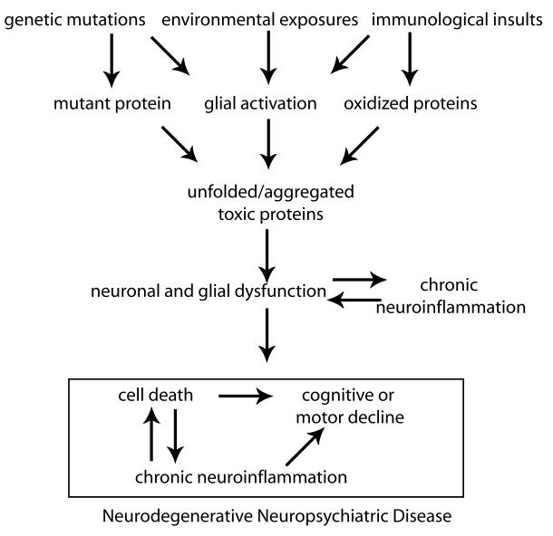
Chronic neuroinflammation is associated with a broad spectrum of neurodegenerative diseases of aging, including Alzheimer’s disease (AD), Parkinson’s disease (PD), amyotrophic lateral sclerosis (ALS), spinal muscular atrophy (SMA), and all of the tauopathies (McGeer and McGeer 2004; Block and Hong 2005; Mrak and Griffin 2005; Nagatsu and Sawada 2006). Neurodegenerative diseases are characterized by the loss of specific neuronal populations and often by intraneuronal as well as extracellular accumulation of fibrillary materials. Formation of intracellular inclusion bodies may result from abnormal protein-protein interactions, aberrant protein folding, and/or dysregulation of the ubiquitin-proteasome system (UPS). These conditions, often referred to as ‘proteinopathies,’ are now thought to play a principal role in neuronal dysfunction and death of neurons that characterizes several common neurodegenerative diseases (Ross and Poirier 2004; Selkoe 2004; Moore 2005; Lansbury and Lashuel 2006). Although the key molecular and cellular events underlying development of AD, PD, HD, and ALS are clearly divergent, it should be pointed out that one common way in which a number of divergent molecular or cellular events (e.g., mutations, oxidation, protein misfolding, truncation, or aggregation) may all contribute over time to death of neurons is via activation of resident microglial populations in specific brain regions. If the initial trigger that elicited microglial activation is not resolved (as in the case of a genetic mutation or a prolonged or repeated environmental exposure), a self-sustaining cycle of neuroinflammation is likely to ensue and contribute to neuronal dysfunction and eventual death of vulnerable neuronal populations leading to motor and/or cognitive decline (Figure 1). In support of this idea, formation of intracellular and extracellular protein aggregates as well as other cellular and molecular processes that activate microglia in the CNS and promote acute or chronic neuroinflammation have been identified in recent years and are characteristic of chronic neurodegenerative conditions. These include accumulation of abnormally modified cellular components, molecules released from or associated with injured neurons or synapses, and deregulation of inflammatory control mechanisms such as those that occur with aging. Thus, experimental, clinical, and epidemiological studies strongly suggest that activation of resident microglial populations may be occurring in parallel with the neuronal dysfunction underlying the neurodegenerative disease process. This Special Issue highlights reviews on a number of different neuropsychiatric conditions characterized by neuroinflammation (Alzheimer’s disease, Parkinson’s disease, Amyotrophic Lateral Sclerosis and Spinal Muscular Atrophy, Human Immunodeficiency Virus, Cytokine-induced depression, and Lyme’s and other tick-borne diseases) in which chronic neuroinflammation and/or neuroimmune dysregulation is believed to play central roles in disease pathophysiology and progression. It is our hope that raising awareness of the known neuroinflammatory features as well as the enigmatic or unresolved issues in each will serve to heighten research interest in these areas with the long-term goal of exploring the opportunity for therapeutic intervention that could ameliorate these diseases or slow down their course in patients afflicted with these conditions.
Figure 1.
References
- Block ML, Hong JS. Microglia and inflammation-mediated neurodegeneration: multiple triggers with a common mechanism. Prog Neurobiol. 2005;76(2):77–98. doi: 10.1016/j.pneurobio.2005.06.004. [DOI] [PubMed] [Google Scholar]
- Lansbury PT, Lashuel HA. A century-old debate on protein aggregation and neurodegeneration enters the clinic. Nature. 2006;443(7113):774–779. doi: 10.1038/nature05290. [DOI] [PubMed] [Google Scholar]
- McGeer PL, McGeer EG. Inflammation and the degenerative diseases of aging. Ann N Y Acad Sci. 2004;1035:104–116. doi: 10.1196/annals.1332.007. [DOI] [PubMed] [Google Scholar]
- Moore DJ, West AB, Dawson VL, Dawson TM. Molecular pathophysiology of Parkinson’s Disease. Annu Rev Neurosci. 2005;28:57–87. doi: 10.1146/annurev.neuro.28.061604.135718. [DOI] [PubMed] [Google Scholar]
- Mrak RE, Griffin WS. Glia and their cytokines in progression of neurodegeneration. Neurobiol Aging. 2005;26(3):349–354. doi: 10.1016/j.neurobiolaging.2004.05.010. [DOI] [PubMed] [Google Scholar]
- Nagatsu T, Sawada M. Cellular and molecular mechanisms of Parkinson’s disease: neurotoxins, causative genes, and inflammatory cytokines. Cell Mol Neurobiol. 2006;26(4-6):781–802. doi: 10.1007/s10571-006-9061-9. [DOI] [PMC free article] [PubMed] [Google Scholar]
- Ross CA, Poirier MA. Protein aggregation and neurodegenerative disease. Nat Med. 2004;10(Suppl):S10–17. doi: 10.1038/nm1066. [DOI] [PubMed] [Google Scholar]
- Selkoe DJ. Cell biology of protein misfolding: the examples of Alzheimer’s and Parkinson’s diseases. Nat Cell Biol. 2004;6(11):1054–1061. doi: 10.1038/ncb1104-1054. [DOI] [PubMed] [Google Scholar]
- Wyss-Coray T, Mucke L. Inflammation in neurodegenerative disease--a double-edged sword. Neuron. 2002;35(3):419–432. doi: 10.1016/s0896-6273(02)00794-8. [DOI] [PubMed] [Google Scholar]


