Abstract
The production of neurons from neural progenitor cells, the growth of axons and dendrites and the formation and reorganization of synapses are examples of neuroplasticity. These processes are regulated by cell-autonomous and intercellular (paracrine and endocrine) programs that mediate responses of neural cells to environmental input. Mitochondria are highly mobile and move within and between subcellular compartments involved in neuroplasticity (synaptic terminals, dendrites, cell body and the axon). By generating energy (ATP and NAD+), and regulating subcellular Ca2+ and redox homoeostasis, mitochondria may play important roles in controlling fundamental processes in neuroplasticity, including neural differentiation, neurite outgrowth, neurotransmitter release and dendritic remodelling. Particularly intriguing is emerging data suggesting that mitochondria emit molecular signals (e.g. reactive oxygen species, proteins and lipid mediators) that can act locally or travel to distant targets including the nucleus. Disturbances in mitochondrial functions and signalling may play roles in impaired neuroplasticity and neuronal degeneration in Alzheimer's disease, Parkinson's disease, psychiatric disorders and stroke.
Keywords: neural progenitor cell, mitochondria biogenesis, mitochondria fission and fusion
Abbreviations: Aβ, amyloid β-peptide; AD, Alzheimer's disease; AP, adaptor protein; APP, amyloid precursor protein; BDNF, brain-derived neurotrophic factor; CaMK, Ca2+/calmodulin-dependent protein kinase; CR, caloric restriction; CREB, cAMP-response-element-binding protein; ES, embryonic stem; ETC, electron transport chain; HD, Huntington's disease; LRRK2, leucine-rich repeat kinase 2; LTP, long-term potentiation; MAPK, mitogen-activated protein kinase; Mn-SOD, manganese superoxide dismutase; NGF, nerve growth factor; NMDA, N-methyl-d-aspartate; Nrf1, nuclear respiratory factor 1; OPA1, Optic Atrophy-1; PD, Parkinson's disease; PGC1α, peroxisome-proliferator-activated receptor γ co-activator 1α; PINK1, PTEN (phosphatase and tensin homologue deleted on chromosome 10)-induced kinase 1; PPAR, peroxisome-proliferator-activated receptor; UCP, uncoupling protein
INTRODUCTION
Neuroplasticity is a term used to describe a range of adaptive changes that occur in the structure and function of cells in the nervous system in response to physiological or pathological perturbations. Examples of neuroplasticity include the sprouting and growth of axons or dendrites, synapse formation, the strengthening of synapses in response to repeated activation and neurogenesis (the production of new neurons from stem cells). The biological basis of this capacity for structural and functional adaptation encompasses a diverse set of cellular and molecular mechanisms, including the pre- and post-synaptic apparatuses for neurotransmission, cytoskeletal remodelling, membrane trafficking, gene transcription, protein synthesis and proteolysis (Shepherd and Huganir, 2007; Bramham, 2008; Greer and Greenberg, 2008; Pak et al., 2008; Tai and Schuman, 2008; Shah et al., 2010). In addition, glial cells (astrocytes, microglia and oligodendrocytes) play important roles in neuroplasticity by producing soluble and surface-bound factors that influence neurite outgrowth, synaptic plasticity and cell survival (Haydon and Carmignoto, 2006; Barres, 2008).
Among the many specialized cell types in the body, neurons are particularly marvellous because they elaborate tree-like shapes, are electrically excitable, and engage in spectacular spatiotemporal displays of signal detection, integration and storage, and generation of adaptive responses to life situations. Newly generated neurons possess an intrinsic pre-ordained sequence of events that determine at least some of their structural and functional phenotypes. For example, the dendritic morphology of cerebellar Purkinje cells is largely established independently of interactions with other cells (Sotelo and Dusart, 2009). Interestingly, the most closely related neuronal cells, mitotic sister neurons arising from a common progenitor, elaborate morphologies more similar to themselves compared with their close neighbours (Mattson et al., 1989). While the cytoarchitecture and functional capabilities of the nervous system are highly complex, several of the signalling mechanisms that control the formation and adaptive plasticity have been elucidated. Neurotransmitters, neurotrophic factors and cell adhesion molecules are three highly conserved classes of intercellular signals that regulate the genesis and adaptive plasticity of nervous systems (Mattson, 1988; Loers and Schachner, 2007; Gottmann et al., 2009). The most intensively studied representatives of these three classes of signals are the excitatory neurotransmitter glutamate (Mattson, 2008; Newpher and Ehlers, 2008), BDNF (brain-derived neurotrophic factor; Mattson et al., 2004; Lipsky and Marini, 2007) and the NCAM (neural cell adhesion molecule; Hildebrandt et al., 2007). Details of the components of these signalling pathways will not be covered in the present paper. Instead, we focus on emerging findings regarding the interactions of the latter signalling pathways with mitochondria, an organelle increasingly recognized as not only a power station, but also as a signalling platform involved in fundamental events in the formation and plasticity of neuronal circuits.
Mitochondria are vital ATP-generating organelles present in all eukaryotic cells. In addition to converting energy substrates into ATP, mitochondria participate in ROS (reactive oxygen species) metabolism, calcium signalling and apoptosis (Mattson et al., 2008). Mitochondria are distributed throughout the length of axons and in presynaptic terminals. Mitochondria in dendrites are located mainly in the dendritic shafts and are occasionally found associated with spines (Cameron et al., 1991; Popov et al., 2005).
The application of novel imaging and molecular biology technologies to studies of mitochondria has revealed several surprising properties and functions of mitochondria in neuroplasticity. For example, we now know that mitochondria (i) move rapidly within and between subcellular compartments (e.g. dendrites, the axon shaft, presynaptic terminals) (Zinsmaier et al., 2009); (ii) undergo fission and fusion (Liesa et al., 2009); (iii) respond (e.g. move, change their energy output, take up or release calcium) to electrical activity and activation of neurotransmitter and growth factor receptors (Macaskill et al., 2009); and (iv) function as signalling outposts that contain kinases, deacetylases and other signal transduction enzymes (Stowe and Camara, 2009). Mitochondrial fission and fusion are mediated by two distinct protein complexes involving GTPase. Key molecular mechanisms involved in mitochondrial fission and fusion have recently been elucidated (Berman et al., 2008). Two key proteins, Drp (dynamin-related protein) and Fis1, mediate mitochondrial fission, whereas mitofusins (Mfn1 and Mfn2) and OPA1 (Optic Atrophy-1) mediate mitochondrial fusion (Karbowski et al., 2002; Shaw and Nunnari, 2002; Koshiba et al., 2004; Detmer and Chan, 2007). Mitochondrial fission and fusion can occur rapidly (within less than 1 min) and remodel individual mitochondria continuously. Mitochondria dynamics are important for neuronal functions since they regulate mitochondrial location, morphology, number and function (Detmer and Chan, 2007).
The notion that mitochondria participate in neuroplasticity is not new. For example, more than four decades ago, Sotelo and Palay (1968) examined electron micrographs of the lateral vestibular nucleus and observed: “The distal segments of some dendrites display broad expansions packed with slender mitochondria and glycogen particles. These distinctive formations are interpreted as being growing tips of dendrites, and the suggestion is advanced that they are manifestations of architectonic plasticity in the mature central nervous system.” In the present review article, we consider the emerging roles of mitochondria in neuroplasticity.
Mitochondria and neurogenesis
Neurogenesis, the birth of new neurons from stem cells, occurs rapidly and globally during development of the nervous system, and to a much more limited extent in some regions of the adult nervous system. Adult neurogenesis is believed to be functionally important as a mechanism of brain plasticity under physiological conditions and in brain repair after injury (Kempermann et al., 2004; Ge et al., 2008; Kitamura et al., 2009). How are mitochondria involved in the process of neurogenesis? First, the mitochondria may play a role in the self-renewal capacity of neural stem cells, a defining property of all stem cells. Studies of mouse ES (embryonic stem) cells suggest that the proliferative capacity of stem cells is correlated with low mitochondrial oxygen consumption and high levels of glycolytic activity (Kondoh et al., 2007). Using typical bicarbonate buffer and a 5% CO2/95% air atmosphere, cells are exposed to much higher levels oxygen compared with what they would be exposed to in vivo. Reducing the oxygen level enhances the proliferation and multipotency of neural stem cells (Rodrigues et al., 2010; Santilli et al., 2010). Human ES cells exhibit an ‘anaerobic’ metabolic profile, and when somatic cells are induced to revert to an ES cell-like phenotype, their mitochondria also revert to an ES cell-like state with respect to their morphology, subcellular distribution, biogenesis, and ROS and ATP production (Prigione et al., 2010). Cultured neural crest stem cells incubated in the presence of bone morphogenetic protein 2 and forskolin can be coaxed, by exposure to mild hypoxia, to differentiate into a relatively pure population of sympathoadrenal neuronal cells that produce adrenaline (norepinephrine) and dopamine (Morrison et al., 2000). A link between mitochondrial Ca handling and neurogenesis is suggested by results showing that, when neuroblastoma cells are induced to stop dividing and differentiate into neuron-like cells, there is an increase of mitochondrial fusion and an increase in intramitochondrial Ca2+ levels (Voccoli and Colombaioni, 2009).
The second step in neurogenesis is the differentiation of neural progenitors into postmitotic neurons. While much has been learned regarding the molecular changes involved in neuronal differentiation (Hamby et al., 2008; Zhang et al., 2008; Cane and Anderson, 2009), very little is known of the roles of mitochondria in these processes. Neuronal differentiation is associated with an increase in mitochondrial mass per cell, and the mitochondrial translation inhibitor chloramphenicol prevents differentiation, indicating participation of the mitochondrial genome and mitochondrial protein synthesis in neuronal differentiation (Vayssière et al., 1992). The increased mitochondrial biogenesis associated with differentiation provides ATP to support fundamental cellular processes involved in neurite outgrowth (cytoskeletal dynamics, membrane turnover and transport of various RNA and protein cargos).
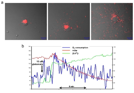
While mitochondrial mass increases on neuronal differentiation to increase ATP production (Figure 1), energy generation is only one mechanism by which mitochondria enable neurite outgrowth. Mitochondrial UCPs (uncoupling proteins) have received increasing attention regarding their roles in physiological processes other than heat generation in brown fat cells (Mattson and Liu, 2003). By leaking protons across the mitochondrial inner membrane, UCPs not only reduce ATP production, but also decrease the generation of ROS and modify mitochondrial and endoplasmic reticulum Ca2+ dynamics (Chan et al., 2006; Liu et al., 2006). UCP4 is expressed preferentially in neurons (Liu et al., 2006) and its developmental expression characteristics suggest a role for UCP4 in neuronal differentiation (Smorodchenko et al., 2009). In addition, the transcription factor NeuroD6 can induce the differentiation of the neuronal progenitor-like PC12 cells and up-regulate the expression of several mitochondria-related genes; NeuroD6-induced expression of mitochondrial genes is maximum at a very early stage of neurite outgrowth (Baxter et al., 2009). One mitochondria-related gene induced by NeuroD6 is KIF5B, a kinesin motor protein involved in mitochondrial transport.
Figure 1. Examples of methods for evaluating mitochondrial morphology, subcellular localization and functional status.
(a) Each panel shows a single live embryonic rat hippocampal neuron at a different stage of development in culture (1, 5 and 14 days). Mitochondria in the neurons were stained with the fluorescent probe MitoTracker Red. Note that the immature neuron has elaborated short processes, and that the vast majority of mitochondria are clustered in a perinuclear location. At 5 days in culture the neuron exhibits longer neurites that contain multiple mitochondria, and perinuclear mitochondria remain abundant. Mitochondrial numbers have increased, indicating that biogenesis has occurred. At 14 days in culture the neuron has elaborated an extensive neuritic network with each neurite containing multiple mitochondria. Relative numbers of mitochondria in a perinuclear location are reduced. (b) Example of the results of an experiment in which oxygen consumption, mitochondrial membrane potential and cytoplasmic Ca2+ levels were monitored in a single cultured embryonic rat hippocampal neuron before and during exposure to the excitatory neurotransmitter glutamate. In response to glutamate receptor activation, oxygen consumption increased and then slowly returned towards baseline levels, intracellular Ca2+ levels rose rapidly and remained elevated, whereas mitochondrial membrane potential declined progressively. Adapted from Gleichmann et al. (2009).
Mitochondria and neurite outgrowth
During their differentiation, neurons extend neurites with one becoming the axon and others becoming dendrites. This polarity is important in the formation of functional neuronal circuits.
During axogenesis, mitochondria congregate at the base of the developing neurites that are destined to become axon (Mattson and Partin, 1999). Once the axon forms and accelerates its growth there is increased entry of mitochondria into the new axon where they are concentrated at the growth cone (Ruthel and Hollenbeck, 2003). Rather strikingly, depletion of mitochondria at or before the stage of axogenesis, under conditions where cellular ATP levels are maintained using pyruvate and uridine as energy sources, prevents axon formation (Mattson and Partin, 1999). Furthermore, in dorsal root ganglion cells isolated from neonatal rats, the mitochondria become reorganized to form clusters in the axonal hillock of regenerating axons (Dedov et al., 2000). These studies suggest that mitochondria play an important role in neural polarization and axonal outgrowth regulation.
The mechanisms by which mitochondria influence the establishment of neuronal polarity remain to be established. It was reported that the cytoplasmic free Ca2+ concentration is lower surrounding the mitochondria at the base of the developing axon in cultured embryonic rat hippocampal neurons, and that exposure of the neurons to a calcium ionophore (which increases the cytoplasmic Ca2+ concentration throughout the cell) prevents axon formation (Mattson and Partin, 1999). One possible explanation is that mitochondria act as a calcium buffering organelle to reduce the Ca2+ concentration at the base of the presumptive axon, thereby promoting polymerization of microtubules and the rapid growth and differentiation of the axon (Figures 2 and 3).
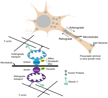
Figure 2. Molecular machinery that actively moves mitochondria to and fro within axons.
A major mechanism by which mitochondria are transported in either anterograde or retrograde directions in axons involves their energy (ATP)-dependent movement along microtubules. ATP-dependent ‘motor’ proteins interact with the microtubules to generate the force that moves the mitochondria in anterograde (kinesin) or retrograde (dynein) directions respectively. Several APs (adaptor proteins) mediate the interaction of mitochondria with motor proteins, including APs that interact with kinesin (Milton, syntabulin and the Rho GTPase Miro) and APs that associate with dynein (dynactin). In addition, in synaptic terminals and growth cones, microtubules may be moved by myosin-mediated interactions with actin filaments. Myosin V can drive short-range movements along F-actin, as well as modulate long-range transport by pulling mitochondria away from microtubules by facilitating anchorage of mitochondria to F-actin by unknown actin–mitochondrion crosslinkers. Adapted from Mattson et al. (2008).
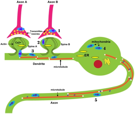
Figure 3. The landscape of mitochondrial involvement in the plasticity of neuronal structure and information processing.
Increasing evidence suggests that mitochondria play active roles in regulating the outgrowth of axons and dendrites, synaptogenesis and morphological and functional responses to synaptic activity. Mitochondria in presynaptic terminals (1) provide the energy for the maintenance and restoration of membrane potential, and may modulate neurotransmitter packaging and release. Mitochondria in postsynaptic spines (2) and dendritic shafts (3) may enable/regulate both structural and functional responses of these compartments to synaptic activity. Mitochondria in the cell body (4) provide the energy required for numerous biochemical processes, and may also serve as signalling platforms involved in information transfer within the neuron. Mitochondria in axons (5) provide the energy necessary for the transport of various proteins and organelles from the axon terminal to the cell body and vice versa.
In addition to playing critical roles in neural polarization and axonal outgrowth regulation, mitochondria may also be required for normal dendrite development, maintenance and plasticity. Studying the correlation between mitochondrial energy status and mitochondrial membrane potential, Overly et al. (1996) found that mitochondria in dendrites are metabolically more active than those in the axons, although the reason for this differential mitochondrial activity is unknown. Recent studies provide experimental evidence that mitochondria are important in regulating dendrite development, maintenance and plasticity. Disruption of mitochondrial protein translation in Drosophila olfactory projection neurons preferentially reduces dendritic arborization, while axon morphology is relatively unaltered (Chihara et al., 2007). Dendritic mitochondria also have essential roles in dendritic spine morphogenesis and plasticity.
Mechanisms by which mitochondria move within neurites are beginning to be understood; their movement is affected by energy-dependent transport along microtubules. Mitochondrial transport can occur bidirectionally; microtubule plus end-directed kinesin moves mitochondria in the anterograde direction, whereas minus end-directed dynein motors move mitochondria retrogradely (Hollenbeck and Saxton, 2005; Zinsmaier et al., 2009; Pathak et al., 2010; Figure 2). Measurements of the membrane potential of individual mitochondria in the growing axons of chick sensory neurons using the dye TMRM (tetramethylrhodamine methyl ester) revealed no major differences among mitochondria along the length of the axon, and no differences in membrane potential in stationary versus moving mitochondria (Verburg and Hollenbeck, 2008). However, the membrane potential of mitochondria in the lamellipodia of growth cones is significantly greater than the membrane potential of mitochondria in the axon shaft. In another study that employed the mitochondrial membrane potential-sensing dye JC-1 to image mitochondria in growing axons of cultured chick sensory neurons, it was found that most of the mitochondria with a high potential were transported towards the growth cone, whereas most mitochondria with a low potential were transported towards the cell body (Miller and Sheetz, 2004).
Using beads coupled with signals for axon outgrowth [NGF (nerve growth factor)] or guidance (semaphoring 3A), it was shown that both of these signals cause an increase in the membrane potential of mitochondria immediately adjacent to the site of the beads (Verburg and Hollenbeck, 2008). Additional data in the latter study provided evidence that PI3K (phosphoinositide 3-kinase) and MAPK (mitogen-activated protein kinase) mediated the effects of NGF and semaphorin 3A on mitochondrial potential. Quantitative analyses of motility show that the accumulation of axonal mitochondria near a focus of NGF stimulation is due to increased movement into bead regions followed by inhibition of movement out of these regions and that anterograde movement and retrograde movement are differentially affected. In axons made devoid of F-actin by latrunculin B treatment, bidirectional transport of mitochondria continues, but they can no longer accumulate in the region of NGF stimulation. Additional experiments provided evidence that the regulation of mitochondrial movement by NGF signalling involves increased transport to the sites of stimulation in combination with retention of the mitochondria by interactions with the actin cytoskeleton (Chada and Hollenbeck, 2004).
Although most of the ATP production by mitochondria occurs in the ETC (electron transport chain), mitochondrial glycogenesis may enable or regulate physiological processes in neurons, including neurite outgrowth. It is clear that neuronal cells can survive without a functioning mitochondrial ETC, as demonstrated in cultured cells in which mitochondria are depleted of their ATP and provided lactate and pyruvate as energy substrates (Miller et al., 1996; Hyun et al., 2007). One example comes from studies in which the activity of hexokinase was manipulated in growing neurons; hexokinase is an enzyme that plays a pivotal role early in the glycolytic pathway in which glucose is metabolized to generate ATP. When hexokinase is selectively inhibited using a hexokinase-binding peptide, the ability of NGF to stimulate neurite outgrowth in cultured adult sensory neurons is impaired (Wang et al., 2008a).
Mitochondria and synaptic plasticity
The behaviour and functional properties of mitochondria differ in axons and dendrites. For example, twice as many mitochondria are motile in the axons compared with the dendrites of cultured hippocampal neurons, and there is a greater proportion of highly charged, more metabolically active mitochondria in dendrites than in axons (Overly et al., 1996). Both presynaptic axon terminals and postsynaptic dendrites experience considerable metabolic and oxidative stress as a result of activation of membrane voltage- and ligand-gated Ca2+ channels (Bezprozvanny and Mattson, 2008). Mitochondrial functions in synaptic plasticity have been mostly studied at glutamatergic synapses, which are the most abundant type of excitatory synapse in the central nervous system. Activation of AMPA (α-amino-3-hydroxy-5-methylisoxazole-4-propionic acid), NMDA (N-methyl-d-aspartate) and metabotropic glutamate receptors results in Ca2+ influx through plasma membrane voltage-dependent channels and NMDA receptor channels, and Ca2+ release from the endoplasmic reticulum (Toresson and Grant, 2005). The mitochondria are essential components in synaptic transmission, since they are a major source of energy (ATP and NAD+) required for maintenance and restoration of ion gradients; mitochondria are also intimately involved in the regulation of Ca2+ homoeostasis and Ca2+ signalling (Duchen, 2000; Nicholls and Budd, 2000; Toescu 2000). Presynaptic terminals typically contain several mitochondria (Sheehan et al., 1997), indicating that the local need for ATP and Ca2+ handling is great in this compartment. In response to neuronal stimulation, synaptic mitochondria redistribute and enhance their activity (Miller and Sheetz, 2004).
Mitochondria in dendrites are located mainly in the dendritic shafts and are occasionally found associated with spines (Cameron et al., 1991; Popov et al., 2005). The dendritic distribution of mitochondria is regulated by neuronal activity and mitochondrial fission/fusion proteins Drp1 and OPA1. Li et al. (2004) showed that the number of dendritic spines with mitochondria increases when the dendritic spines are actively growing (embryonic rat hippocampal neurons 11–14 days in culture), or when the neurons are subjected to repeated depolarization with KCl (Li et al., 2004). Overexpressing a dominant-negative form of Drp1 (A38K) reduces the number of dendritic mitochondria and decreases the number of synapses, whereas overexpressing wild-type Drp or treating cells with creatine increases the number and/or activity of dendritic mitochondria and nearly doubles the number of synapses. Therefore mitochondria are important in dendritic spine morphogenesis and plasticity.
Changes in mitochondria have been associated with LTP (long-term potentiation) of synaptic transmission, a key event in the learning and memory process (Williams et al., 1998; Calabresi et al., 2001; Li et al., 2010). On the presynaptic side there is evidence that the protein syntaphilin is a negative regulator of mitochondrial motility in axons where it may modulate mitochondria-mediated Ca2+ signalling at presynaptic terminals (Kang et al., 2008). Kinesin-mediated transport of mitochondria to presynaptic terminals was demonstrated to be important for synaptic transmission as demonstrated in studies of the effects of Milton-deficient photoreceptors in Drosophila (Stowers et al., 2002). The GTPase dMiro is also required for axonal transport of mitochondria to Drosophila synapses (Guo et al., 2005). Further work will be required to establish specific and essential roles for mitochondrial recruitment to synapses in LTP and other forms of synaptic plasticity.
While changes in the location of mitochondria within axons and dendrites may play roles in synaptic plasticity, even more rapid functional changes in mitochondria are increasingly implicated. These changes may include mitochondrial Ca2+ uptake or release, the production of superoxide and other ROS, and the release of proteins and other factors. Post-tetanic potentiation, a form of plasticity that arises from a persistent presynaptic Ca2+ elevation after tetanic stimulation, is blocked by inhibitors of mitochondrial Ca2+ uptake and release (Tang and Zucker, 1997). Chemical blockade of mitochondrial permeability transition pores results in a reduction in mitochondrial superoxide production (Wang et al., 2008b) and an increase of basal synaptic transmission and impaired synaptic plasticity (Levy et al., 2003). Other findings suggest that presynaptic mitochondria play a role in the maintenance of synaptic transmission by sequestering Ca2+ and thereby accelerating recovery from synaptic transmission during periods of moderate to high synaptic activity (Billups and Forsythe, 2002). Neurons in Drosophila Drp1 mutants exhibit synapses devoid of mitochondria and elevations in resting cytoplasmic Ca2+ levels at neuromuscular junctions (Verstreken et al., 2005). Basal synaptic transmission is largely normal in Drp1 mutant flies, but during intense stimulation mutants fail to maintain normal neurotransmission. Although exo- and endo-cytosis are normal in the mutants, the ability to mobilize reserve pool vesicles is impaired as the result of reduced ATP availability. Moreover, age-related cognitive impairment in rats, and presumably the synaptic plasticity subserving learning and memory, is associated with structural abnormalities in mitochondria and oxidation of RNA and DNA (Liu et al., 2002).
Emerging findings suggest roles for mitochondria as mediators of at least some of the effects of glutamate and BDNF on synaptic plasticity. An increasing number of signalling functions for mitochondria are being discovered. For example, glutamate receptor-mediated patterned synaptic activity, such as occurs during stimulus train-induced bursting, results in slow and prolonged changes in mitochondrial potential that exhibit both temporal and spatial correlations with the intensity of the electrical activity (Bindokas et al., 1998). The patterned changes in mitochondrial membrane potential involve glutamate receptor-mediated Ca2+ influx. Synaptic activation of glutamate receptors may also affect mitochondrial bioenergetics independently of Ca2+ influx as suggested by studies of stimulus-evoked changes in NAD(P)H fluorescence at CA1 synapses in hippocampal slices (Shuttleworth et al., 2003). Several neurotrophic factors have been shown to modify synaptic plasticity including BDNF, which plays a pivotal role in hippocampus-dependent learning and memory (Lu et al., 2009). BDNF promotes synaptic plasticity, in part, by enhancing mitochondrial energy production because it increases glucose utilization in cultured cortical neurons in response to enhanced energy demand (Burkhalter et al., 2003) and increases mitochondrial respiratory coupling at Complex I (Markham et al., 2004). Consistent with this possibility, BDNF expression and signalling is increased in response to environmental factors such as exercise, cognitive stimulation and dietary energy restriction that increase cellular energy demand (Mattson et al., 2004).
Many questions remain unanswered concerning the roles of mitochondria in synaptic plasticity. Are mitochondria in presynaptic terminals phenotypically different from those in dendrites? If and how does synaptic activity affect mitochondrial function and motility? This could be determined by combining manipulations of specific signalling components (glutamate receptors, Ca2+-dependent and other kinases, cyclic nucleotides, nitric oxide etc.) with high-resolution imaging-based measurements of mitochondrial motility and functional status (membrane potential, ROS and Ca2+ levels etc.). Behavioural and electrophysiological evaluation of synaptic plasticity in mice with a conditional knockdown of proteins with specific roles in mitochondrial motility (Drp1, Fis1, Mfn and OPA) and function [components of the mitochondrial ETC (electron transport chain), PTP (protein tyrosine phosphatase), Ca2+ handling systems, for example] should help establish the roles of these proteins in synaptic plasticity.
Mitochondrial biogenesis and neuroplasticity
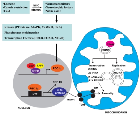
Mitochondrial biogenesis is defined simply as the growth and division of mitochondria. The human mitochondrial genome contains 37 genes that encode 13 polypeptides, 22 tRNAs and two rRNAs. All 13 polypeptides are subunits of electron transport protein complexes in the inner membrane of mitochondria. The vast majority of mitochondrial proteins (∼1000 proteins) are encoded by nuclear genes and include proteins involved in controlling mitochondrial membrane potential and ion fluxes, ATP production, and fission and fusion. These nuclear DNA-encoded mitochondrial proteins are imported into mitochondria, wherein they are sorted to their sites of action (Figure 4). In addition, the growth of mitochondria requires the synthesis of lipid bilayers of the inner and outer mitochondrial membranes. Mitochondrial DNA replication and mitochondrial fission and fusion mechanisms must also be co-ordinated.
Figure 4. Mechanisms that regulate the growth and function of mitochondria.
The vast majority of mitochondrial proteins are encoded by nuclear genes, whereas only 13 proteins that are all components of the ETC are encoded by mitochondrial genes. Proteins are imported into mitochondria by translocases of the outer membrane (TOM) and inner membrane (TIM) in a mitochondrial membrane potential-dependent manner. Mitochondrial DNA transcription is mediated by an RNA polymerase, the transcription factor A (TFAM), TFB1M or TFB2M for transcription initiation, and mTERE for transcription termination. Neurons contain particularly high levels of PGC1α, apparently because of their high energy utilization. PGC1α is a transcriptional co-activator that interacts with various transcription factors, including the nuclear receptors PPARγ, PPARα, oestrogen, thyroid hormone and retinoid receptors and Nrf1/2. Nrf1/2 induces the transcription of nuclear genes that encode ETC proteins, TFAM and proteins critical for mitochondrial biogenesis. Environmental factors (exercise, energy restriction and cold temperature, for example) activate signalling pathways involving neurotransmitters, neurotrophic factors and nitric oxide (for example), and intracellular cascades involving Ca2+, CaMKII (Ca2+/calmodulin-dependent protein kinase II), the phosphatase calcineurin, and the transcription factor CREB (cAMP-response-element-binding protein). In this manner, PGC1α is activated and mitochondrial biogenesis is stimulated. Modified from Mattson et al. (2008).
PGC1α (peroxisome-proliferator-activated receptor γ co-activator 1α) is a master regulator of mitochondrial biogenesis (Figure 4). PGC1α is expressed at high levels in mitochondria-rich cells with high energy demands, including cardiac myocytes, skeletal-muscle cells and neurons (Andersson and Scarpulla, 2001). It is a transcriptional co-activator that lacks DNA-binding activity; instead it interacts with and co-activates numerous transcription factors, including nuclear receptors such as PPARγ (peroxisome-proliferator-activated receptor γ), PPARα, oestrogen, thyroid hormone and retinoid receptors, and other nuclear transcription factors such as Nrf1 (nuclear respiratory factor 1). Nrf1 can activate the expression of mitochondrial target genes, including the mitochondrial transcription factor (TFAM), a factor critical for the initiation of mitochondrial DNA transcription and replication. Nrf1 can further regulate the transcription of nuclear genes encoding respiratory complex subunits and other mitochondrial proteins.
An interesting aspect of mitochondrial biogenesis is that it is influenced by dietary energy intake and expenditure. CR (caloric restriction) and exercise stimulate mitochondrial biogenesis in muscle and nerve cells by a PGC1α-mediated mechanism (Puigserver et al., 1998; Yoon et al., 2001; Baar et al., 2002; Lin et al., 2004; Norrbom et al., 2004; Nisoli et al., 2005; López-Lluch et al., 2006). As described above, PGC1α activates several transcription factors involved in the regulation of mitochondrial biogenesis, lipid metabolism and antioxidant response. PGC-lα protein is very abundant in neural progenitor cells and newly generated neurons in the embryonic and early postnatal brain, suggesting a role for PGC-lα in the dynamic processes of neuroplasticity occurring during this time period (Cowell et al., 2007). PGC1α-knockout mice exhibit behavioral abnormalities and progressive cellular vacuolization in various brain regions (Lin et al., 2004). Both CR and exercise can stimulate adult neurogenesis and enhance synaptic plasticity, which may enhance cognitive function and the ability of the brain to resist aging (Ingram et al., 1987; Pitsikas and Algeri, 1992; van Praag et al., 1999; Eckles-Smith et al., 2000; Kempermann et al., 2000; Mattson et al., 2003). It is conceivable that CR can enhance neural plasticity during aging by maintaining PGC-1α-and mitochondrial turnover, as has been described in muscle cells of animals maintained on CR (Hepple et al., 2006). CR can enhance mitochondrial bioenergetics in neurons by increasing cellular NAD+ levels and increasing the activity of the NAD+-dependent histone deacetylase sirtuin-1 (Liu et al., 2008). Mild mitochondrial uncoupling and CR exert hormetic effects by stimulating bioenergetics in neurons, thereby increasing tolerance of neurons to metabolic stress and facilitating plasticity.
Several signalling molecules known to regulate neural plasticity also modify mitochondrial biogenesis; examples include nitric oxide, oestrogen, and growth factors, including BDNF, EGF (epidermal growth factor) and bFGF (basic fibroblast growth factor; Barsoum et al., 2006; Gutsaeva et al., 2008; Klinge, 2008; Hebert et al., 2009; Renton et al., 2010). Oestrogen, BDNF and nitric oxide each play critical roles in modulating neurogenesis, hippocampal synaptic plasticity and learning and memory (Cheng et al., 2003; Foy et al., 2008; Hojo et al., 2008; Nikonenko et al., 2008; Brinton, 2009; Harooni et al., 2009; Minichiello, 2009; Vincent, 2009). Thus there are several prominent signalling pathways that stimulate mitochondrial biogenesis and energy metabolism, while simultaneously regulating neuroplasticity. However, in most cases the data linking mitochondrial biogenesis to neuronal plasticity are associational in nature, and cause–effect relationships remain to be established. It will be of considerable interest to selectively inhibit mitochondrial biogenesis using molecular genetic approaches that target PGC-1α to establish roles for mitochondrial biogenesis in experimental models of neuroplasticity.
Inherited neurodegenerative disorders provide clues to the functions of mitochondria in neuroplasticity
PD (Parkinson's disease) is characterized by the progressive degeneration of dopaminergic and noradrenergic neurons that control motor and autonomic nervous system function respectively (Halliday et al., 1990). Mitochondrial dysfunction is believed to be an early and pivotal event in PD, because (i) the activities of enzymes involved in mitochondrial energy production are reduced early in the disease process (Schulz and Beal, 1994); (ii) toxins that selectively inhibit mitochondrial Complex I can induce PD-like pathology and symptoms in rodents, monkeys and humans (Greenamyre et al., 1999); and (iii) some cases of inherited PD result from mutations in genes encoding proteins involved in mitochondrial physiology (Nuytemans et al., 2010). Recessively inherited loss-of-function mutations in three such genes, Parkin, DJ-1 and Pink1, compromise mitochondrial biogenesis and function (Figure 5). Parkin mutations result in impaired mitochondrial Complex I activity and abnormalities in mitochondrial structure and fusion (Mortiboys et al., 2008; Park et al., 2009). Studies of Parkin-deficient neurons suggest that this E3 ubiquitin ligase plays a role in synaptic plasticity, possibly by suppressing neurotransmitter release. Studies of nigrostriatal neurons in Parkin-null mice suggest that Parkin is a negative regulator of dopamine release (Goldberg et al., 2003). Knockdown of endogenous Parkin in cultured hippocampal neurons enhances glutamatergic transmission and increases neuronal vulnerability to synaptic excitotoxicity (Helton et al., 2008). DJ-1, which localizes to mitochondria in neurons (Zhang et al., 2005), plays an essential role in long-term decrease of synaptic transmission (Wang et al., 2008c), suggesting that impaired synaptic plasticity plays an early role in the pathogenesis of PD.
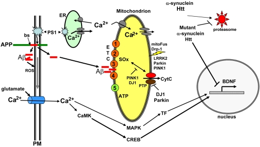
Figure 5. Influence of neurodegenerative disease-related proteins proteins on mitochondrial function and plasticity.
The AD APP is a single-pass transmembrane protein that includes the 42-amino-acid Aβ (red). Aβ is liberated upon sequential cleavages of APP by β-secretase (bs) at the N-terminus of Aβ and γ-secretase at the C-terminus of Aβ; the latter cleavage is executed by presenilin-1 (PS1; an integral membrane protein that is the enzymatic component of the γ-secretase protein complex). An alternative cleavage of APP within the Aβ sequence by the α-secretase enzyme produces a secreted form of APP that is believed to play important roles in developmental and synaptic plasticity, and neuron survival. Whereas Aβ is normally cleared from the brain, in AD it self-aggregates to form oligomers and during this process ROS are generated and propagated to the cell membrane, resulting in lipid peroxidation, impairment of membrane ion (Na+ and Ca2+) transporters and excessive Ca2+ influx through glutamate receptor (NMDA) channels. In addition to increasing Aβ production, mutations in PS1 that cause AD perturb endoplasmic reticulum (ER) Ca2+, resulting in excessive Ca2+ release. Mitochondrial Ca2+ homoeostasis and energy production may be impaired in neurons in AD as the result of cellular Ca2+ overload and increased oxidative stress possibly involving direct actions of Aβ on mitochondrial membranes. The pathological scenario just described involves perturbations in signalling pathways normally involved in adaptive neuroplasticity, with glutamate signalling being a well-established example. Glutamate-induced Ca2+ is normally transient with the Ca2+ activating kinases such as CaMK and MAPK. The kinases in turn activate transcription factors (TF) that induce the expression of nuclear genes that encode proteins involved in effecting adaptive morphological and functional responses of the neurons. CREB is one such synaptic activity-responsive transcription factor that induces the expression of BDNF, a protein critically involved in synaptic plasticity and neuronal survival. The successive discoveries of genes-linked inherited forms of PD placed mitochondrial alterations at centre stage in the pathogenesis of PD, and also revealed novel proteins that regulate mitochondrial functional and structural plasticity. A feature of all cases of PD is the intracellular accumulation of α-synuclein, apparently as the result of impaired degradation by the proteasome. Parkin (an E3 ubiquitin ligase), DJ-1, PINK1 and LRRK2 have each been shown to modify mitochondrial function (ATP and ROS production), and some findings suggest that Parkin, PINK1 and LRRK2 also influence mitochondrial fission and fusion. PINK1 and DJ-1 may suppress intramitochondrial oxidative stress, whereas Parkin and DJ-1 inhibit the opening of mitochondrial permeability transition pores, thereby protecting neurons against apoptotic cell death that can be triggered by release of cytochrome c (CytC) from the mitochondria. Finally, in HD, mutant huntingtin (Htt) self-aggregates and impairs CREB-mediated transcription of BDNF, thereby compromising a pathway critical for neuronal plasticity and survival. See the text of this paper and the following references for additional information and discussion (Mattson, 1997; Mattson, 2004; Mattson et al., 2004; Cattaneo et al., 2005; Cookson, 2005; Nuytemans et al., 2010).
Further evidence that perturbed mitochondrial function is sufficient to cause synaptic dysfunction and PD comes from studies of PINK1 [PTEN (phosphatase and tensin homologue deleted on chromosome 10)-induced kinase 1; Figure 5]. PINK1 deficiency or PD-causing PINK1 mutations impair mitochondrial Complex I activity, resulting in mitochondrial membrane depolarization and increased vulnerability to stress (Morais et al., 2009). An early functional abnormality in PINK1 mutant animals is impaired neurotransmitter release from presynaptic terminals, a problem that can be corrected by increasing cellular ATP levels (Morais et al., 2009).
Mutations in LRRK2 (leucine-rich repeat kinase 2) are responsible for some cases of autosomal dominantly inherited PD; LRRK2 associates with the mitochondrial outer membrane and interacts with Parkin (Mata et al., 2006). LRRK2 has been shown to modify the vulnerability of cells to mitochondrial dysfunction, with PD mutant forms of LRRK2 promoting mitochondrial dysfunction (Saha et al., 2009). Primary cultured neurons derived from LRRK2 G2019S mutant transgenic mice exhibit increased growth cone size and reduced neurite outgrowth characterized by increased levels of F-actin in the growth cones (Parisiadou et al., 2009), suggesting an adverse gain-of-function effect of this LRRK2 mutation on neuronal plasticity. The evidence that multiple genetic aberrancies cause PD by adversely affecting mitochondrial function and biogenesis suggests that the earliest impairments of synaptic plasticity in this disease are the result of mitochondrial abnormalities.
Some cases of AD (Alzheimer's disease) are inherited in an autosomal dominant manner as the result of mutations in APP (amyloid precursor protein) or presenilin-1 (Mattson, 2004). Both APP and presenilin-1 are believed to play roles in developmental and synaptic plasticity, and mutations in these proteins have been reported to alter neurogenesis and synaptic plasticity (Mattson, 1997). There is evidence that the secreted form of APP generated by α-secretase cleavage of APP enhances neurogenesis (Caillé et al., 2004) and synaptic plasticity (Ishida et al., 1997). On the other hand, the Aβ (amyloid β-peptide) generated by β- and γ-secretases can impair neurogenesis (Haughey et al., 2002a, 2002b) and synaptic plasticity (Chapman et al., 1999). Inasmuch as Aβ impairs mitochondrial function (Keller et al., 1998), there may be a role for perturbed mitochondrial function in the adverse effects of Aβ on neural plasticity (Mattson et al., 1998), although this remains to be established. In the case of presenilin-1 there is a well-established mechanism by which this protease regulates neurogenesis and adult neuroplasticity, namely presenilin-1 cleaves Notch to generate the NICD (Notch intracellular domain), which then translocates to the nucleus where it modulates the transcription of genes involved in regulating neuroplasticity (Lathia et al., 2008). Studies of Drosophila (Presente et al., 2004) and mice with reduced Notch levels (Costa et al., 2003; Wang et al., 2004) have demonstrated a critical role for Notch signalling in synaptic plasticity and learning and memory. Presenilin-1 mutations that cause AD have been reported to impair neurogenesis by perturbing Notch signalling (Veeraraghavalu et al., 2010) and may also impair some forms of synaptic plasticity (Auffret et al., 2010). Whether there is a role for mitochondrial changes in the actions of Notch signalling on neuroplasticity is unknown; however, it has been reported that Notch signalling interacts with mitochondrial remodelling proteins (Perumalsamy et al., 2010), suggesting a potential role for mitochondria in some biological effects of Notch signalling.
Perturbed mitochondrial behaviours are also implicated in the impaired synaptic plasticity, and motor and cognitive deficits, in HD (Huntington's disease). Polyglutamine repeat expansions in the huntingtin protein cause HD, and several lines of transgenic mice expressing such mutant huntingtin proteins develop neurodegenerative changes in the striatum and cerebral cortex, and motor and cognitive deficits similar to HD. In one HD mouse model, layer II/III neurons in the perirhinal cortex exhibit impaired LTP, which is associated with nuclear accumulation of mutant huntingtin and membrane depolarization (e.g. Cummings et al., 2006). LTP at hippocampal synapses is impaired in HD mutant mice (Usdin et al., 1999). Accumulating data point to mitochondrial abnormalities as an early consequence of mutant huntingtin that contributes to impaired neural plasticity. Early events may include defective mitochondrial calcium handling, ATP production and trafficking (Oliveira, 2010). Mutant huntingtin can impair transcription of nuclear genome-encoded mitochondrial proteins, and may also inhibit biogenesis and subcellular trafficking of mitochondria.
The kinds of findings described in the preceding paragraphs suggest that impaired neural plasticity occurs in the earliest stages of neurodegenerative disorders, and abnormalities in mitochondrial function, motility and biogenesis may contribute to the early dysfunction of neurons. The complexity of the architecture and signalling processes in neuronal circuits provides the basis for sophisticated behaviours including higher cognitive functions (language, decision making, creativity etc.). However, this cellular complexity increases the demands on the networks and the potential for ‘weak links’ (neurons that are particularly prone to faltering under adverse conditions such as traumatic injury, stroke, diabetes and aging). The development of therapeutic interventions aimed at enhancing mitochondrial function and plasticity in vulnerable neurons is an active area for preclinical and translational research for many neurological disorders (Mattson et al., 2008). Indeed, preclinical studies have demonstrated beneficial effects of creatine (a chemical that facilitates regeneration of ATP in mitochondria) in animal models of neurodegenerative disorders (Adhihetty and Beal, 2008) and traumatic brain injury (Sullivan et al., 2000). Additional mitochondria-focused treatments that show promise in preclinical studies include Mn-SOD (manganese superoxide dismutase) mimetics (Brazier et al., 2008), mitochondrial uncoupling agents (Liu et al., 2006; Pandya et al., 2007) and coenzyme Q10 (Chaturvedi and Beal, 2008).
FUTURE DIRECTIONS
While considerable data have accrued regarding the localization of mitochondria at subcellular sites where plasticity occurs and the movement and fission or fusion of mitochondria in response to signals that induce plasticity, questions remain just correlative and/or unanswered concerning the roles of mitochondria in neuroplasticity. To establish roles of mitochondrial energy production, redox and Ca2+ signalling, motility and biogenesis in neuroplasticity, several approaches may be employed: (i) use RNA interference technology to knock down the expression or function of proteins involved in specific mitochondrial events such as fission/fusion, biogenesis or targeted inhibition of mitochondrial mobility and functions selectively in subcellular compartments such as synapse, dendrites or axons. This could be accomplished in both cell culture and in vivo models. (ii) Perform time lapse analysis of mitochondrial movements within subcellular compartments in response to physiological stimuli, and in combination with selective ablation and/or artificial movement of mitochondria may establish the requirement of mitochondria for specific physiological events (neurite outgrowth and dendritic remodelling). (iii) Behavioural and electrophysiological evaluation of synaptic plasticity in mice with conditional knockout of proteins with specific roles in mitochondrial motility (Drp1, Mfn1/2 and OPA1) and biogenesis (PGC1α and TFAM). (iv) Utilization of novel fluorescent probes for mitochondrial superoxide, in combination with superoxide scavengers (Mn-SOD mimetics, for example). As such technologies are becoming widely available, it is very likely that there will be a rapid acceleration of our knowledge of how, where and why mitochondria respond to and regulate neural plasticity and how abnormalities in such mechanisms contribute to the pathogenesis of neurological disorders.
REFERENCES
- Adhihetty PJ, Beal MF. Creatine and its potential therapeutic value for targeting cellular energy impairment in neurodegenerative diseases. Neuromolecular Med. 2008;10:275–290. doi: 10.1007/s12017-008-8053-y. [DOI] [PMC free article] [PubMed] [Google Scholar]
- Andersson U, Scarpulla RC. Pgc-1-related coactivator, a novel, serum-inducible coactivator of nuclear respiratory factor 1-dependent transcription in mammalian cells. Mol Cell Biol. 2001;21:3738–3749. doi: 10.1128/MCB.21.11.3738-3749.2001. [DOI] [PMC free article] [PubMed] [Google Scholar]
- Auffret A, Gautheron V, Mattson MP, Mariani J, Rovira C. Progressive age-related impairment of the late long-term potentiation in Alzheimer's disease presenilin-1 mutant knock-in mice. J Alzheimers Dis. 2010;19:1021–1033. doi: 10.3233/JAD-2010-1302. [DOI] [PMC free article] [PubMed] [Google Scholar]
- Baar K, Wende AR, Jones TE, Marison M, Nolte LA, Chen M, Kelly DP, Holloszy JO. Adaptations of skeletal muscle to exercise: rapid increase in the transcriptional coactivator PGC-1. FASEB J. 2002;16:1879–1886. doi: 10.1096/fj.02-0367com. [DOI] [PubMed] [Google Scholar]
- Barres BA. The mystery and magic of glia: a perspective on their roles in health and disease. Neuron. 2008;60:430–440. doi: 10.1016/j.neuron.2008.10.013. [DOI] [PubMed] [Google Scholar]
- Barsoum MJ, Yuan H, Gerencser AA, Liot G, Kushnareva Y, Gräber S, Kovacs I, Lee WD, Waggoner J, Cui J, White AD, Bossy B, Martinou JC, Youle RJ, Lipton SA, Ellisman MH, Perkins GA, Bossy-Wetzel E. Nitric oxide-induced mitochondrial fission is regulated by dynamin-related GTPases in neurons. EMBO J. 2006;25:3900–3911. doi: 10.1038/sj.emboj.7601253. [DOI] [PMC free article] [PubMed] [Google Scholar]
- Baxter KK, Uittenbogaard M, Yoon J, Chiaramello A. The neurogenic basic helix–loop–helix transcription factor neuroD6 concomitantly increases mitochondrial mass and regulates cytoskeletal organization in the early stages of neuronal differentiation. ASN NEURO. 2009;1((4)) doi: 10.1042/AN20090036. art:e00016doi:10.1042/ AN20090036. [DOI] [PMC free article] [PubMed] [Google Scholar]
- Berman SB, Pineda FJ, Hardwick JM. Mitochondrial fission and fusion dynamics: the long and short of it. Cell Death Differ. 2008;15:1147–1152. doi: 10.1038/cdd.2008.57. [DOI] [PMC free article] [PubMed] [Google Scholar]
- Bezprozvanny I, Mattson MP. Neuronal calcium mishandling and the pathogenesis of Alzheimer's disease. Trends Neurosci. 2008;31:454–463. doi: 10.1016/j.tins.2008.06.005. [DOI] [PMC free article] [PubMed] [Google Scholar]
- Billups B, Forsythe ID. Presynaptic mitochondrial calcium sequestration influences transmission at mammalian central synapses. J Neurosci. 2002;22:5840–5847. doi: 10.1523/JNEUROSCI.22-14-05840.2002. [DOI] [PMC free article] [PubMed] [Google Scholar]
- Bindokas VP, Lee CC, Colmers WF, Miller RJ. Changes in mitochondrial function resulting from synaptic activity in the rat hippocampal slice. J Neurosci. 1998;18:4570–4587. doi: 10.1523/JNEUROSCI.18-12-04570.1998. [DOI] [PMC free article] [PubMed] [Google Scholar]
- Bramham CR. Local protein synthesis, actin dynamics, and LTP consolidation. Curr Opin Neurobiol. 2008;18:524–531. doi: 10.1016/j.conb.2008.09.013. [DOI] [PubMed] [Google Scholar]
- Brazier MW, Doctrow SR, Masters CL, Collins SJ. A manganese-superoxide dismutase/catalase mimetic extends survival in a mouse model of human prion disease. Free Radical Biol Med. 2008;45:184–192. doi: 10.1016/j.freeradbiomed.2008.04.006. [DOI] [PubMed] [Google Scholar]
- Brinton RD. Estrogen-induced plasticity from cells to circuits: predictions for cognitive function. Trends Pharmacol Sci. 2009;30:212–222. doi: 10.1016/j.tips.2008.12.006. [DOI] [PMC free article] [PubMed] [Google Scholar]
- Burkhalter J, Fiumelli H, Allaman I, Chatton JY, Martin JL. Brain-derived neurotrophic factor stimulates energy metabolism in developing cortical neurons. J Neurosci. 2003;23:8212–8220. doi: 10.1523/JNEUROSCI.23-23-08212.2003. [DOI] [PMC free article] [PubMed] [Google Scholar]
- Caillé I, Allinquant B, Dupont E, Bouillot C, Langer A, Müller U, Prochiantz A. Soluble form of amyloid precursor protein regulates proliferation of progenitors in the adult subventricular zone. Development. 2004;131:2173–2181. doi: 10.1242/dev.01103. [DOI] [PubMed] [Google Scholar]
- Calabresi P, Gubellini P, Picconi B, Centonze D, Pisani A, Bonsi P, Greengard P, Hipskind RA, Borrelli E, Bernardi G. Inhibition of mitochondrial complex II induces a long-term potentiation of NMDA-mediated synaptic excitation in the striatum requiring endogenous dopamine. J Neurosci. 2001;21:5110–5120. doi: 10.1523/JNEUROSCI.21-14-05110.2001. [DOI] [PMC free article] [PubMed] [Google Scholar]
- Cameron HA, Kaliszewski CK, Greer CA. Organization of mitochondria in olfactory bulb granule cell dendritic spines. Synapse. 1991;8:107–118. doi: 10.1002/syn.890080205. [DOI] [PubMed] [Google Scholar]
- Cane KN, Anderson CR. Generating diversity: Mechanisms regulating the differentiation of autonomic neuron phenotypes. Auton Neurosci. 2009;151:17–29. doi: 10.1016/j.autneu.2009.08.010. [DOI] [PubMed] [Google Scholar]
- Cattaneo E, Zuccato C, Tartari M. Normal huntingtin function: an alternative approach to Huntington's disease. Nat Rev Neurosci. 2005;6:919–930. doi: 10.1038/nrn1806. [DOI] [PubMed] [Google Scholar]
- Chada SR, Hollenbeck PJ. Nerve growth factor signaling regulates motility and docking of axonal mitochondria. Curr Biol. 2004;14:1272–1276. doi: 10.1016/j.cub.2004.07.027. [DOI] [PubMed] [Google Scholar]
- Chan SL, Liu D, Kyriazis GA, Bagsiyao P, Ouyang X, Mattson MP. Mitochondrial uncoupling protein-4 regulates calcium homeostasis and sensitivity to store depletion-induced apoptosis in neural cells. J Biol Chem. 2006;281:37391–37403. doi: 10.1074/jbc.M605552200. [DOI] [PubMed] [Google Scholar]
- Chapman PF, White GL, Jones MW, Cooper-Blacketer D, Marshall VJ, Irizarry M, Younkin L, Good MA, Bliss TV, Hyman BT, Younkin SG, Hsiao KK. Impaired synaptic plasticity and learning in aged amyloid precursor protein transgenic mice. Nat Neurosci. 1999;2:271–276. doi: 10.1038/6374. [DOI] [PubMed] [Google Scholar]
- Chaturvedi RK, Beal MF. Mitochondrial approaches for neuroprotection. Ann NY Acad Sci. 2008;1147:395–412. doi: 10.1196/annals.1427.027. [DOI] [PMC free article] [PubMed] [Google Scholar]
- Cheng A, Wang S, Cai J, Rao MS, Mattson MP. Nitric oxide acts in a positive feedback loop with BDNF to regulate neural progenitor cell proliferation and differentiation in the mammalian brain. Dev Biol. 2003;258:319–333. doi: 10.1016/s0012-1606(03)00120-9. [DOI] [PubMed] [Google Scholar]
- Chihara T, Luginbuhl D, Luo L. Cytoplasmic and mitochondrial protein translation in axonal and dendritic terminal arborization. Nat Neurosci. 2007;10:828–837. doi: 10.1038/nn1910. [DOI] [PubMed] [Google Scholar]
- Cookson MR. The biochemistry of Parkinson's disease. Annu Rev Biochem. 2005;74:29–52. doi: 10.1146/annurev.biochem.74.082803.133400. [DOI] [PubMed] [Google Scholar]
- Costa RM, Honjo T, Silva AJ. Learning and memory deficits in Notch mutant mice. Curr Biol. 2003;13:1348–1354. doi: 10.1016/s0960-9822(03)00492-5. [DOI] [PubMed] [Google Scholar]
- Cowell RM, Blake KR, Russell JW. Localization of the transcriptional coactivator PGC-1α to GABAergic neurons during maturation of the rat brain. J Comp Neurol. 2007;502:1–18. doi: 10.1002/cne.21211. [DOI] [PubMed] [Google Scholar]
- Cummings DM, Milnerwood AJ, Dallérac GM, Waights V, Brown JY, Vatsavayai SC, Hirst MC, Murphy KP. Aberrant cortical synaptic plasticity and dopaminergic dysfunction in a mouse model of Huntington's disease. Hum Mol Genet. 2006;15:2856–2868. doi: 10.1093/hmg/ddl224. [DOI] [PubMed] [Google Scholar]
- Dedov VN, Armati PJ, Roufogalis BD. Three-dimensional organisation of mitochondrial clusters in regenerating dorsal root ganglion (DRG) neurons from neonatal rats: evidence for mobile mitochondrial pools. J Peripher Nerv Syst. 2000;5((1)):3–10. doi: 10.1046/j.1529-8027.2000.00153.x. [DOI] [PubMed] [Google Scholar]
- Detmer SA, Chan DC. Functions and dysfunctions of mitochondrial dynamics. Nat Rev Mol Cell Biol. 2007;8:870–879. doi: 10.1038/nrm2275. [DOI] [PubMed] [Google Scholar]
- Duchen MR. Mitochondria and Ca2+ in cell physiology and pathophysiology. Cell Calcium. 2000;28:339–348. doi: 10.1054/ceca.2000.0170. [DOI] [PubMed] [Google Scholar]
- Eckles-Smith K, Clayton D, Bickford P, Browning MD. Caloric restriction prevents age-related deficits in LTP and in NMDA receptor expression. Mol Brain Res. 2000;78:154–162. doi: 10.1016/s0169-328x(00)00088-7. [DOI] [PubMed] [Google Scholar]
- Foy MR, Baudry M, Diaz Brinton R, Thompson RF. Estrogen and hippocampal plasticity in rodent models. J Alzheimers Dis. 2008;15:589–603. doi: 10.3233/jad-2008-15406. [DOI] [PMC free article] [PubMed] [Google Scholar]
- Ge S, Sailor KA, Ming GL, Song H. Synaptic integration and plasticity of new neurons in the adult hippocampus. J Physiol. 2008;586:3759–3765. doi: 10.1113/jphysiol.2008.155655. [DOI] [PMC free article] [PubMed] [Google Scholar]
- Gleichmann M, Collis LP, Smith PJ, Mattson MP. Simultaneous single neuron recording of O2 consumption, [Ca2+]i and mitochondrial membrane potential in glutamate toxicity. J Neurochem. 2009;109:644–655. doi: 10.1111/j.1471-4159.2009.05997.x. [DOI] [PMC free article] [PubMed] [Google Scholar]
- Goldberg MS, Fleming SM, Palacino JJ, Cepeda C, Lam HA, Bhatnagar A, Meloni EG, Wu N, Ackerson LC, Klapstein GJ, Gajendiran M, Roth BL, Chesselet MF, Maidment NT, Levine MS, Shen J. Parkin-deficient mice exhibit nigrostriatal deficits but not loss of dopaminergic neurons. J Biol Chem. 2003;278:43628–43635. doi: 10.1074/jbc.M308947200. [DOI] [PubMed] [Google Scholar]
- Gottmann K, Mittmann T, Lessmann V. BDNF signaling in the formation, maturation and plasticity of glutamatergic and GABAergic synapses. Exp Brain Res. 2009;199:203–234. doi: 10.1007/s00221-009-1994-z. [DOI] [PubMed] [Google Scholar]
- Greenamyre JT, MacKenzie G, Peng TI, Stephans SE. Mitochondrial dysfunction in Parkinson's disease. Biochem Soc Symp. 1999;66:85–97. doi: 10.1042/bss0660085. [DOI] [PubMed] [Google Scholar]
- Greer PL, Greenberg ME. From synapse to nucleus: calcium-dependent gene transcription in the control of synapse development and function. Neuron. 2008;59:846–860. doi: 10.1016/j.neuron.2008.09.002. [DOI] [PubMed] [Google Scholar]
- Guo X, Macleod GT, Wellington A, Hu F, Panchumarthi S, Schoenfield M, Marin L, Charlton MP, Atwood HL, Zinsmaier KE. The GTPase dMiro is required for axonal transport of mitochondria to Drosophila synapses. Neuron. 2005;47:379–393. doi: 10.1016/j.neuron.2005.06.027. [DOI] [PubMed] [Google Scholar]
- Gutsaeva DR, Carraway MS, Suliman HB, Demchenko IT, Shitara H, Yonekawa H, Piantadosi CA. Transient hypoxia stimulates mitochondrial biogenesis in brain subcortex by a neuronal nitric oxide synthase-dependent mechanism. J Neurosci. 2008;28:2015–2024. doi: 10.1523/JNEUROSCI.5654-07.2008. [DOI] [PMC free article] [PubMed] [Google Scholar]
- Halliday GM, Li YW, Blumbergs PC, Joh TH, Cotton RG, Howe PR, Blessing WW, Geffen LB. Neuropathology of immunohistochemically identified brainstem neurons in Parkinson's disease. Ann Neurol. 1990;27:373–385. doi: 10.1002/ana.410270405. [DOI] [PubMed] [Google Scholar]
- Hamby ME, Coskun V, Sun YE. Transcriptional regulation of neuronal differentiation: the epigenetic layer of complexity. Biochim Biophys Acta. 2008;1779:432–437. doi: 10.1016/j.bbagrm.2008.07.006. [DOI] [PMC free article] [PubMed] [Google Scholar]
- Harooni HE, Naghdi N, Sepehri H, Rohani AH. The role of hippocampal nitric oxide (NO) on learning and immediate, short- and long-term memory retrieval in inhibitory avoidance task in male adult rats. Behav Brain Res. 2009;201:166–172. doi: 10.1016/j.bbr.2009.02.011. [DOI] [PubMed] [Google Scholar]
- Haughey NJ, Nath A, Chan SL, Borchard AC, Rao MS, Mattson MP. Disruption of neurogenesis by amyloid beta-peptide, and perturbed neural progenitor cell homeostasis, in models of Alzheimer's disease. J Neurochem. 2002a;83:1509–1524. doi: 10.1046/j.1471-4159.2002.01267.x. [DOI] [PubMed] [Google Scholar]
- Haughey NJ, Liu D, Nath A, Borchard AC, Mattson MP. Disruption of neurogenesis in the subventricular zone of adult mice, and in human cortical neuronal precursor cells in culture, by amyloid beta-peptide: implications for the pathogenesis of Alzheimer's disease. Neuromolecular Med. 2002b;1:125–135. doi: 10.1385/NMM:1:2:125. [DOI] [PubMed] [Google Scholar]
- Haydon PG, Carmignoto G. Astrocyte control of synaptic transmission and neurovascular coupling. Physiol Rev. 2006;86:1009–1031. doi: 10.1152/physrev.00049.2005. [DOI] [PubMed] [Google Scholar]
- Hebert TL, Wu X, Yu G, Goh BC, Halvorsen YD, Wang Z, Moro C, Gimble JM. Culture effects of epidermal growth factor (EGF) and basic fibroblast growth factor (bFGF) on cryopreserved human adipose-derived stromal/stem cell proliferation and adipogenesis. J Tissue Eng Regen Med. 2009;3:553–561. doi: 10.1002/term.198. [DOI] [PMC free article] [PubMed] [Google Scholar]
- Helton TD, Otsuka T, Lee MC, Mu Y, Ehlers MD. Pruning and loss of excitatory synapses by the parkin ubiquitin ligase. Proc Natl Acad Sci USA. 2008;105:19492–19497. doi: 10.1073/pnas.0802280105. [DOI] [PMC free article] [PubMed] [Google Scholar]
- Hepple RT, Baker DJ, McConkey M, Murynka T, Norris R. Caloric restriction protects mitochondrial function with aging in skeletal and cardiac muscles. Rejuvenation Res. 2006;9:219–222. doi: 10.1089/rej.2006.9.219. [DOI] [PubMed] [Google Scholar]
- Hildebrandt H, Mühlenhoff M, Weinhold B, Gerardy-Schahn R. Dissecting polysialic acid and NCAM functions in brain development. J Neurochem. 2007;103:S56–S64. doi: 10.1111/j.1471-4159.2007.04716.x. [DOI] [PubMed] [Google Scholar]
- Hojo Y, Murakami G, Mukai H, Higo S, Hatanaka Y, Ogiue-Ikeda M, Ishii H, Kimoto T, Kawato S. Estrogen synthesis in the brain–role in synaptic plasticity and memory. Mol Cell Endocrinol. 2008;290:31–43. doi: 10.1016/j.mce.2008.04.017. [DOI] [PubMed] [Google Scholar]
- Hollenbeck PJ, Saxton WM. The axonal transport of mitochondria. J Cell Sci. 2005;118:5411–5419. doi: 10.1242/jcs.02745. [DOI] [PMC free article] [PubMed] [Google Scholar]
- Hyun DH, Hunt ND, Emerson SS, Hernandez JO, Mattson MP, de Cabo R. Up-regulation of plasma membrane-associated redox activities in neuronal cells lacking functional mitochondria. J Neurochem. 2007;100:1364–1374. doi: 10.1111/j.1471-4159.2006.04411.x. [DOI] [PubMed] [Google Scholar]
- Ingram DK, Weindruch R, Spangler EL, Freeman JR, Walford RL. Dietary restriction benefits learning and motor performance of aged mice. J Gerontol. 1987;42:78–81. doi: 10.1093/geronj/42.1.78. [DOI] [PubMed] [Google Scholar]
- Ishida A, Furukawa K, Keller JN, Mattson MP. Secreted form of beta-amyloid precursor protein shifts the frequency dependency for induction of LTD, and enhances LTP in hippocampal slices. Neuroreport. 1997;8:2133–2137. doi: 10.1097/00001756-199707070-00009. [DOI] [PubMed] [Google Scholar]
- Kang JS, Tian JH, Pan PY, Zald P, Li C, Deng C, Sheng ZH. Docking of axonal mitochondria by syntaphilin controls their mobility and affects short-term facilitation. Cell. 2008;132:137–148. doi: 10.1016/j.cell.2007.11.024. [DOI] [PMC free article] [PubMed] [Google Scholar]
- Karbowski M, Lee YJ, Gaume B, Jeong SY, Frank S, Nechushtan A, Santel A, Fuller M, Smith CL, Youle RJ. Spatial and temporal association of Bax with mitochondrial fission sites, Drp1, and Mfn2 during apoptosis. J Cell Biol. 2002;159:931–938. doi: 10.1083/jcb.200209124. [DOI] [PMC free article] [PubMed] [Google Scholar]
- Keller JN, Kindy MS, Holtsberg FW, St Clair DK, Yen HC, Germeyer A, Steiner SM, Bruce-Keller AJ, Hutchins JB, Mattson MP. Mitochondrial manganese superoxide dismutase prevents neural apoptosis and reduces ischemic brain injury: suppression of peroxynitrite production, lipid peroxidation, and mitochondrial dysfunction. J Neurosci. 1998;18:687–697. doi: 10.1523/JNEUROSCI.18-02-00687.1998. [DOI] [PMC free article] [PubMed] [Google Scholar]
- Kempermann G, van Praag H, Gage FH. Activity-dependent regulation of neuronal plasticity and self repair. Prog Brain Res. 2000;127:35–48. doi: 10.1016/s0079-6123(00)27004-0. [DOI] [PubMed] [Google Scholar]
- Kempermann G, Wiskott L, Gage FH. Functional significance of adult neurogenesis. Curr Opin Neurobiol. 2004;14:186–191. doi: 10.1016/j.conb.2004.03.001. [DOI] [PubMed] [Google Scholar]
- Kitamura T, Saitoh Y, Takashima N, Murayama A, Niibori Y, Ageta H, Sekiguchi M, Sugiyama H, Inokuchi K. Adult neurogenesis modulates the hippocampus-dependent period of associative fear memory. Cell. 2009;139:814–827. doi: 10.1016/j.cell.2009.10.020. [DOI] [PubMed] [Google Scholar]
- Klinge CM. Estrogenic control of mitochondrial function and biogenesis. J Cell Biochem. 2008;105:1342–1351. doi: 10.1002/jcb.21936. [DOI] [PMC free article] [PubMed] [Google Scholar]
- Kondoh H, Lleonart ME, Nakashima Y, Yokode M, Tanaka M, Bernard D, Gil J, Beach D. A high glycolytic flux supports the proliferative potential of murine embryonic stem cells. Antioxid Redox Signal. 2007;9:293–299. doi: 10.1089/ars.2006.1467. [DOI] [PubMed] [Google Scholar]
- Koshiba T, Detmer SA, Kaiser JT, Chen H, McCaffery JM, Chan DC. Structural basis of mitochondrial tethering by mitofusin complexes. Science. 2004;305:858–862. doi: 10.1126/science.1099793. [DOI] [PubMed] [Google Scholar]
- Lathia JD, Mattson MP, Cheng A. Notch: from neural development to neurological disorders. J Neurochem. 2008;107:1471–1481. doi: 10.1111/j.1471-4159.2008.05715.x. [DOI] [PMC free article] [PubMed] [Google Scholar]
- Levy M, Faas GC, Saggau P, Craigen WJ, Sweatt JD. Mitochondrial regulation of synaptic plasticity in the hippocampus. J Biol Chem. 2003;278:17727–17734. doi: 10.1074/jbc.M212878200. [DOI] [PubMed] [Google Scholar]
- Li Z, Okamoto K, Hayashi Y, Sheng M. The importance of dendritic mitochondria in the morphogenesis and plasticity of spines and synapses. Cell. 2004;119:873–887. doi: 10.1016/j.cell.2004.11.003. [DOI] [PubMed] [Google Scholar]
- Li Z, Jo J, Jia JM, Lo SC, Whitcomb DJ, Jiao S, Cho K, Sheng M. Caspase-3 activation via mitochondria is required for long-term depression and AMPA receptor internalization. Cell. 2010;141:859–871. doi: 10.1016/j.cell.2010.03.053. [DOI] [PMC free article] [PubMed] [Google Scholar]
- Lin J, Wu PH, Tarr PT, Lindenberg KS, St-Pierre J, Zhang CY, Mootha VK, Jäger S, Vianna CR, Reznick RM, Cui L, Manieri M, Donovan MX, Wu Z, Cooper MP, Fan MC, Rohas LM, Zavacki AM, Cinti S, Shulman GI, Lowell BB, Krainc D, Spiegelman BM. Defects in adaptive energy metabolism with CNS-linked hyperactivity in PGC-1alpha null mice. Cell. 2004;119:121–135. doi: 10.1016/j.cell.2004.09.013. [DOI] [PubMed] [Google Scholar]
- Liesa M, Palacín M, Zorzano A. Mitochondrial dynamics in mammalian health and disease. Physiol Rev. 2009;89:799–845. doi: 10.1152/physrev.00030.2008. [DOI] [PubMed] [Google Scholar]
- Lipsky RH, Marini AM. Brain-derived neurotrophic factor in neuronal survival and behavior-related plasticity. Ann NY Acad Sci. 2007;1122:130–143. doi: 10.1196/annals.1403.009. [DOI] [PubMed] [Google Scholar]
- Liu D, Chan SL, de Souza-Pinto NC, Slevin JR, Wersto RP, Zhan M, Mustafa K, de Cabo R, Mattson MP. Mitochondrial UCP4 mediates an adaptive shift in energy metabolism and increases the resistance of neurons to metabolic and oxidative stress. Neuromolecular Med. 2006;8:389–414. doi: 10.1385/NMM:8:3:389. [DOI] [PubMed] [Google Scholar]
- Liu D, Pitta M, Mattson MP. Preventing NAD+ depletion protects neurons against excitotoxicity: bioenergetic effects of mild mitochondrial uncoupling and caloric restriction. Ann NY Acad Sci. 2008;1147:275–282. doi: 10.1196/annals.1427.028. [DOI] [PMC free article] [PubMed] [Google Scholar]
- Liu J, Head E, Gharib AM, Yuan W, Ingersoll RT, Hagen TM, Cotman CW, Ames BN. Memory loss in old rats is associated with brain mitochondrial decay and RNA/DNA oxidation: partial reversal by feeding acetyl-l-carnitine and/or R-alpha-lipoic acid. Proc Natl Acad Sci USA. 2002;99:2356–2361. doi: 10.1073/pnas.261709299. [DOI] [PMC free article] [PubMed] [Google Scholar]
- Loers G, Schachner M. Recognition molecules and neural repair. J Neurochem. 2007;101:865–882. doi: 10.1111/j.1471-4159.2006.04409.x. [DOI] [PubMed] [Google Scholar]
- López-Lluch G, Hunt N, Jones B, Zhu M, Jamieson H, Hilmer S, Cascajo MV, Allard J, Ingram DK, Navas P, de Cabo R. Calorie restriction induces mitochondrial biogenesis and bioenergetic efficiency. Proc Natl Acad Sci USA. 2006;103:1768–1773. doi: 10.1073/pnas.0510452103. [DOI] [PMC free article] [PubMed] [Google Scholar]
- Lu B, Wang KH, Nose A. Molecular mechanisms underlying neural circuit formation. Curr Opin Neurobiol. 2009;19:162–167. doi: 10.1016/j.conb.2009.04.004. [DOI] [PMC free article] [PubMed] [Google Scholar]
- Macaskill AF, Rinholm JE, Twelvetrees AE, Arancibia-Carcamo IL, Muir J, Fransson A, Aspenstrom P, Attwell D, Kittler JT. Miro1 is a calcium sensor for glutamate receptor-dependent localization of mitochondria at synapses. Neuron. 2009;61:541–555. doi: 10.1016/j.neuron.2009.01.030. [DOI] [PMC free article] [PubMed] [Google Scholar]
- Markham JA, Greenough WT. Experience-driven brain plasticity: beyond the synapse. Neuron Glia Biol. 2004;1:351–363. doi: 10.1017/s1740925x05000219. [DOI] [PMC free article] [PubMed] [Google Scholar]
- Mata IF, Wedemeyer WJ, Farrer MJ, Taylor JP, Gallo KA. LRRK2 in Parkinson's disease: protein domains and functional insights. Trends Neurosci. 2006;29:286–293. doi: 10.1016/j.tins.2006.03.006. [DOI] [PubMed] [Google Scholar]
- Mattson MP. Neurotransmitters in the regulation of neuronal cytoarchitecture. Brain Res. 1988;472:179–212. doi: 10.1016/0165-0173(88)90020-3. [DOI] [PubMed] [Google Scholar]
- Mattson MP. Cellular actions of beta-amyloid precursor protein and its soluble and fibrillogenic derivatives. Physiol Rev. 1997;77:1081–1132. doi: 10.1152/physrev.1997.77.4.1081. [DOI] [PubMed] [Google Scholar]
- Mattson MP, Guthrie PB, Hayes BC, Kater SB. Roles for mitotic history in the generation and degeneration of hippocampal neuroarchitecture. J Neurosci. 1989;9:1223–1232. doi: 10.1523/JNEUROSCI.09-04-01223.1989. [DOI] [PMC free article] [PubMed] [Google Scholar]
- Mattson MP, Partin J, Begley JG. Amyloid beta-peptide induces apoptosis-related events in synapses and dendrites. Brain Res. 1998;807:167–176. doi: 10.1016/s0006-8993(98)00763-x. [DOI] [PubMed] [Google Scholar]
- Mattson MP, Partin J. Evidence for mitochondrial control of neuronal polarity. J Neurosci Res. 1999;56:8–20. doi: 10.1002/(SICI)1097-4547(19990401)56:1<8::AID-JNR2>3.0.CO;2-G. [DOI] [PubMed] [Google Scholar]
- Mattson MP, Liu D. Mitochondrial potassium channels and uncoupling proteins in synaptic plasticity and neuronal cell death. Biochem Biophys Res Commun. 2003;304:539–549. doi: 10.1016/s0006-291x(03)00627-2. [DOI] [PubMed] [Google Scholar]
- Mattson MP, Duan W, Guo Z. Meal size and frequency affect neuronal plasticity and vulnerability to disease: cellular and molecular mechanisms. J Neurochem. 2003;84:417–431. doi: 10.1046/j.1471-4159.2003.01586.x. [DOI] [PubMed] [Google Scholar]
- Mattson MP. Pathways towards and away from Alzheimer's disease. Nature. 2004;430:631–639. doi: 10.1038/nature02621. [DOI] [PMC free article] [PubMed] [Google Scholar]
- Mattson MP, Maudsley S, Martin B. BDNF and 5-HT: a dynamic duo in age-related neuronal plasticity and neurodegenerative disorders. Trends Neurosci. 2004;27:589–594. doi: 10.1016/j.tins.2004.08.001. [DOI] [PubMed] [Google Scholar]
- Mattson MP. Glutamate and neurotrophic factors in neuronal plasticity and disease. Ann NY Acad Sci. 2008;1144:97–112. doi: 10.1196/annals.1418.005. [DOI] [PMC free article] [PubMed] [Google Scholar]
- Mattson MP, Gleichmann M, Cheng A. Mitochondria in neuroplasticity and neurological disorders. Neuron. 2008;60:748–766. doi: 10.1016/j.neuron.2008.10.010. [DOI] [PMC free article] [PubMed] [Google Scholar]
- Miller KE, Sheetz MP. Axonal mitochondrial transport and potential are correlated. J Cell Sci. 2004;117:2791–2804. doi: 10.1242/jcs.01130. [DOI] [PubMed] [Google Scholar]
- Miller SW, Trimmer PA, Parker WD Jr, Davis RE. Creation and characterization of mitochondrial DNA-depleted cell lines with ‘neuronal-like’ properties. J Neurochem. 1996;67:1897–1907. doi: 10.1046/j.1471-4159.1996.67051897.x. [DOI] [PubMed] [Google Scholar]
- Minichiello L. TrkB signalling pathways in LTP and learning. Nat Rev Neurosci. 2009;10:850–860. doi: 10.1038/nrn2738. [DOI] [PubMed] [Google Scholar]
- Morais VA, Verstreken P, Roethig A, Smet J, Snellinx A, Vanbrabant M, Haddad D, Frezza C, Mandemakers W, Vogt-Weisenhorn D, Van Coster R, Wurst W, Scorrano L, De Strooper B. Parkinson's disease mutations in PINK1 result in decreased Complex I activity and deficient synaptic function. EMBO Mol Med. 2009;1:99–111. doi: 10.1002/emmm.200900006. [DOI] [PMC free article] [PubMed] [Google Scholar]
- Morrison SJ, Csete M, Groves AK, Melega W, Wold B, Anderson DJ. Culture in reduced levels of oxygen promotes clonogenic sympathoadrenal differentiation by isolated neural crest stem cells. J Neurosci. 2000;20:7370–7376. doi: 10.1523/JNEUROSCI.20-19-07370.2000. [DOI] [PMC free article] [PubMed] [Google Scholar]
- Mortiboys H, Thomas KJ, Koopman WJ, Klaffke S, Abou-Sleiman P, Olpin S, Wood NW, Willems PH, Smeitink JA, Cookson MR, Bandmann O. Mitochondrial function and morphology are impaired in parkin-mutant fibroblasts. Ann Neurol. 2008;64:555–565. doi: 10.1002/ana.21492. [DOI] [PMC free article] [PubMed] [Google Scholar]
- Newpher TM, Ehlers MD. Glutamate receptor dynamics in dendritic microdomains. Neuron. 2008;58:472–497. doi: 10.1016/j.neuron.2008.04.030. [DOI] [PMC free article] [PubMed] [Google Scholar]
- Nicholls DG, Budd SL. Mitochondria and neuronal survival. Physiol Rev. 2000;80:315–60. doi: 10.1152/physrev.2000.80.1.315. [DOI] [PubMed] [Google Scholar]
- Nikonenko I, Boda B, Steen S, Knott G, Welker E, Muller D. PSD-95 promotes synaptogenesis and multiinnervated spine formation through nitric oxide signaling. J Cell Biol. 2008;183:1115–1127. doi: 10.1083/jcb.200805132. [DOI] [PMC free article] [PubMed] [Google Scholar]
- Nisoli E, Tonello C, Cardile A, Cozzi V, Bracale R, Tedesco L, Falcone S, Valerio A, Cantoni O, Clementi E, Moncada S, Carruba MO. Calorie restriction promotes mitochondrial biogenesis by inducing the expression of eNOS. Science. 2005;310:314–317. doi: 10.1126/science.1117728. [DOI] [PubMed] [Google Scholar]
- Norrbom J, Sundberg CJ, Ameln H, Kraus WE, Jansson E, Gustafsson T. PGC-1alpha mRNA expression is influenced by metabolic perturbation in exercising human skeletal muscle. J Appl Physiol. 2004;96:189–194. doi: 10.1152/japplphysiol.00765.2003. [DOI] [PubMed] [Google Scholar]
- Nuytemans K, Theuns J, Cruts M, Van Broeckhoven C. Genetic etiology of Parkinson disease associated with mutations in the SNCA, PARK2, PINK1, PARK7, and LRRK2 genes: a mutation update. Hum Mutat. 2010;31:763–780. doi: 10.1002/humu.21277. [DOI] [PMC free article] [PubMed] [Google Scholar]
- Oliveira JM. Nature and cause of mitochondrial dysfunction in Huntington's disease: focusing on huntingtin and the striatum. J Neurochem. 2010;114:1–12. doi: 10.1111/j.1471-4159.2010.06741.x. [DOI] [PubMed] [Google Scholar]
- Overly CC, Rieff HI, Hollenbeck PJ. Organelle motility and metabolism in axons vs dendrites of cultured hippocampal neurons. J Cell Sci. 1996;109:971–980. doi: 10.1242/jcs.109.5.971. [DOI] [PubMed] [Google Scholar]
- Pak CW, Flynn KC, Bamburg JR. Actin-binding proteins take the reins in growth cones. Nat Rev Neurosci. 2008;9:136–147. doi: 10.1038/nrn2236. [DOI] [PubMed] [Google Scholar]
- Pandya JD, Pauly JR, Nukala VN, Sebastian AH, Day KM, Korde AS, Maragos WF, Hall ED, Sullivan PG. Post-injury administration of mitochondrial uncouplers increases tissue sparing and improves behavioral outcome following traumatic brain injury in rodents. J Neurotrauma. 24:798–811. doi: 10.1089/neu.2006.3673. [DOI] [PubMed] [Google Scholar]
- Parisiadou L, Xie C, Cho HJ, Lin X, Gu XL, Long CX, Lobbestael E, Baekelandt V, Taymans JM, Sun L, Cai H. Phosphorylation of ezrin/radixin/moesin proteins by LRRK2 promotes the rearrangement of actin cytoskeleton in neuronal morphogenesis. J Neurosci. 2009;29:13971–13980. doi: 10.1523/JNEUROSCI.3799-09.2009. [DOI] [PMC free article] [PubMed] [Google Scholar]
- Park J, Lee G, Chung J. The PINK1-Parkin pathway is involved in the regulation of mitochondrial remodeling process. Biochem Biophys Res Commun. 2009;378:518–523. doi: 10.1016/j.bbrc.2008.11.086. [DOI] [PubMed] [Google Scholar]
- Pathak D, Sepp KJ, Hollenbeck PJ. Evidence that myosin activity opposes microtubule-based axonal transport of mitochondria. J Neurosci. 2010;30((26)):8984–8992. doi: 10.1523/JNEUROSCI.1621-10.2010. [DOI] [PMC free article] [PubMed] [Google Scholar]
- Perumalsamy LR, Nagala M, Sarin A. Notch-activated signaling cascade interacts with mitochondrial remodeling proteins to regulate cell survival. Proc Natl Acad Sci USA. 2010;107:6882–6887. doi: 10.1073/pnas.0910060107. [DOI] [PMC free article] [PubMed] [Google Scholar]
- Pitsikas N, Algeri S. Deterioration of spatial and nonspatial reference and working memory in aged rats: protective effect of life-long calorie restriction. Neurobiol Aging. 1992;13:369–373. doi: 10.1016/0197-4580(92)90110-j. [DOI] [PubMed] [Google Scholar]
- Popov V, Medvedev NI, Davies HA, Stewart MG. Mitochondria form a filamentous reticular network in hippocampal dendrites but are present as discrete bodies in axons: a three-dimensional ultrastructural study. J Comp Neurol. 2005;492:50–65. doi: 10.1002/cne.20682. [DOI] [PubMed] [Google Scholar]
- Presente A, Boyles RS, Serway CN, de Belle JS, Andres AJ. Notch is required for long-term memory in Drosophila. Proc Natl Acad Sci USA. 2004;101:1764–1768. doi: 10.1073/pnas.0308259100. [DOI] [PMC free article] [PubMed] [Google Scholar]
- Prigione A, Fauler B, Lurz R, Lehrach H, Adjaye J. The senescence-related mitochondrial/oxidative stress pathway is repressed in human induced pluripotent stem cells. Stem Cells. 2010;28:721–733. doi: 10.1002/stem.404. [DOI] [PubMed] [Google Scholar]
- Puigserver P, Wu Z, Park CW, Graves R, Wright M, Spiegelman BM. A cold-inducible coactivator of nuclear receptors linked to adaptive thermogenesis. Cell. 1998;92:829–839. doi: 10.1016/s0092-8674(00)81410-5. [DOI] [PubMed] [Google Scholar]
- Renton JP, Xu N, Clark JJ, Hansen MR. Interaction of neurotrophin signaling with Bcl-2 localized to the mitochondria and endoplasmic reticulum on spiral ganglion neuron survival and neurite growth. J Neurosci Res. 2010;88:2239–2251. doi: 10.1002/jnr.22381. [DOI] [PMC free article] [PubMed] [Google Scholar]
- Rodrigues CA, Diogo MM, da Silva CL, Cabral JM. Hypoxia enhances proliferation of mouse embryonic stem cell-derived neural stem cells. Biotechnol Bioeng. 2010;106:260–270. doi: 10.1002/bit.22648. [DOI] [PubMed] [Google Scholar]
- Ruthel G, Hollenbeck PJ. Response of mitochondrial traffic to axon determination and differential branch growth. J Neurosci. 2003;23:8618–8624. doi: 10.1523/JNEUROSCI.23-24-08618.2003. [DOI] [PMC free article] [PubMed] [Google Scholar]
- Saha S, Guillily MD, Ferree A, Lanceta J, Chan D, Ghosh J, Hsu CH, Segal L, Raghavan K, Matsumoto K, Hisamoto N, Kuwahara T, Iwatsubo T, Moore L, Goldstein L, Cookson M, Wolozin B. LRRK2 modulates vulnerability to mitochondrial dysfunction in Caenorhabditis elegans. J Neurosci. 2009;29:9210–9218. doi: 10.1523/JNEUROSCI.2281-09.2009. [DOI] [PMC free article] [PubMed] [Google Scholar]
- Santilli G, Lamorte G, Carlessi L, Ferrari D, Rota Nodari L, Binda E, Delia D, Vescovi AL, De Filippis L. Mild hypoxia enhances proliferation and multipotency of human neural stem cells. PLoS One. 2010;51:e8575. doi: 10.1371/journal.pone.0008575. [DOI] [PMC free article] [PubMed] [Google Scholar]
- Schulz JB, Beal MF. Mitochondrial dysfunction in movement disorders. Curr Opin Neurol. 1994;7:333–339. doi: 10.1097/00019052-199408000-00010. [DOI] [PubMed] [Google Scholar]
- Shah MM, Hammond RS, Hoffman DA. Dendritic ion channel trafficking and plasticity. Trends Neurosci. 2010;33:307–316. doi: 10.1016/j.tins.2010.03.002. [DOI] [PMC free article] [PubMed] [Google Scholar]
- Shaw JM, Nunnari J. Mitochondrial dynamics and division in budding yeast. Trends Cell Biol. 2002;12:178–184. doi: 10.1016/s0962-8924(01)02246-2. [DOI] [PMC free article] [PubMed] [Google Scholar]
- Sheehan JP, Swerdlow RH, Miller SW, Davis RE, Parks JK, Parker WD, Tuttle JB. Calcium homeostasis and reactive oxygen species production in cells transformed by mitochondria from individuals with sporadic Alzheimer's disease. J Neurosci. 1997;17:4612–4622. doi: 10.1523/JNEUROSCI.17-12-04612.1997. [DOI] [PMC free article] [PubMed] [Google Scholar]
- Shepherd JD, Huganir RL. The cell biology of synaptic plasticity: AMPA receptor trafficking. Annu Rev Cell Dev Biol. 2007;23:613–643. doi: 10.1146/annurev.cellbio.23.090506.123516. [DOI] [PubMed] [Google Scholar]
- Shuttleworth CW, Brennan AM, Connor JA. NAD(P)H fluorescence imaging of postsynaptic neuronal activation in murine hippocampal slices. J Neurosci. 2003;23:3196–208. doi: 10.1523/JNEUROSCI.23-08-03196.2003. [DOI] [PMC free article] [PubMed] [Google Scholar]
- Smorodchenko A, Rupprecht A, Sarilova I, Ninnemann O, Bräuer AU, Franke K, Schumacher S, Techritz S, Nitsch R, Schuelke M, Pohl EE. Comparative analysis of uncoupling protein 4 distribution in various tissues under physiological conditions and during development. Biochim Biophys Acta. 2009;1788:2309–2319. doi: 10.1016/j.bbamem.2009.07.018. [DOI] [PubMed] [Google Scholar]
- Sotelo C, Palay SL. The fine structure of the lateral vestibular nucleus in the rat: I. Neurons and neuroglial cells. J Cell Biol. 1968;36:151–179. [PubMed] [Google Scholar]
- Sotelo C, Dusart I. Intrinsic versus extrinsic determinants during the development of Purkinje cell dendrites. Neuroscience. 2009;162:589–600. doi: 10.1016/j.neuroscience.2008.12.035. [DOI] [PubMed] [Google Scholar]
- Stowe DF, Camara AK. Mitochondrial reactive oxygen species production in excitable cells: modulators of mitochondrial and cell function. Antioxid Redox Signalling. 2009;11:1373–1414. doi: 10.1089/ars.2008.2331. [DOI] [PMC free article] [PubMed] [Google Scholar]
- Stowers RS, Megeath LJ, Górska-Andrzejak J, Meinertzhagen IA, Schwarz TL. Axonal transport of mitochondria to synapses depends on Milton, a novel Drosophila protein. Neuron. 2002;36:1063–1077. doi: 10.1016/s0896-6273(02)01094-2. [DOI] [PubMed] [Google Scholar]
- Sullivan PG, Geiger JD, Mattson MP, Scheff SW. Dietary supplement creatine protects against traumatic brain injury. Ann Neurol. 2000;48:723–729. [PubMed] [Google Scholar]
- Tai HC, Schuman EM. Ubiquitin, the proteasome and protein degradation in neuronal function and dysfunction. Nat Rev Neurosci. 2008;9:826–838. doi: 10.1038/nrn2499. [DOI] [PubMed] [Google Scholar]
- Tang Y, Zucker RS. Mitochondrial involvement in post-tetanic potentiation of synaptic transmission. Neuron. 1997;18((3)):483–491. doi: 10.1016/s0896-6273(00)81248-9. [DOI] [PubMed] [Google Scholar]
- Toescu EC. Mitochondria and Ca2+ signaling. J Cell Mol Med. 2000;4:164–175. doi: 10.1111/j.1582-4934.2000.tb00114.x. [DOI] [PMC free article] [PubMed] [Google Scholar]
- Toresson H, Grant SG. Dynamic distribution of endoplasmic reticulum in hippocampal neuron dendritic spines. Eur J Neurosci. 2005;22:1793–1798. doi: 10.1111/j.1460-9568.2005.04342.x. [DOI] [PubMed] [Google Scholar]
- Usdin MT, Shelbourne PF, Myers RM, Madison DV. Impaired synaptic plasticity in mice carrying the Huntington's disease mutation. Hum Mol Genet. 1999;8:839–846. doi: 10.1093/hmg/8.5.839. [DOI] [PubMed] [Google Scholar]
- van Praag H, Christie BR, Sejnowski TJ, Gage FH. Running enhances neurogenesis, learning, and long-term potentiation in mice. Proc Natl Acad Sci USA. 1999;96:13427–13431. doi: 10.1073/pnas.96.23.13427. [DOI] [PMC free article] [PubMed] [Google Scholar]
- Vayssière JL, Cordeau-Lossouarn L, Larcher JC, Basseville M, Gros F, Croizat B. Participation of the mitochondrial genome in the differentiation of neuroblastoma cells. In Vitro Cell Dev Biol. 1992;28A:763–772. doi: 10.1007/BF02631065. [DOI] [PubMed] [Google Scholar]
- Veeraraghavalu K, Choi SH, Zhang X, Sisodia SS. Presenilin 1 mutants impair the self-renewal and differentiation of adult murine subventricular zone-neuronal progenitors via cell-autonomous mechanisms involving notch signaling. J Neurosci. 2010;30:6903–6915. doi: 10.1523/JNEUROSCI.0527-10.2010. [DOI] [PMC free article] [PubMed] [Google Scholar]
- Verburg J, Hollenbeck PJ. Mitochondrial membrane potential in axons increases with local nerve growth factor or semaphorin signaling. J Neurosci. 2008;28:8306–8315. doi: 10.1523/JNEUROSCI.2614-08.2008. [DOI] [PMC free article] [PubMed] [Google Scholar]
- Verstreken P, Ly CV, Venken KJ, Koh TW, Zhou Y, Bellen HJ. Synaptic mitochondria are critical for mobilization of reserve pool vesicles at Drosophila neuromuscular junctions. Neuron. 2005;47:365–378. doi: 10.1016/j.neuron.2005.06.018. [DOI] [PubMed] [Google Scholar]
- Vincent JL. Learning and memory: while you rest, your brain keeps working. Curr Biol. 2009;19:R484–R486. doi: 10.1016/j.cub.2009.05.024. [DOI] [PubMed] [Google Scholar]
- Voccoli V, Colombaioni L. Mitochondrial remodeling in differentiating neuroblasts. Brain Res. 2009;1252:15–29. doi: 10.1016/j.brainres.2008.11.026. [DOI] [PubMed] [Google Scholar]
- Wang Y, Chan SL, Miele L, Yao PJ, Mackes J, Ingram DK, Mattson MP, Furukawa K. Involvement of Notch signaling in hippocampal synaptic plasticity. Proc Natl Acad Sci USA. 2004;101:9458–9462. doi: 10.1073/pnas.0308126101. [DOI] [PMC free article] [PubMed] [Google Scholar]
- Wang Z, Gardiner NJ, Fernyhough P. Blockade of hexokinase activity and binding to mitochondria inhibits neurite outgrowth in cultured adult rat sensory neurons. Neurosci Lett. 2008a;434:6–11. doi: 10.1016/j.neulet.2008.01.057. [DOI] [PubMed] [Google Scholar]
- Wang W, Fang H, Groom L, Cheng A, Zhang W, Liu J, Wang X, Li K, Han P, Zheng M, Yin J, Wang W, Mattson MP, Kao JP, Lakatta EG, Sheu SS, Ouyang K, Chen J, Dirksen RT, Cheng H. Superoxide flashes in single mitochondria. Cell. 2008b;134:279–290. doi: 10.1016/j.cell.2008.06.017. [DOI] [PMC free article] [PubMed] [Google Scholar]
- Wang Y, Chandran JS, Cai H, Mattson MP. DJ-1 is essential for long-term depression at hippocampal CA1 synapses. Neuromol Med. 2008c;10:40–45. doi: 10.1007/s12017-008-8023-4. [DOI] [PubMed] [Google Scholar]
- Williams JM, Thompson VL, Mason-Parker SE, Abraham WC, Tate WP. Synaptic activity-dependent modulation of mitochondrial gene expression in the rat hippocampus. Mol Brain Res. 1998;60:50–56. doi: 10.1016/s0169-328x(98)00165-x. [DOI] [PubMed] [Google Scholar]
- Yoon JC, Puigserver P, Chen G, Donovan J, Wu Z, Rhee J, Adelmant G, Stafford J, Kahn CR, Granner DK, Newgard CB, Spiegelman BM. Control of hepatic gluconeogenesis through the transcriptional coactivator PGC-1. Nature. 2001;413:131–138. doi: 10.1038/35093050. [DOI] [PubMed] [Google Scholar]
- Zhang L, Shimoji M, Thomas B, Moore DJ, Yu SW, Marupudi NI, Torp R, Torgner IA, Ottersen OP, Dawson TM, Dawson VL. Mitochondrial localization of the Parkinson's disease related protein DJ-1: implications for pathogenesis. Hum Mol Genet. 2005;14:2063–2073. doi: 10.1093/hmg/ddi211. [DOI] [PubMed] [Google Scholar]
- Zhang P, Pazin MJ, Schwartz CM, Becker KG, Wersto RP, Dilley CM, Mattson MP. Nontelomeric TRF2-REST interaction modulates neuronal gene silencing and fate of tumor and stem cells. Curr Biol. 2008;18:1489–1494. doi: 10.1016/j.cub.2008.08.048. [DOI] [PMC free article] [PubMed] [Google Scholar]
- Zinsmaier KE, Babic M, Russo GJ. Mitochondrial transport dynamics in axons and dendrites. Results Probl Cell Differ. 2009;48:107–139. doi: 10.1007/400_2009_20. [DOI] [PubMed] [Google Scholar]


