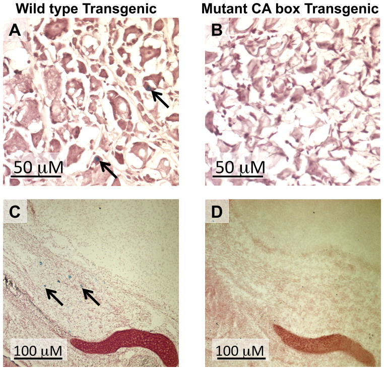
Fig. 4.
nAChR β4 subunit promoter activity of in the DRG of PD30 transgenic mice and in the Trigeminal ganglion of ED18.5 transgenic mice. Sections of WT transgenic line 54 (A) and mutant CA box transgenic line 28 (B) PD30 DRG are shown as well as WT transgenic line 54 (C) and mutant CA box transgenic line 28 (d) ED18.5 trigeminal ganglion. These sections were simultaneously stained for β-Gal activity and then counter-stained with neutral red. Arrows in panels A and C indicate β-Gal-expressing cells.

