Abstract
With the advent of functional brain imaging techniques and recent developments in the analysis of cortical connectivity, the focus of mental imagery studies has shifted from a semi-modular approach to an integrated cortical network perspective. Functional magnetic resonance imaging studies of visual imagery of faces and objects show that activation of content-specific representations stored in the ventral visual stream is top-down-modulated by parietal and frontal regions. Recent findings in patients with conscious awareness disorders reveal that mental imagery can be used to map patterns of residual cognitive function in their brain and to provide diagnostic and prognostic indicators.
Introduction and context
Visual imagery is the ability to generate percept-like images in the absence of retinal input and is therefore a vivid demonstration of retrieving pictorial information from memory. Psychophysical and brain imaging studies have demonstrated functional similarities between visual perception and visual imagery to the extent that common mechanisms appear to be activated by both [1-4]. Numerous neuroimaging studies have shown that visual imagery, like visual perception, evokes activation in occipito-parietal and occipito-temporal visual association areas [5,6]. In some studies, the primary visual cortex [7,8] was activated during imagery, suggesting that the generation of mental images may involve sensory representations at the earlier processing stages in the visual pathway. Studies of patients with brain damage have demonstrated a dissociation of visual-object and visual-spatial imagery [9], indicating that different parts of the visual system mediate ‘where’ and ‘what’ imagery, a dissociation that parallels the two anatomically distinct visual systems proposed for visual perception [10]. Single-unit recordings in epileptic patients revealed that neurons in the human medial temporal lobe fired selectively during both visual perception and visual imagery, suggesting that the neurons activated during storage of incoming visual input are later reactivated during the mnemonic retrieval [11].
Although many studies have focused on the overlap and similarities between perception and imagery, the subjective experiences of imagining and seeing are clearly very different. The intriguing case study of CK, a patient with severe visual agnosia who cannot recognize objects but can draw them with considerable details from memory, suggests that visual imagery can be dissociated from visual perception [12]. It has also been shown that during visual imagery, deactivation in auditory cortex is negatively correlated with activation in visual cortex and with the score of subjective vividness of visual imagery, suggesting that in order to generate vivid mental images, the brain needs to filter out irrelevant sensory information [13].
Where bottom-up meets top-down
Functional magnetic resonance imaging (fMRI) studies have reported that within the ventral pathway, faces and other objects, such as outdoor scenes, houses, animals, and tools, have distinct representations [14,15]. In particular, it has been shown that faces, houses, and chairs evoke highly consistent patterns of neural responses in occipital and temporal cortices [16,17]. Similar activation is observed in extrastriate cortex during visual and tactile recognition of faces and man-made objects in sighted subjects and during tactile recognition in blind subjects [18], suggesting that more abstract features of object form are represented in the visual ventral stream. Inspired by the consistent topology of the response to faces, houses, and chairs, a series of studies were conducted to investigate whether visual imagery of these objects would evoke content-related activation within the same ventral regions that are activated during perception [19,20]. Content-related activation during imagery was found in temporal cortex (e.g., imagery of faces activated the lateral fusiform gyrus, whereas imagery of houses activated the medial fusiform gyrus), but this activity was restricted to small subsets of the regions that were activated during perception [19,20]. Moreover, visual imagery of faces and objects evoked activity in parietal and frontal cortex, suggesting that content-related activation during imagery is mediated by the retrieval of face and object representations from long-term memory and their maintenance in the ‘mind’s eye’ [19,20]. Analysis of effective connectivity revealed that during visual perception, category-selective patterns of activation in extrastriate cortex are mediated by content-sensitive forward connections from early visual areas [21,22]. In contrast, during visual imagery, category-selective activation is mediated by content-sensitive backward connections from prefrontal cortex, suggesting that neuronal interactions between occipito-temporal, parietal, and frontal regions are task- and stimulus-dependent. Additionally, non-selective, top-down processes, originating in superior parietal areas, contribute to the generation of mental images, regardless of their content, and their maintenance through visual imagery [22]. Similar findings were reported in a study of motion imagery, which found activation in a network composed of motion-sensitive regions (human middle temporal/V5 complex) and prefrontal areas (frontal eye field and Brodmann area [BA] 9/46) [23]. It therefore seems that mental imagery is a dynamic cognitive function that engages distributed cortical networks activated during the allocation of attention and retrieval from memory. Interestingly, recent studies of art perception have shown that when confronted with abstract and indeterminate paintings, viewers use mental imagery and contextual associations to understand the content of these art compositions [24,25]. These novel findings indicate that visual imagery is an essential cognitive ability required not only for anticipating and retrieving information from memory but, importantly, for comprehending the world around us.
Major recent advances
Imagery in patients with conscious awareness disorders
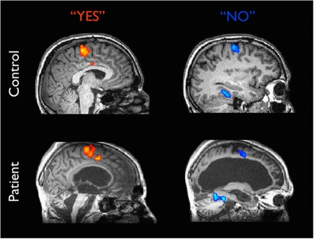
Recent studies in vegetative state patients have discovered normal patterns of activation in their brain during mental imagery. When a young woman in a vegetative state was asked to imagine playing tennis or to navigate her way around her house, significant activity was observed in her brain, similar to the activity in the brain of healthy volunteers performing the same tasks (Figure 1) [26,27]. These surprising findings suggest that vegetative state patients retain the ability to understand verbal instructions and to carry out mental tasks in response to those instructions, namely to exhibit willed, voluntary behavior in the absence of any overt action. Mental imagery can therefore be used as a neural proxy for behavior in order to assess the degree of consciousness in non-communicative brain-damaged patients and perhaps predict their recovery [28]. A recent study has shown that the precuneus, which mediates memory-related imagery [19,29], is also activated during hypnosis, suggesting that such a state of enhanced self-monitoring is achieved by control of motor responses by internal representations [30]. Thus, understanding the neural mechanisms of mental imagery could have far-reaching implications for understanding conscious awareness and its various disorders.
Figure 1. Brain activity in a healthy subject and a vegetative state patient.
Neural activation during imagining playing tennis (to convey ‘Yes’ responses) and imagining moving from room to room in the home (to convey ‘No’ responses). The similar patterns of neural activation suggest that some patients in a vegetative or minimally conscious state have activity that reflects awareness and cognition. Adapted from Monti et al., 2010 [36], courtesy of Adrian Owen.
Future directions
Recent developments in deciphering the mental chronometry [31] and functional and effective connectivity during spatial imagery [32] suggest hierarchical temporal dynamics within the imagery network. The use of classifiers for decoding mental states [33] and predictive coding models [34] would enable additional insights into the neural mechanisms of both perception and mental imagery. Understanding the neuronal interactions among visual, parietal, and prefrontal regions has far-reaching clinical implications, reinforced by the fact that vegetative state patients show healthy patterns of brain activation during mental imagery which reflect will and intention, the hallmark of conscious awareness. Identifying residual cognitive function in such patients by means of fMRI and electroencephalogram can be used not only for diagnosis and prognosis but perhaps also as a form of communication with patients who lack speech or the motor act of expression [35,36]. Hopefully, in the near future, combining cutting-edge analytic techniques will enable clinicians to make accurate predictions about the recovery of non-communicative brain-damaged patients.
Acknowledgments
The author is supported by Swiss National Science Foundation grant 3200B0-105278 and by the Swiss National Center for Competence in Research: Neural Plasticity and Repair.
Abbreviation
- fMRI
functional magnetic resonance imaging
Competing Interests
The author declares that she has no competing interests.
The electronic version of this article is the complete one and can be found at: http://f1000.com/reports/b/2/34
References
- 1.Roland PE, Eriksson L, Stone-Elander S, Widen L. Does mental activity change the oxidative metabolism of the brain? J Neurosci. 1987;7:2373–89. [PMC free article] [PubMed] [Google Scholar]
- 2.Farah M, Peronnet F, Gonon MA, Giard MH. Electrophysiological evidence for a shared representational medium for visual images and visual percepts. J Exp Psychol Gen. 1988;117:248–57. doi: 10.1037/0096-3445.117.3.248. [DOI] [PubMed] [Google Scholar]
- 3.Goldenberg G, Poderka I, Steiner M, Willmes K, Suess E, Deecke L. Regional cerebral blood flow patterns in visual imagery. Neuropsychologia. 1989;27:641–64. doi: 10.1016/0028-3932(89)90110-3. [DOI] [PubMed] [Google Scholar]
- 4.Ishai A, Sagi D. Common mechanisms of visual imagery and perception. Science. 1995;268:1772–4. doi: 10.1126/science.7792605. [DOI] [PubMed] [Google Scholar]
- 5.Mellet E, Tzourio N, Crivello F, Joliot M, Denis M, Mazoyer B. Functional anatomy of spatial mental imagery generated from verbal instructions. J Neurosci. 1996;16:6504–12. doi: 10.1523/JNEUROSCI.16-20-06504.1996. [DOI] [PMC free article] [PubMed] [Google Scholar]
- 6.D’Esposito M, Deter JA, Aguirre GK, Stallcup M, Alsop DC, Tippet LJ, Farah MJ. A functional MRI study of mental image generation. Neuropsychologia. 1997;35:725–30. doi: 10.1016/S0028-3932(96)00121-2. [DOI] [PubMed] [Google Scholar]
- 7.Le Bihan D, Turner R, Zeffiro TA, Cuénod CA, Jezzard P, Bonnerot V. Activation of human primary visual cortex during visual recall: a magnetic resonance imaging study. Proc Natl Acad Sci U S A. 1993;90:11802–5. doi: 10.1073/pnas.90.24.11802. [DOI] [PMC free article] [PubMed] [Google Scholar]
- 8.Kosslyn SM, Alpert NM, Thompson WL, Maljkovic V, Weise SB, Chabris CF, Hamilton SE, Rauch SL, Buonanno FS. Visual mental imagery activates topographically organized visual cortex: PET investigations. J Cogn Neurosci. 1993;5:263–87. doi: 10.1162/jocn.1993.5.3.263. [DOI] [PubMed] [Google Scholar]
- 9.Levine DN, Warach J, Farah M. Two visual systems in mental imagery: dissociation of ‘what’ and ‘where’ in imagery disorders due to bilateral posterior cerebral lesions. Neurology. 1985;35:1010–8. doi: 10.1212/wnl.35.7.1010. [DOI] [PubMed] [Google Scholar]
- 10.Ungerleider LG, Mishkin M. Two cortical visual systems. In: Ingle DJ, Goodale MA, Mansfield RJW, editors. Analysis of Visual Behavior. Cambridge, MA: MIT Press; 1982. [Google Scholar]
- 11.Kreiman G, Koch C, Fried I. Imagery neurons in the human brain. Nature. 2000;408:357–61. doi: 10.1038/35042575. [DOI] [PubMed] [Google Scholar]
- 12.Behrmann M, Winocur G, Moscovitch M. Dissociation between mental imagery and object recognition in a brain-damaged patient. Nature. 1992;359:636–7. doi: 10.1038/359636a0. [DOI] [PubMed] [Google Scholar]
- 13.Amedi A, Malach R, Pascual-Leone A. Negative BOLD differentiates visual imagery and perception. Neuron. 2005;48:859–72. doi: 10.1016/j.neuron.2005.10.032. [DOI] [PubMed] [Google Scholar]
- 14.Epstein R, Kanwisher N. A cortical representation of the local visual environment. Nature. 1998;392:598–601. doi: 10.1038/33402. [DOI] [PubMed] [Google Scholar]
- 15.Chao LL, Haxby JV, Martin A. Attribute-based neural substrates in posterior temporal cortex for perceiving and knowing about objects. Nat Neurosci. 1999;2:913–9. doi: 10.1038/13217. [DOI] [PubMed] [Google Scholar]
- 16.Ishai A, Ungerleider LG, Martin A, Schouten JL, Haxby JV. Distributed representation of objects in the human ventral visual pathway. Proc Natl Acad Sci U S A. 1999;96:9379–84. doi: 10.1073/pnas.96.16.9379. [DOI] [PMC free article] [PubMed] [Google Scholar]
- 17.Ishai A, Ungerleider LG, Martin A, Haxby JV. The representation of objects in the human occipital and temporal cortex. J Cogn Neurosci. 2000;12:35–51. doi: 10.1162/089892900564055. [DOI] [PubMed] [Google Scholar]
- 18.Pietrini P, Furey ML, Ricciardi E, Gobbini MI, Wu WH, Cohen L, Guazzelli M, Haxby JV. Beyond sensory images: object-based representation in the human ventral pathway. Proc Natl Acad Sci U S A. 2004;101:5658–63. doi: 10.1073/pnas.0400707101. [DOI] [PMC free article] [PubMed] [Google Scholar]; F1000 Factor 3.0 RecommendedEvaluated by Susan Courtney 27 May 2004
- 19.Ishai A, Ungerleider LG, Haxby JV. Distributed neural systems for the generation of visual images. Neuron. 2000;28:979–90. doi: 10.1016/S0896-6273(00)00168-9. [DOI] [PubMed] [Google Scholar]
- 20.Ishai A, Haxby JV, Ungerleider LG. Visual imagery of famous faces: effects of memory and attention revealed by fMRI. NeuroImage. 2002;17:1729–41. doi: 10.1006/nimg.2002.1330. [DOI] [PubMed] [Google Scholar]
- 21.Mechelli A, Price CJ, Noppeney U, Friston KJ. A dynamic causal modeling study on category effects: bottom-up or top-down mediation? J Cogn Neurosci. 2003;15:925–34. doi: 10.1162/089892903770007317. [DOI] [PubMed] [Google Scholar]
- 22.Mechelli A, Price CJ, Friston KJ, Ishai A. Where bottom-up meets top-down: neuronal interactions during perception and imagery. Cereb Cortex. 2004;14:1256–65. doi: 10.1093/cercor/bhh087. [DOI] [PubMed] [Google Scholar]
- 23.Goebel R, Khorram-Sefat D, Muckli L, Hacker H, Singer W. The constructive nature of vision: direct evidence from functional magnetic resonance imaging studies of apparent motion and motion imagery. Eur J Neurosci. 1998;10:1563–73. doi: 10.1046/j.1460-9568.1998.00181.x. [DOI] [PubMed] [Google Scholar]
- 24.Fairhall SL, Ishai A. Neural correlates of object indeterminacy in art compositions. Conscious Cogn. 2008;17:923–32. doi: 10.1016/j.concog.2007.07.005. [DOI] [PubMed] [Google Scholar]
- 25.Wiesmann M, Ishai A. Training facilitates object recognition in cubist paintings. Front Hum Neurosci. 2010;4:11. doi: 10.3389/neuro.09.011.2010. [DOI] [PMC free article] [PubMed] [Google Scholar]
- 26.Owen AM, Coleman MR, Boly M, Davis MH, Laureys S, Pickard JD. Detecting awareness in the vegetative state. Science. 2006;313:1402. doi: 10.1126/science.1130197. [DOI] [PubMed] [Google Scholar]; F1000 Factor 3.0 RecommendedEvaluated by Kevan A Martin 14 Sep 2006
- 27.Boly M, Coleman MR, Davis MH, Hampshire A, Bor D, Moonen G, Maquet PA, Pickard JD, Laureys S, Owen AM. When thoughts become action: an fMRI paradigm to study volitional brain activity in non-communicative brain injured patients. NeuroImage. 2007;36:979–92. doi: 10.1016/j.neuroimage.2007.02.047. [DOI] [PubMed] [Google Scholar]
- 28.Di H, Boly M, Weng X, Ledoux D, Laureys S. Neuroimaging activation studies in the vegetative state: predictors of recovery? Clin Med. 2008;8:502–7. doi: 10.7861/clinmedicine.8-5-502. [DOI] [PMC free article] [PubMed] [Google Scholar]
- 29.Fletcher PC, Frith CD, Baker SC, Shallice T, Frackowiak RSJ, Dolan RJ. The mind’s eye - precuneus activation in memory-related imagery. NeuroImage. 1995;2:195–200. doi: 10.1006/nimg.1995.1025. [DOI] [PubMed] [Google Scholar]
- 30.Cojan Y, Waber L, Schwartz S, Rossier L, Forster A, Vuilleumier P. The brain under self-control: modulation of inhibitory and monitoring cortical networks during hypnotic paralysis. Neuron. 2009;62:862–75. doi: 10.1016/j.neuron.2009.05.021. [DOI] [PubMed] [Google Scholar]
- 31.Formisano E, Goebel R. Tracking cognitive processes with functional MRI mental chronometry. Curr Opin Neurobiol. 2003;13:174–81. doi: 10.1016/S0959-4388(03)00044-8. [DOI] [PubMed] [Google Scholar]
- 32.Sack AT, Jacobs C, De Martino F, Staeren N, Goebel R, Formisano E. Dynamic premotor-to-parietal interactions during spatial imagery. J Neurosci. 2008;28:8417–29. doi: 10.1523/JNEUROSCI.2656-08.2008. [DOI] [PMC free article] [PubMed] [Google Scholar]
- 33.Reddy L, Tsuchiya N, Serre T. Reading the mind’s eye: decoding category information during mental imagery. NeuroImage. 2010;50:818–25. doi: 10.1016/j.neuroimage.2009.11.084. [DOI] [PMC free article] [PubMed] [Google Scholar]
- 34.Hohwy J, Roepstorff A, Friston K. Predictive coding explains binocular rivalry: an epistemological review. Cognition. 2008;108:687–701. doi: 10.1016/j.cognition.2008.05.010. [DOI] [PubMed] [Google Scholar]
- 35.Owen AM, Schiff ND, Laureys S. A new era of coma and consciousness science. Prog Brain Res. 2009;177:399–411. doi: 10.1016/S0079-6123(09)17728-2. [DOI] [PubMed] [Google Scholar]
- 36.Monti MM, Vanhaudenhuyse A, Coleman MR, Boly M, Pickard JD, Tshibanda L, Owen AM, Laureys S. Willful modulation of brain activity in disorders of consciousness. N Engl J Med. 2010;362:579–89. doi: 10.1056/NEJMoa0905370. [DOI] [PubMed] [Google Scholar]; F1000 Factor 9.9 ExceptionalEvaluated by Laurie Zoloth 10 Mar 2010, Tarek Sharshar 12 Mar 2010, Thomas Milhorat 12 Mar 2010


