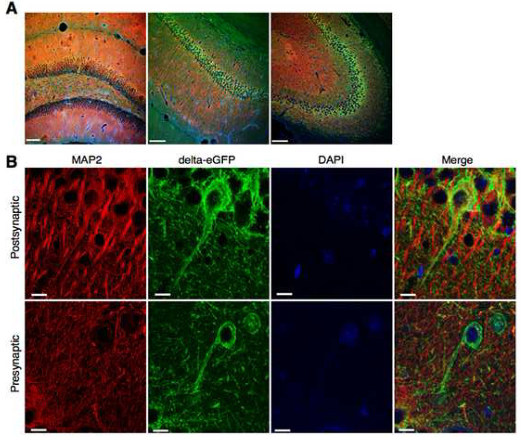
Fig. 3. Delta opioid receptors are localized both pre- and post-synaptically in the hippocampus (D. Massotte and B. Kieffer, unpublished).
Confocal imaging of brain sections from delta-eGFP mice, labeled with a MAP2 antibody that labels somatodendritic, but not axonal, compartments. MAP immunostaining is shown in red, fluorescently labeled delta receptors are shown in green, and cell nuclei in blue (DAPI). (A) A general view at the level of the hippocampus. Dentate gyrus (left panel), CA1 region (central panel) and CA3 region (right panel). Scale bars 100 µm. (B) Top panels: the delta receptor is expressed in dendrites. In this neuron MAP2 and delta-eGFP are co-localized (merged image on the right), suggesting a postsynaptic localization of the receptor. Scale bar 10 µm. Bottom panels: the delta receptor is also expressed in axons. In this neuron, lack of MAP2 and delta-eGFP co-staining (merged image on the right) indicates a presynaptic receptor localization. Scale bar 10µm.

