

Abstract
The highly potent botulinum neurotoxins are responsible for botulism, a severe neuroparalytic disease. Strains of nonproteolytic Clostridium botulinum form neurotoxins of types B, E, and F and are the main hazard associated with minimally heated refrigerated foods. Recent developments in quantitative microbiological risk assessment (QMRA) and food safety objectives (FSO) have made food safety more quantitative and include, as inputs, probability distributions for the contamination of food materials and foods. A new method that combines a selective enrichment culture with multiplex PCR has been developed and validated to enumerate specifically the spores of nonproteolytic C. botulinum. Key features of this new method include the following: (i) it is specific for nonproteolytic C. botulinum (and does not detect proteolytic C. botulinum), (ii) the detection limit has been determined for each food tested (using carefully structured control samples), and (iii) a low detection limit has been achieved by the use of selective enrichment and large test samples. The method has been used to enumerate spores of nonproteolytic C. botulinum in 637 samples of 19 food materials included in pasta-based minimally heated refrigerated foods and in 7 complete foods. A total of 32 samples (5 egg pastas and 27 scallops) contained spores of nonproteolytic C. botulinum type B or F. The majority of samples contained <100 spores/kg, but one sample of scallops contained 444 spores/kg. Nonproteolytic C. botulinum type E was not detected. Importantly, for QMRA and FSO, the construction of probability distributions will enable the frequency of packs containing particular levels of contamination to be determined.
Food-borne botulism is a severe and deadly intoxication caused by the consumption of food containing as little as 30 to 100 ng of preformed botulinum neurotoxin (45). More than 2,500 cases of botulism were reported in Europe in 1999 and 2000, with the majority of cases in the east of the continent (44). Currently, 25 to 50 food-borne botulism cases are diagnosed annually in the United States (27). There are seven distinct botulinum neurotoxins (types A to G) and a number of subtypes (6, 26, 45). In view of the potency of the botulinum neurotoxin and the severity of botulism, four phylogenetically distinct bacteria are grouped together as the Clostridium botulinum species, solely on the basis of their ability to form botulinum neurotoxin. The divergence between these four distinct bacteria is strong enough to merit their classification as distinct species and in some cases is significantly greater than that between bacteria belonging to different genera, e.g., Bacillus subtilis and Staphylococcus aureus (7). Two of these bacteria (proteolytic C. botulinum and nonproteolytic C. botulinum) are responsible for the majority of cases of food-borne botulism. Strains of proteolytic C. botulinum produce neurotoxins of type A, B, or F, form spores of high heat resistance, and have a minimum growth temperature of approximately 12°C (39). Strains of nonproteolytic C. botulinum produce neurotoxins of type B, E, or F, form spores of moderate heat resistance, and are able to grow and form toxin at 3°C (18, 48) and are recognized as the major hazard associated with minimally heated refrigerated foods (4, 37, 43, 44, 48). These new foods meet consumer demand for high-quality, convenient foods that are low in preservatives, and sales are presently increasing by about 10% per annum in many countries (3, 47).
Quantitative microbiological risk assessment (QMRA) is now established as an important microbiology food safety tool (42). Process risk models have been used to assess the safety of specific foods with respect to nonproteolytic C. botulinum and the food-borne botulism hazard (e.g., 2, 41). These process risk models benefit from high-quality information, including that on the incidence of spores of nonproteolytic C. botulinum spores in food materials. The implementation of food safety objectives (FSO) also benefits from the availability of high-quality information on the microbial contamination of foods and food materials (24). This information is most effective in the form of probability distributions rather than as average spore concentrations or other statistics.
The difficulty with enumerating nonproteolytic C. botulinum in foods is that there is no effective selective culture medium available. Surveys of the extent of contamination of foods and food materials have used a nonselective enrichment followed by either testing for neurotoxin using a mouse test or enzyme-linked immunosorbent assay (ELISA) or testing for the presence of neurotoxin genes using a PCR test (3, 10, 13, 35, 38, 39). This approach, however, is not optimized for nonproteolytic C. botulinum or proteolytic C. botulinum (therefore potentially failing to recover all spores of either organism) and may also not distinguish nonproteolytic C. botulinum from proteolytic C. botulinum. Heating at 80°C for 10 min followed by incubation at 35°C (54) may be reasonably selective for proteolytic C. botulinum, but there is no similar approach for nonproteolytic C. botulinum, although incubation at 28°C (54) may offer an element of selection. It is necessary, therefore, to develop a method to enumerate spores of nonproteolytic C. botulinum in food materials that is robust and optimized, as well as sensitive and specific for this particular pathogen (and does not also detect proteolytic C. botulinum). When enumerating bacteria in foods, it is essential to demonstrate the efficiency of the method by verifying that small concentrations (in the present study, spores of nonproteolytic C. botulinum) can be detected following addition to test samples.
This paper describes the development, validation, and application of a new method to enumerate spores of nonproteolytic C. botulinum in foods and in food materials. This method has been designed to generate data for the construction of probability distributions that can be used in QMRA and FSO settings. Most of the effort has been dedicated to the development and evaluation of the enrichment procedure rather than the PCR test, as the PCR test has received much attention from others (e.g., 3, 10, 16, 36, 38). A low-temperature selective-enrichment procedure is described that has been optimized specifically for nonproteolytic C. botulinum over proteolytic C. botulinum and other bacteria. In order to detect low concentrations of spores, large quantities (200 g) of food materials and foods have been tested. Specific detection of neurotoxin genes is achieved by the use of an established multiplex PCR (36), with an internal amplification control now included (25). By the use of a set of control samples inoculated with defined concentrations of spores of nonproteolytic C. botulinum, the detection limit has been estimated for each food material and food tested. The method has been used in an extensive survey of raw materials intended for use in pasta ready meals, as well as the final meals themselves. The implications for risk assessment and risk management of chilled foods are discussed.
MATERIALS AND METHODS
Preparation of spores of nonproteolytic C. botulinum and proteolytic C. botulinum.
Spores of nonproteolytic C. botulinum were produced using a two-phase medium of water over cooked meat agar (46), and four spore suspensions were prepared. Type B spore mixture 1 contained spores of strains Colworth 151, CDC 3875, CDC 5900, and Eklund 2B (used in all tests except the scallop most probable number [MPN] test); type B spore mixture 2 contained spores of strains Hobbs FT50, Eklund 17B, Eklund 2B, and Kapchunka B2 (used only in the scallop MPN test); the type E spore mixture contained spores of strains Hazen 36208, Dolman VH, CDC 7854, and Foster B96; and the type F spore mixture contained spores of strains Eklund 202F and Craig 610. Spores of three strains of proteolytic C. botulinum (type A [strain 62A], type B [strain 213B], and type F [strain Walls 8G]) were prepared as described previously (9) and included in a single spore mixture. Spores were harvested, washed, and separated from cell debris by discontinuous density gradient centrifugation (52) and stored in water at 2°C until required. The proteolytic activity of strains was assessed on reinforced clostridial medium agar (Oxoid, Basingstoke, United Kingdom) containing 5% (wt/vol) skim milk incubated at 30°C for 48 h, under a headspace of CO2-H2 (10:90) (49). Spores were enumerated and purity was confirmed on Trypticase peptone glucose yeast extract (TPGY) agar (comprising TPGY broth [54] plus agar [Oxoid] at 20 g/liter) incubated under the same conditions. All spore mixtures were prepared containing an equal concentration of spores of each strain and were held at 2°C prior to use.
Optimized protocol for determining the presence of nonproteolytic C. botulinum in food materials: food sample preparation.
The food materials tested were potential ingredients of pasta-based chilled ready meals and the assembled meals. Each food material was sourced globally from a range of locations and seasons and, in the case of the vegetables, included different cultivars. Where spores of C. botulinum were likely to be present on the surface of the food material (e.g., vegetable tissue), the food was sliced or chopped. For some food materials (e.g., cheese, pasta) or entire meals, spores may be incorporated in the food during processing. These food samples were cut or diced into small pieces and where appropriate further reduced in size in the grinder unit of a food processor to ensure that any potential spores were exposed to the enrichment medium. Entire meals were homogenized in a food blender. These steps were carried out using aseptic techniques.
Preparation of anaerobic TPGY medium.
Two methods of preparing anaerobic TPGY medium were compared. In method I, anaerobic medium was prepared using strict anaerobic technique (i.e., boiled, cooled, and sparged under a headspace of anaerobic gas [CO2-N2-H2, 10:85:5]) prior to autoclaving (51), and in method II, it was prepared by boiling for 15 min prior to autoclaving. Because of the large number of samples to be tested, it was not possible to boil or steam the autoclaved medium immediately before use. The redox potential of the media prepared using the two different methods was measured relative to a standard hydrogen electrode (Eh) as described previously and calculated using a Eref value of +207 mV (17). In order to achieve a lower detection limit than procedures that test a 25-g food sample, it was necessary to test larger food samples (200 g was most frequently used). Samples were cultured with 500 ml TPGY medium in a 1-liter bottle. Since many of the foods were acidic, the effectiveness of adding sodium phosphate buffer (final concentration, 0.1 M) for maintaining an optimum pH of the TPGY medium was evaluated. It should be noted that foods with a pH lower than approximately 5.0 will not support growth and neurotoxin formation by nonproteolytic C. botulinum but may contain such spores and were included here to investigate their possible contribution of spores to nonacid processed foods.
Addition of food materials to the TPGY enrichment medium and heat treatment.
The prepared food samples (200 g, except for semolina, where 70 g was tested) were weighed into 1-liter bottles and degassed in the interchange of an anaerobic cabinet before 500 ml of TPGY medium was added inside the cabinet (anaerobic gas mixture in cabinet [CO2-N2-H2, 10:85:5]). To prevent germination of spores of nonproteolytic C. botulinum between this addition and the subsequent heat treatment, the TPGY medium was preheated to 65°C and added hot to the food samples. The bottles were sealed with airtight caps and immediately transferred from the cabinet to a water bath at 67°C for 60 min, with gentle shaking (100 rpm). The bottles were then cooled rapidly to approximately 15°C in running cold water.
Detection limit determination.
The minimum concentration of spores that could be detected (detection limit) was calculated for each food material. Parallel samples of each food were inoculated with spores of mixtures of strains of nonproteolytic C. botulinum (each toxin type was tested separately). In most cases additions were at approximate concentrations of 4, 10, 30 and 100 spores per bottle. The actual concentrations for the control inocula were estimated from plate counts. In food materials with a poor detection limit (e.g., mushrooms, zucchini), higher concentrations of spores were also added to control bottles (up to 105 spores/bottle). A maximum likelihood detection limit and 95% confidence intervals were calculated as described in the Appendix.
Incubation temperature for primary enrichment culture.
Incubation temperature was used as a selective factor for growth of nonproteolytic C. botulinum over proteolytic C. botulinum and other competing bacteria. The minimum and optimum growth temperatures for nonproteolytic C. botulinum are reported as 3°C and 25°C, while proteolytic C. botulinum has a minimum growth temperature of about 12°C and an optimum of 37°C (44). Preliminary tests with food materials evaluated the effectiveness of incubation temperature (10°C, 12°C, and 15°C) on promoting selective growth of nonproteolytic C. botulinum after 7 days' incubation.
Optimization of the subculture prior to PCR.
It was necessary to include a subculture step prior to the PCR test, as preliminary work had shown that some foods (e.g., basil) were otherwise inhibitory to the PCR test. An aliquot (0.2 ml) of the primary enrichment culture was added to 19.8 ml TPGY medium. This subculture was incubated at 20°C for 24 h in order to select for nonproteolytic C. botulinum over proteolytic C. botulinum. ComBase Predictor (www.Combase.cc) predicts a 106-fold increase in the viable count of nonproteolytic C. botulinum under these conditions but less than a 2-fold increase in proteolytic C. botulinum. As the minimum detectable limit (MDL) of the PCR is about 1 × 10⁵ cells/ml of the original culture when a 1-μl PCR sample (i.e., 102 cells per reaction mixture [36]) is used, the subculture was sufficient to allow a single spore/cell of nonproteolytic C. botulinum, but not a single spore/cell of proteolytic C. botulinum, to multiply to a detectable concentration.
Optimization of the PCR procedure.
The PCR test used in the present study was based on the multiplex PCR test originally described by Lindström et al. (36). Since this method was first described, a number of different neurotoxin subtypes have been described (45). By alignment of available sequences for subtypes of type B, E, and F neurotoxin genes, it was confirmed that the existing PCR probes would detect genes encoding all subtypes of these three toxins known at this time. A proprietary reagent (PrepMan Ultra; Applied Biosystems, Warrington, United Kingdom) was used to extract DNA from the subculture, as initial tests had shown it to be superior to the boiling step originally described (36). Culture aliquots (1 ml) were extracted according to the manufacturer's instructions, and 1-μl aliquots were used in a 25-μl PCR mix. The primers, their concentrations, and the thermocycling profile were as originally described (36). Oligonucleotides were synthesized by Sigma-Genosys (Haverhill, United Kingdom). The “Titanium” Taq PCR kit (BD Biosciences, Cowley, United Kingdom) was used to amplify target DNA, as a preliminary evaluation demonstrated that it gave a more consistent amplification than two other polymerase systems (PCR master mix [Promega, Southampton, United Kingdom] and HotStarTaq master mix [Qiagen, Crawley, United Kingdom]). PCR products were detected using agarose gel electrophoresis (36).
It has been recommended (25) that an internal positive amplification control (IAC) be included when performing diagnostic PCR. IACs have been previously included in some tests for botulinum neurotoxin genes (see, e.g., references 3 and 10). In the present study, calf thymus DNA (included in the “Titanium” Taq PCR kit) was used as a template. Primers (5′-CATTGCCACTGTCGTAGCCA-3′; 5′-GCAACTTCTCTTGGGAGGCAA-3′) with melting temperatures similar to those given by Lindström et al. (36) were included and gave an IAC band of 945 bp. Since smaller products are often amplified more readily than larger products, it was important that the IAC product was larger than the products for the four neurotoxin genes. Template (50 ng) and primers (0.2 μM) were incorporated into the PCR reagent mix. Additional controls comprised DNA extracted from C. botulinum strains forming type A, B, E, and F neurotoxins.
Isolation of nonproteolytic C. botulinum.
A medium selective for C. botulinum and a nonselective medium were used (54). Samples from PCR-positive enrichment cultures were streaked onto plates of TPGY agar containing 10% (vol/vol) egg yolk emulsion with and without the antibiotics described previously (11) and cultured anaerobically (CO2-H2, 10:90) at 30°C. From each sample, five colonies showing typical characteristics (e.g., lipase positive) were selected and purity confirmed. Cultures were then tested for the presence of neurotoxin genes using the PCR procedure, while supernatants were tested for neurotoxin using an ELISA. PCR products from amplifications were purified using a commercial spin column method (Qiagen, United Kingdom). The concentration of this DNA was estimated by agarose gel electrophoresis, and the sequence was then determined.
ELISAs.
Culture fluids were tested using a commercial botulinum toxin ELISA (Metabiologics, Madison, WI) for the appropriate toxin type. The culture fluids were diluted 1:1 with double-strength casein buffer before testing using the manufacturer's protocol.
Optimized protocol for enumeration of nonproteolytic C. botulinum in food materials and complete meals.
Samples shown to contain spores of nonproteolytic C. botulinum in the initial presence tests were examined further to determine the concentration of spores present. The concentration of spores of nonproteolytic C. botulinum was determined using a three-tube MPN, with six 10-fold dilutions. In the case of scallops, 200 g, 20 g, and 2 g, etc., were tested, while for egg pastas only 10 to 30 g was available for the first sample. Each sample was enriched and PCR performed as described above. Controls were prepared from uncontaminated samples of the same food type inoculated at concentrations of 1 × 10³ spores/kg and 1 × 10⁵ spores/kg (egg pastas) or 3 × 101 spores/kg, 1 × 10³ spores/kg, and 6 × 10⁴ spores/kg (scallops). The MPN and 95% confidence intervals were calculated as previously described (21).
RESULTS
Optimization of protocol for determining the presence of nonproteolytic C. botulinum in food materials.
In order to achieve a low detection limit, 200-g samples of food materials were cultured with 500 ml TPGY medium. However, since many of the foods were themselves acidic (e.g., parmesan cheese, pH 5.2; tomato, pH 4.2), sodium phosphate (final concentration, 0.1 M) was added to the TPGY medium to give an optimal starting pH and maintain a higher pH during growth. This addition maintained TPGY medium (500 ml) to which tomato (200 g) or parmesan cheese (200 g) were added at pH >6.0 after incubation for 21 h at 12°C. The pH of the TPGY medium to which additional buffer was not added fell as low as pH 5.4. At the end of most enrichments (7 days at 12°C), the phosphate-buffered TPGY had a pH of between 6.2 and 6.9; however, for some food materials the final pH was lower: a pH of between 5.7 and 6.1 (zucchini, mozzarella cheese, pecorillo cheese, celery, parsley) or pH 5.2 to 5.6 (carrot, red pepper, peas).
Two methods of preparing the anaerobic medium were evaluated. Autoclaved TPGY medium prepared using strict anaerobic technique had a redox potential approximately 250 mV lower than that of TPGY medium prepared by boiling for 15 min prior to autoclaving. When samples of tomato (250 g) were deoxygenated by passage through the interchange of an anaerobic cabinet and added to the medium (500 ml) in the cabinet, the redox potential was −158 mV in medium prepared using strict anaerobic technique and +86 mV in medium prepared by boiling alone. Similarly, medium prepared using strict anaerobic technique to which 250 g of potato or salmon was added also had a low redox potential (−170 mV). In view of these effects, all subsequent media were prepared using strict anaerobic technique, since it was reported that approximately 20 times more spores were required to give growth of nonproteolytic C. botulinum in a medium at +83 mV than in the same medium at −190 mV (40).
To create conditions favorable for nonproteolytic C. botulinum, a heat treatment was applied that would reduce the concentration of non-spore-forming bacteria but not the viability of spores of nonproteolytic C. botulinum. It was important to ensure that spores of nonproteolytic C. botulinum did not germinate prior to the heat treatment. Prewarmed (65°C) TPGY medium was added to the food in an anaerobic cabinet, and the mixture was then quickly transferred to a water bath at 67°C for application of the heat treatment. The temperature of the food-containing TPGY medium did not fall below 46°C during this transfer, a temperature at which germination of spores of nonproteolytic C. botulinum is negligible (49). The heat treatment was applied by immersing bottles for 60 min in a gently shaking (100 rpm) water bath preset to 67°C. The bottles were at ≥64.5°C for 19 min, and the total heat treatment was equivalent to 65°C for 32 min (using a z value of 7°C). In control tests it was established that heating at 65°C for 60 min in TPGY medium brought about less than a 0.1 decimal reduction in the concentration of spores of nonproteolytic C. botulinum types B and E. Also, this heat treatment had no significant effect on the concentration of spores of nonproteolytic C. botulinum types B, E, and F that were recovered when added to penne pasta/TPGY medium.
Following heat treatment, cooled bottles were incubated at 12°C for 7 days. Tests with food materials showed that incubation at 12°C for 7 days provided better selective enrichment of nonproteolytic C. botulinum than incubation at 10°C or 15°C, with growth of competing bacteria, including proteolytic C. botulinum, minimized. The PCR subculture involved a hundredfold dilution of the 7-day primary enrichment culture (as direct addition of the primary enrichment of basil, and a 10-fold dilution subculture, inhibited the subsequent PCR). This subculture involved the addition of a 0.2-ml aliquot from the primary enrichment culture to 19.8 ml fresh TPGY medium and incubation at 20°C for 24 h. This stage was selective for nonproteolytic C. botulinum over proteolytic C. botulinum (as outlined in Materials and Methods). DNA was then extracted, and the presence of neurotoxin genes was detected using PCR. The final procedure is summarized in Table 1.
TABLE 1.
Summary of optimized protocol for determining the presence of nonproteolytic C. botulinum in food materialsa
| Day | Operation |
|---|---|
| 1 | Outside anaerobic cabinet: aseptically cut, slice, and/or grind food materials, weigh 200-g aliquots into sterile 1-liter bottle, store overnight at +2°C |
| ↓ | |
| 2 | Transfer bottle containing food sample to anaerobic cabinet (deoxygenate in the interchange of the cabinet). Inside anaerobic cabinet, add 500 ml preheated (65°C) sterile anaerobic TPGY broth (CO2-N2-H2, 10:85:5) to deoxygenated food sample. Seal bottle. Transfer out of anaerobic cabinet |
| ↓ | |
| 2 | Outside anaerobic cabinet, quickly immerse the bottles in a water bath at 67°C for 60 min (to deliver heat treatment equivalent to approximately 65°C/30 min), with gentle shaking (100 rpm). Cool to <15°C and incubate at 12°C for 7 days (where appropriate add spores to control samples) |
| ↓ | |
| 9 | Remove subsample and subculture in fresh TPGY broth and incubate at 20°C for 24 h |
| ↓ | |
| 10/11 | Extract DNA from cultures. Amplify neurotoxin genes by PCR; determine PCR product size by agarose gel electrophoresis |
Further details are given in Materials and Methods.
Detection limits for proteolytic C. botulinum and nonproteolytic C. botulinum.
This method was designed to be highly selective for nonproteolytic C. botulinum over proteolytic C. botulinum. In a preliminary test, six spores of nonproteolytic C. botulinum (mixture of strains forming type B, E, and F neurotoxins) could be detected per bottle containing 500 ml medium only or with the addition of 200 g of basil, prawns, bacon, or penne pasta, while the addition of 3 × 10⁶ spores of proteolytic C. botulinum (mixture of strains forming type A, B, and F neurotoxins) per bottle yielded a negative result. Proteolytic C. botulinum was detected, however, when the primary enrichment was at 15°C rather than 12°C (data not shown).
The detection limit for each food or food material was calculated using data from the control bottles. Detection of strains forming type B, E, or F toxin was similar, allowing calculation of an overall maximum likelihood detection limit for nonproteolytic C. botulinum (Table 2). For example, for asparagus, 21 negative results were recorded for nonproteolytic C. botulinum from 21 tests each corresponding to 200 g of material. Control tests using 200 g of asparagus indicated that, according to a maximum likelihood method, the detection limit was ∼6 spores per 200-g sample with a 95% confidence interval of 4 and 8 spores per sample. Thus, we estimate that for asparagus, spore loads of nonproteolytic C. botulinum were <30 spores/kg. For most of the tested foods, similar conclusions can be established, and the results are included in Table 2. A maximum likelihood detection limit of 1 spore per bottle (200-g sample) was achieved for nonegg pasta, chives, and some complete meals. For most of the foods tested the detection limit was less than 10 spores per bottle (50 spores/kg).
TABLE 2.
Summary of initial screening of food and food materials for the presence of spores of nonproteolytic Clostridium botulinum
| Raw material | Total no. of samples tested | No. of negative samples | Estimated detection limit (no. of spores/200 g)a | No. of positive samples |
|---|---|---|---|---|
| Food materials | ||||
| Nonegg pasta | 48 | 48 | 1 (1, 2) | 0 |
| Egg pasta | 47 | 42 | 15 (12, 18) | 5 |
| Pasta sheets | 18 | 18 | 2 (1, 3) | 0 |
| Semolina | 13 | 13 | 8 (5, 13) | 0 |
| Asparagus | 21 | 21 | 6 (4, 8) | 0 |
| Basil | 18 | 18 | 14 (11, 102) | 0 |
| Carrots | 21 | 21 | 12 (10, 15) | 0 |
| Chives | 3 | 3 | 1 (1, 96)b | 0 |
| Green pepper | 26 | 26 | 11 (9, 14) | 0 |
| Red pepper | 21 | 21 | 104 (89, 123) | 0 |
| Zucchini | 43 | 43 | 1,269 (1,002, 4,994) | 0 |
| Mushrooms | 45 | 45 | 207 (113, 1,032) | 0 |
| Mozzarella | 22 | 22 | 3 (2, 5) | 0 |
| Ham/bacon | 45 | 45 | 5 (3, 6) | 0 |
| Shrimps/prawns | 42 | 42 | 14 (11, 17) | 0 |
| Scallops | 44 | 17 | 3 (2, 4) | 27 |
| Uncooked béchamel sauce | 23 | 23 | 5 (3, 6) | 0 |
| Cooked béchamel sauce | 12 | 12 | 5 (4, 7) | 0 |
| Assorted mixed toppings | 30 | 30 | 3 (2, 5) | 0 |
| Chilled ready meals | ||||
| Fusilli con pollo | 17 | 17 | 79 (61, 97)b | 0 |
| Penne arrabbiata | 19 | 19 | NDc | 0 |
| Penne vegetariana | 16 | 16 | 4 (2, 6) | 0 |
| Fettucine | 13 | 13 | 1 (1, 79)b | 0 |
| Tagliatelle frutti di mare | 14 | 14 | 2 (1, 4) | 0 |
| Spaghetti ragu | 10 | 10 | 1 (1, 79)b | 0 |
| Other ready meals | 6 | 6 | 5 (4, 7) | 0 |
Maximum likelihood estimate (95% confidence interval).
The confidence interval is wide, as only a limited number of control samples were tested.
ND, not determined.
Enumeration of nonproteolytic C. botulinum in food materials and complete meals.
A total of 637 samples of 26 different foods were tested for the presence of spores of nonproteolytic C. botulinum. Of these, 605 samples were negative and 32 samples were positive (Table 2). Only two food types gave PCR-positive samples. These were egg pasta, where five samples were positive for the type B neurotoxin gene, and scallops, where 27 samples were positive, 25 for the type B neurotoxin gene and two for the type F neurotoxin gene. All 95 samples of chilled ready meals were negative (Table 2).
The five egg pasta samples that tested positive for the type B neurotoxin gene in the initial PCR test were examined further. Two of these samples yielded bacterial colonies that were positive for the type B neurotoxin gene in the PCR test. On milk-containing agar, the organisms were nonproteolytic and were thus designated nonproteolytic C. botulinum type B. A failure to isolate nonproteolytic C. botulinum from the other three egg pasta samples, using either plating medium, was due to overgrowth by other spore-forming bacteria. The concentration of spores in the five samples that gave a PCR-positive result from the initial series was determined using an MPN method. The control samples inoculated with nonproteolytic C. botulinum type B, 103 spores/kg and 105 spores/kg, gave MPNs of 9 × 10² spores/kg and 2 × 10⁵ spores/kg, respectively, indicating good recovery. The concentration of spores in five egg pasta samples was estimated [mean (95% confidence intervals)] as follows: trucioli, 85 (20, 373) spores/kg; fettucine d'Emilia, <38 (5, 272) spores/kg; tagliatelle all'uovo, <13 (2, 91) spores/kg and <19 (3, 133) spores/kg; and Larghe, <11 (2, 79) spores/kg. The different detection limits and confidence intervals for the five samples result partly from the fact that only small amounts of sample materials remained for testing. Nonproteolytic C. botulinum type B was isolated from the positive sample. Although four of the egg pasta samples showed >75 spores/kg (15 spores/200 g) in the initial test but <38 spores/kg in the MPN test, the difference between the two results is not statistically significant (the 95% confidence intervals overlap in each case). It seems reasonable to assume that these four samples contained spore concentrations in the vicinity of the detection limit of the initial test, ∼75 spores/kg. Thus, a majority (42 samples) of the egg pasta samples were negative for spores of nonproteolytic C. botulinum (detection limit, 75 spores/kg), 4 samples contained ∼75 spores/kg, and 1 sample contained 85 spores/kg. The variability in contamination probably reflects different batches of raw materials used in the preparation of the pasta by the different producers.
The 25 scallop samples that gave a positive result for the type B neurotoxin gene were examined further. The culture supernatant of 21 of these samples was also positive for type B neurotoxin in the ELISA. All samples negative in the PCR test were also negative in the ELISA. Suspected colonies of nonproteolytic C. botulinum were isolated from 3 of 13 samples, characterized further, and confirmed as nonproteolytic C. botulinum type B. The concentration of spores in six of the type B neurotoxin PCR-positive samples was determined using an MPN method. The control samples inoculated with 3 × 101 spores/kg, 1 × 10³ spores/kg, and 6 × 10⁴ spores/kg of nonproteolytic C. botulinum type B gave MPNs of 8 × 100 spores/kg, 5 × 10² spores/kg, and 7 × 10⁴ spores/kg, respectively. Thus, the efficiencies for recovery were 27%, 50%, and 116%, respectively. All six scallop samples had an MPN <2 spores/kg (i.e., all bottles were negative). By taking the low recovery efficiency at low concentration into account, we may revise the MPN estimate to <7 spores/kg. The difference between the results from the MPN tests and the initial test is not statistically significant; the 95% confidence intervals (spores/kg) for the initial test (interval, 10, 20) are contained within the 95% confidence intervals for the MPN test (interval, 0, 48). Thus, for these six scallop samples, the contamination is low, and from the initial screening tests it might be reasonable to assume a concentration close to the detection limit of ∼15 spores/kg. Two scallop samples that gave a positive result for the type F neurotoxin gene were also tested further using the MPN test and found to be contaminated at 19 spores/kg and 444 spores/kg (assuming a recovery efficiency of 27%, as above). PCR products from two tests for each sample were purified and sequenced (data not shown). Following a BLAST search, the four sequenced fragments were found to be identical to the type F toxin gene of nonproteolytic C. botulinum strain Eklund 202F and shared 91 to 95% and 83 to 85% identity with the type F neurotoxin genes of proteolytic C. botulinum strain Langeland and Clostridium baratii, respectively. Representative samples from PCR-positive scallop tests yielded suspect colonies that were confirmed as nonproteolytic C. botulinum type F. Culture supernatants tested positive for type F neurotoxin using an ELISA. In summary, 17 of the scallop samples were negative for spores of nonproteolytic C. botulinum (detection limit, 15 spores/kg), 26 samples contained 15 to 20 spores/kg of nonproteolytic C. botulinum, and 1 sample contained 444 spores/kg of nonproteolytic C. botulinum type F.
DISCUSSION
A new, robust method has been developed and validated for the specific and sensitive enumeration of spores of nonproteolytic C. botulinum in foods. This method has been used to determine the incidence and levels of contamination in 19 types of raw food materials and seven types of complete pasta-based ready meals. One key feature of the new approach is the use of an enrichment method, which is a combination of a strictly anaerobic medium, heat treatment at 65°C, and incubation at 12°C and which is selective for nonproteolytic C. botulinum over proteolytic C. botulinum. The testing of large quantities of foods and food materials has enabled a low detection limit to be achieved. The Lindström multiplex PCR test (36) with internal amplification control (25) has been used for specific detection of the type B, E, and F botulinum neurotoxin genes.
The sensitivity of the method (limit of detection) for each type of food sample has been determined by testing the recovery of known inocula of spores. Where relatively high limits of detection occurred in some foods, this may have been due to growth of associated, inhibitory microflora and/or to factors in the food that inhibited germination/growth of nonproteolytic C. botulinum. In assessing the incidence of nonproteolytic C. botulinum in foods it is essential to determine the limit of detection in samples of that food. The lowest (i.e., best) detection limits were achieved with nonegg pasta, where a detection limit of one spore per bottle (five spores/kg food) was achieved. For 16 of the foods tested the detection limit was less than 10 spores per bottle (50 spores/kg food). Five others had a detection limit of 10 to 20 spores per bottle (50 to 100 spores/kg food), and three (red peppers, mushrooms, and zucchini) had a detection limit of more than 20 spores per bottle (100 spores/kg food). Interestingly, autoclaved mushroom puree and zucchini puree previously supported growth and toxin formation by nonproteolytic C. botulinum (red pepper was not tested) (5). A failure to detect one spore per bottle for all foods was attributed to a limitation of the enrichment procedure rather than the PCR test (as shown by the controls). Other clostridia, lactic acid bacteria, and Bacillus species were identified in the enrichments (data not shown), and previous studies have reported other species to be inhibitory to strains of nonproteolytic C. botulinum (50, 53).
Food materials are often contaminated with spores of nonproteolytic C. botulinum and/or proteolytic C. botulinum, albeit generally at a low concentration (e.g., 13, 35, 39). For example, Carlin et al. (3) found 28 out of 423 samples of food materials used in the production of chilled meals in France to be contaminated. In the present study, spores of nonproteolytic C. botulinum were detected in 32 out of 637 samples of raw materials used in the production of chilled meals and in the complete meals themselves. A total of 5 samples of egg pasta (out of 47 tested) were contaminated with spores of nonproteolytic C. botulinum type B, but the concentration was <100 spores/kg. There appear to be no previously published reports on the incidence of nonproteolytic C. botulinum spores in pasta, but testing of a related product, gnocchi and its ingredients (including flour), failed to detect such spores (9), although the method was not as sensitive as that used in the present study. The heat treatment delivered to pasta prior to its addition to a chilled meal (or when in the meal) could reduce the viability of spores of nonproteolytic C. botulinum, but any surviving spores may germinate in response to the associated rise in water activity and lead to growth and toxin formation if appropriate controls are not in place.
Fish and shellfish (e.g., crustaceans, mollusks) are among the food materials most frequently and most heavily contaminated with spores of nonproteolytic C. botulinum and/or proteolytic C. botulinum (13, 20, 37, 39), with more than 103 spores/kg of fish reported on several occasions (1, 22, 23, 34). Strains of nonproteolytic C. botulinum, in particular type E strains, are generally prevalent in fish and shellfish from temperate waters (8, 14, 19, 20, 22, 33, 35, 37, 39). However, there may be some regional variation, as surveys of fish/shellfish used in the production of chilled foods in France and from northern France found that only 2 of 38 samples contaminated with spores of nonproteolytic C. botulinum or proteolytic C. botulinum contained nonproteolytic C. botulinum type E (3, 16). Surveys of fish, shellfish, and sediments from tropical waters reported strains forming neurotoxin of types A, B, C, and D but not type E (28, 29). Of 44 samples of scallops tested in the present study, 27 were contaminated with spores of nonproteolytic C. botulinum, 25 samples with strains forming type B toxin, and 2 samples with strains forming type F toxin. It is likely that all these samples contained <20 spores/kg, except for one sample that contained 444 spores/kg of nonproteolytic C. botulinum type F. The presence of strains of nonproteolytic C. botulinum types B and F in scallops, but not strains of nonproteolytic C. botulinum type E, is unexpected given that the scallops originated from the north Atlantic Ocean (Canadian waters) and that type E strains are more prevalent in Canadian waters (12, 30, 31, 32). One possible explanation is that the detected strains inhibited growth of nonproteolytic C. botulinum type E (15). Spores of nonproteolytic C. botulinum were not detected in 42 samples of shrimps/prawns (detection limit, 70 spores/kg). Several types of shrimps and prawns from various sources in North America and Asia were tested, in some cases in the form in which they are added to the final dish (i.e., cooked). The failure to detect spores of nonproteolytic C. botulinum may also be because some samples of shrimps/prawns originated from warmer waters, where this bacterium is less prevalent (14, 28, 29).
While data on the mean contamination of foods and food materials with spores of nonproteolytic C. botulinum are valuable for quantitative microbiological risk assessments and the implementation of food safety objectives (FSO), what is more useful is a description of beliefs concerning the distribution of this contamination. In particular, data are needed on the frequency and concentration of spores in more heavily contaminated samples. The literature indicates that food materials show a pattern of low background contamination with spores of C. botulinum, with the occasional moderately heavily contaminated sample and the rare heavily contaminated sample (13, 39). Soil and sediment samples show a greater extent of contamination than food samples (12). The method used in the present study has been designed specifically to detect nonproteolytic C. botulinum, the detection limit has been reported for food materials and foods tested, and it has been optimized to determine the frequency and magnitude of more heavily contaminated samples. The spore loading of the final pasta meals can be calculated based on the relative contribution of the various ingredients, with a majority of packs of these minimally heated refrigerated pasta meals and other similar foods that include these ingredients containing <100 spores/kg of nonproteolytic C. botulinum. The construction of probability distributions will enable the frequency of packs containing different spore loadings to be determined and will enable the risk to be established (and then managed/controlled) in a more accountable fashion. Such a risk management strategy requires extensive surveillance, and the testing needs to be carefully monitored for quality, especially in determination of the detection limit for nonproteolytic C. botulinum in each of the foods under examination (including whether changes that occur [e.g., source, supplier, season] have a significant effect).
Acknowledgments
We are grateful to Marjon Wells-Bennik and Rob Foxall for their contribution to the development of this method and to Barbara Lund for her comments on the manuscript.
This research was carried out in collaboration with Nestlé Research Centre.
APPENDIX
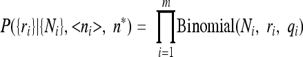
Maximum likelihood estimation of the limit of detection for spores of nonproteolytic Clostridium botulinum in foods and food materials. A standard method for estimating the limit of detection in food microbiology is to observe the presence or absence of bacteria, by a prescribed protocol, in a series of test samples with a range of known bacterial loads. We assume that each test is independent and that a well-defined load, n*, in a test volume, v, partitions the outcomes (i.e., for all loads in which n > n* the test indicates the presence of bacteria). The limit of detection, n*, in volume v can be estimated using a maximum likelihood method based on a series of test results which indicate ri positive results from ni tests with a nominal load <ni> for i = 1, m.
We assume that the actual loads, n, in the test materials are Poisson distributed with expectation <ni>. Then in a test with nominal load <ni> the probability that the actual load exceeds the detection limit is
 |
In turn, the likelihood of the test results is given as
 |
where each test at level i is considered a Bernoulli trial. The logarithm of the likelihood has m contributions and can be maximized with respect to the threshold value n*.
 |
The constant, which depends only on the data and not the threshold, is irrelevant for maximum likelihood determination. The analysis uses a single test volume, v, and a corresponding threshold load, n*, but could be extended to heterogeneous tests.
A confidence interval can be established from the likelihood ratio (difference in log likelihood around the maximum value). The ratio
 |
is assumed to be distributed as χ2 with 1 df so that γ > χ 2α,1 defines a 100·(1 − α) percentage confidence interval.
Footnotes
Published ahead of print on 13 August 2010.
REFERENCES
- 1.Baker, D. A., C. Genigeorgis, and G. Garcia. 1990. Prevalence of Clostridium botulinum in seafood and significance of multiple incubation temperatures for determination of its presence and type in fresh retail fish. J. Food Prot. 53:668-673. [DOI] [PubMed] [Google Scholar]
- 2.Barker, G. C., P. K. Malakar, M. Del Torre, M. L. Stecchini, and M. W. Peck. 2005. Probabilistic representation of the exposure of consumers to Clostridium botulinum neurotoxin in a minimally processed potato product. Int. J. Food Microbiol. 100:345-357. [DOI] [PubMed] [Google Scholar]
- 3.Carlin, F., V. Broussolle, S. Perelle, S. Litman, and P. Fach. 2004. Prevalence of Clostridium botulinum in food raw materials used in REPFEDs manufactured in France. Int. J. Food Microbiol. 91:141-145. [DOI] [PubMed] [Google Scholar]
- 4.Carlin, F., H. Girardin, M. W. Peck, S. C. Stringer, G. C. Barker, A. Martinez, A. Fernandez, P. Fernandez, W. M. Waites, S. Movahedi, F. van Leusden, M. Nauta, R. Moezelaar, M. Del Torre, and S. Litman. 2000. Research on factors allowing a risk assessment of spore forming pathogenic bacteria in cooked chilled foods containing vegetables: a FAIR collaborative project. Int. J. Food Microbiol. 60:117-135. [DOI] [PubMed] [Google Scholar]
- 5.Carlin, F., and M. W. Peck. 1995. Growth and toxin production by non-proteolytic and proteolytic Clostridium botulinum in cooked vegetables. Lett. Appl. Microbiol. 20:152-156. [DOI] [PubMed] [Google Scholar]
- 6.Carter, A. T., C. J. Paul, D. R. Mason, S. M. Twine, M. J. Alston, S. M. Logan, J. W. Austin, and M. W. Peck. 2009. Independent evolution of neurotoxin and flagellar genetic loci in proteolytic Clostridium botulinum. BMC Genomics 10:115. [DOI] [PMC free article] [PubMed] [Google Scholar]
- 7.Collins, M. D., and A. K. East. 1998. Phylogeny and taxonomy of the food-borne pathogen Clostridium botulinum and its neurotoxins. J. Appl. Microbiol. 84:5-17. [DOI] [PubMed] [Google Scholar]
- 8.Craig, J. M., S. Hayes, and K. S. Pilcher. 1968. Incidence of Clostridium botulinum type E in salmon and other marine fish in the Pacific Northwest. Appl. Microbiol. 16:553-557. [DOI] [PMC free article] [PubMed] [Google Scholar]
- 9.Del Torre, M., M. L. Stecchini, A. Braconnier, and M. W. Peck. 2004. Prevalence of Clostridium species and behaviour of Clostridium botulinum in gnocchi, a REPFED of Italian origin. Int. J. Food Microbiol. 96:115-131. [DOI] [PubMed] [Google Scholar]
- 10.De Medici, D., F. Anniballi, G. M. Wyatt, M. Lindström, U. Messelhauser, C. F. Aldus, E. Delibato, H. Korkeala, M. W. Peck, and L. Fenicia. 2009. Multiplex PCR for detection of botulinum neurotoxin-producing clostridia in clinical, food and environmental samples. Appl. Environ. Microbiol. 75:6457-6461. [DOI] [PMC free article] [PubMed] [Google Scholar]
- 11.Dezfulian, M., L. M. McCroskey, C. L. Hatheway, and V. R. Dowell. 1981. Selective medium for isolation of Clostridium botulinum from human feces. J. Clin. Microbiol. 13:526-531. [DOI] [PMC free article] [PubMed] [Google Scholar]
- 12.Dodds, K. L. 1993. Clostridium botulinum in the environment, p. 21-51. In A. H. W. Hauschild and K. L. Dodds (ed.), Clostridium botulinum. Ecology and control in foods. Marcel Dekker, New York, NY.
- 13.Dodds, K. L. 1993. Clostridium botulinum in foods, p. 53-68. In A. H. W. Hauschild and K. L. Dodds (ed.), Clostridium botulinum. Ecology and control in foods. Marcel Dekker, New York, NY.
- 14.Eklund, M. W., and F. T. Poysky. 1970. Distribution of Clostridium botulinum on the Pacific coast of the United States, p. 304-308. In M. Herzberg (ed.), Toxic microorganisms. U.S. Department of Interior, Washington, DC.
- 15.Eklund, M. W., F. T. Poysky, M. E. Peterson, R. N. Paranjpye, and G. A. Pelroy. 2004. Competitive inhibition between different Clostridium botulinum types and strains. J. Food Prot. 67:2682-2687. [DOI] [PubMed] [Google Scholar]
- 16.Fach, P., S. Perelle, F. Dilasser, J. Grout, C. Dargaignaratz, L. Bottela, J. M. Gourreau, F. Carlin, M. R. Popoff, and V. Broussolle. 2002. Detection by PCR-enzyme-linked immunosorbent assay of Clostridium botulinum in fish and environmental samples from a coastal area in northern France. Appl. Environ. Microbiol. 68:5870-5876. [DOI] [PMC free article] [PubMed] [Google Scholar]
- 17.George, S. M., L. C. C. Richardson, I. E. Pol, and M. W. Peck. 1998. Effect of oxygen concentration and redox potential on recovery of sublethally heat-damaged cells of Escherichia coli O157:H7, Salmonella enteritidis and Listeria monocytogenes. J. Appl. Microbiol. 84:903-909. [DOI] [PubMed] [Google Scholar]
- 18.Graham, A. F., D. R. Mason, F. J. Maxwell, and M. W. Peck. 1997. Effect of pH and NaCl on growth from spores of non-proteolytic Clostridium botulinum at chill temperatures. Lett. Appl. Microbiol. 24:95-100. [DOI] [PubMed] [Google Scholar]
- 19.Hielm, S., K. Björkroth, E. Hyytia, and H. Korkeala. 1998. Prevalence of Clostridium botulinum in Finnish trout farms: pulsed-field gel electrophoresis typing reveals extensive genetic diversity among type E isolates. Appl. Environ. Microbiol. 64:4161-4167. [DOI] [PMC free article] [PubMed] [Google Scholar]
- 20.Hielm, S., M. Lindström, and H. Korkeala. 2002. Food safety in seafood; epidemiological concerns related to the geography of fishing grounds, p. 229-246. In F. J. M. Smulders and J. D. Collins (ed.), Food safety assurance in the pre-harvest phase, vol. 1. Food safety assurance and veterinary public health. Wageningen Academic Press, Wageningen, Netherlands. [Google Scholar]
- 21.Hurley, M. A., and M. E. Roscoe. 1983. Automated statistical analysis of microbial enumeration by dilution series. J. Appl. Bacteriol. 55:159-164. [Google Scholar]
- 22.Huss, H. H., A. Pedersen, and D. C. Cann. 1974. The incidence of Clostridium botulinum in Danish trout farms. I. Distribution in fish and their environment. J. Food Technol. 9:445-450. [Google Scholar]
- 23.Hyytia, E., S. Hielm, and H. Korkeala. 1998. Prevalence of Clostridium botulinum type E in Finnish fish and fishery product. Epidemiol. Infect. 120:245-250. [DOI] [PMC free article] [PubMed] [Google Scholar]
- 24.International Commission on Microbiological Specifications for Foods. 2002. Microorganisms in foods 7. Microbiological testing in food safety management. Plenum Press, New York, NY.
- 25.International Organization for Standardization. 2002. Draft International Standard ISO/DIS 22174, Microbiology of food and animal feeding stuffs—PCR for the detection of foodborne pathogens—general specific method requirements. International Organization for Standardization, Geneva, Switzerland.
- 26.Johnson, E. A. 2005. Clostridial neurotoxins, p. 491-525. In P. Durre (ed.), Handbook on clostridia. CRC, Boca Raton, FL.
- 27.Johnson, E. A. 2007. Clostridium botulinum, p. 401-421. In M. P. Doyle and L. R. Beuchat (ed.), Food microbiology: fundamentals and frontiers, 3rd ed. ASM Press, Washington, DC.
- 28.Lalitha, K. V., and K. Gopakumar. 2000. Distribution and ecology of Clostridium botulinum in fish and aquatic environments of a tropical region. Food Microbiol. 17:535-541. [Google Scholar]
- 29.Lalitha, K. V., and P. K. Surendran. 2002. Occurrence of Clostridium botulinum in fresh and cured fish in retail trade in Cochin (India). Int. J. Food Microbiol. 72:169-174. [DOI] [PubMed] [Google Scholar]
- 30.Laycock, R. A., and A. A. Longard. 1972. Clostridium botulinum in sediments from the Canadian Atlantic Seaboard. J. Fish. Res. Board Can. 29:443-446. [Google Scholar]
- 31.Laycock, R. A., and D. H. Loring. 1972. Distribution of Clostridium botulinum type E in the Gulf of St. Lawrence in relation to the physical environment. Can. J. Microbiol. 18:763-773. [DOI] [PubMed] [Google Scholar]
- 32.Leclair, D., F. Pagotto, J. M. Farber, B. Cadieux, and J. W. Austin. 2006. Comparison of DNA fingerprinting methods for use in investigation of type E botulism outbreaks in the Canadian Arctic. J. Clin. Microbiol. 44:1635-1644. [DOI] [PMC free article] [PubMed] [Google Scholar]
- 33.Lilly, T., S. M. Harmon, D. A. Kautter, H. M. Solomon, and R. K. Lynt. 1971. An improved medium for detection of Clostridium botulinum type E. J. Milk Food Technol. 34:492-497. [Google Scholar]
- 34.Lindroth, S., and C. Genigeorgis. 1986. Probability of growth and toxin production by nonproteolytic Clostridium botulinum in rockfish stored under modified atmospheres. Int. J. Food Microbiol. 3:167-181. [Google Scholar]
- 35.Lindström, M., M. Fredriksson-Ahomaa, and H. Korkeala. 2009. Molecular epidemiology of group I and group II Clostridium botulinum, p. 103-130. In H. Bruggemann and G. Gottschalk (ed.), Clostridia, molecular biology in the post-genomic era. Caister Academic Press, Norfolk, United Kingdom.
- 36.Lindström, M., R. Keto, A. Markkula, M. Nevas, S. Hielm, and H. Korkeala. 2001. Multiplex PCR assay for detection and identification of Clostridium botulinum types A, B, E, and F in food and fecal material. Appl. Environ. Microbiol. 67:5694-5699. [DOI] [PMC free article] [PubMed] [Google Scholar]
- 37.Lindström, M., K. Kiviniemi, and H. Korkeala. 2006. Hazard and control of group II (non-proteolytic) Clostridium botulinum in modern food processing. Int. J. Food Microbiol. 108:92-104. [DOI] [PubMed] [Google Scholar]
- 38.Lindström, M., and H. Korkeala. 2006. Laboratory diagnostics of botulism. Clin. Microbiol. Rev. 19:298-314. [DOI] [PMC free article] [PubMed] [Google Scholar]
- 39.Lund, B. M., and M. W. Peck. 2000. Clostridium botulinum, p. 1057-1109. In B. M. Lund, A. C. Baird-Parker, and G. W. Gould (ed.), The microbiological safety and quality of food. Aspen, Gaithersburg, MD.
- 40.Lund, B. M., and G. M. Wyatt. 1984. The effect of redox potential, and its interaction with sodium chloride concentration, on the probability of growth of Clostridium botulinum type E from spore inocula. Food Microbiol. 1:49-65. [Google Scholar]
- 41.Malakar, P. K., G. C. Barker, and M. W. Peck. 2010. Quantitative risk assessment for hazards that arise from non-proteolytic Clostridium botulinum in minimally processed chilled dairy-based foods. Food Microbiol. doi: 10.1016/j.fm.2010.04.004. [DOI] [PubMed]
- 42.National Research Council of the National Academies. 2009. Science and decisions. The National Academies Press, Washington, DC.
- 43.Peck, M. W. 1997. Clostridium botulinum and the safety of refrigerated processed foods of extended durability. Trends Food Sci. Technol. 8:186-192. [Google Scholar]
- 44.Peck, M. W. 2006. Clostridium botulinum and the safety of minimally heated chilled foods: an emerging issue? J. Appl. Microbiol. 101:556-570. [DOI] [PubMed] [Google Scholar]
- 45.Peck, M. W. 2009. Biology and genomic analysis of Clostridium botulinum. Adv. Microb. Physiol. 55:183-265. [DOI] [PubMed] [Google Scholar]
- 46.Peck, M. W., D. A. Fairbairn, and B. M. Lund. 1992. The effect of recovery medium on the estimated heat-inactivation of spores of non-proteolytic Clostridium botulinum. Lett. Appl. Microbiol. 15:146-151. [DOI] [PubMed] [Google Scholar]
- 47.Peck, M. W., K. E. Goodburn, R. P. Betts, and S. C. Stringer. 2008. Assessment of the potential for growth and neurotoxin formation by non-proteolytic Clostridium botulinum in short shelf-life commercial foods designed to be stored chilled. Trends Food Sci. Technol. 19:207-216. [Google Scholar]
- 48.Peck, M. W., and S. C. Stringer. 2005. The safety of pasteurised in-pack chilled meat products with respect to the foodborne botulism hazard. Meat Sci. 70:461-475. [DOI] [PubMed] [Google Scholar]
- 49.Plowman, J., and M. W. Peck. 2002. Use of a novel method to characterise the response of spores of non-proteolytic Clostridium botulinum types B, E and F to a wide range of germinants and conditions. J. Appl. Microbiol. 92:681-694. [DOI] [PubMed] [Google Scholar]
- 50.Smith, L. D. S. 1975. Inhibition of Clostridium botulinum by strains of Clostridium perfringens isolated from soil. Appl. Microbiol. 30:319-323. [DOI] [PMC free article] [PubMed] [Google Scholar]
- 51.Stringer, S. C., N. Haque, and M. W. Peck. 1999. Growth from spores of nonproteolytic Clostridium botulinum in heat-treated vegetable juice. Appl. Environ. Microbiol. 65:2136-2142. [DOI] [PMC free article] [PubMed] [Google Scholar]
- 52.Stringer, S. C., M. D. Webb, S. M. George, C. Pin, and M. W. Peck. 2005. Heterogeneity of times required for germination and outgrowth from single spores of nonproteolytic Clostridium botulinum. Appl. Environ. Microbiol. 71:4998-5003. [DOI] [PMC free article] [PubMed] [Google Scholar]
- 53.Sugiyama, H., T. L. Bott, and E. M. Foster. 1970. Clostridium botulinum type E in an inland bay (Green Bay of Lake Michigan), p. 287-291. In M. Herzberg (ed.), Toxic microorganisms. U.S. Department of Interior, Washington, DC.
- 54.U.S. Food and Drug Administration. 2001. Bacteriological analytical manual. Center for Food Safety and Applied Nutrition, U.S. Food and Drug Administration, College Park, MD. http://www.cfsan.fda.gov/∼ebam/bam-toc.html.