Abstract
Triggering of the Hendra virus fusion (F) protein is required to initiate the conformational changes which drive membrane fusion, but the factors which control triggering remain poorly understood. Mutation of a histidine predicted to lie near the fusion peptide to alanine greatly reduced fusion despite wild-type cell surface expression levels, while asparagine substitution resulted in a moderate restoration in fusion levels. Slowed kinetics of six-helix bundle formation, as judged by sensitivity to heptad repeat B-derived peptides, was observed for all H372 mutants. These data suggest that side chain packing beneath the fusion peptide is an important regulator of Hendra virus F triggering.
Hendra virus and Nipah virus are highly pathogenic paramyxoviruses infecting humans. They were identified in 1994 and 1999, respectively, as the etiological agents behind cases of severe encephalitis and respiratory disease in Australia and Malaysia (7, 10, 17-18). Owing to their unusually high virulence, broad host range, and genetic similarity, Hendra virus and Nipah virus (NiV) have been classified into the new genus Henipavirus (31). Henipavirus membrane fusion requires the concerted effort of two viral surface glycoproteins (3-4, 30): the attachment protein (G), which binds receptor, and the fusion (F) protein, which drives membrane merger through vast conformational changes. Paramyxovirus F proteins are synthesized as inactive F0 precursors which are subsequently cleaved into fusogenic disulfide-linked heterodimers, F1+F2. Despite a conserved requirement for cleavage, protease usage varies among paramyxoviruses, with henipavirus F being cleaved by the endosomal/lysosomal cysteine protease cathepsin L (19-20). This cleavage event positions the fusion peptide (FP) at the newly created N terminus and acts to prime the F protein. Following cleavage, the primed F protein must be triggered to begin the sequence of conformational changes required for membrane fusion. Like most F proteins, triggering of the henipavirus F proteins likely involves the henipavirus attachment proteins, though the mechanism remains poorly understood (reviewed in reference 28). F triggering facilitates refolding and extension of heptad repeat A (HRA) toward the target cell membrane, resulting in FP insertion into the bilayer (2). Further rearrangement brings HRA and HRB into close proximity, resulting in the formation of a stable six-helix bundle and culminating in a fully formed fusion pore (reviewed in reference 32).
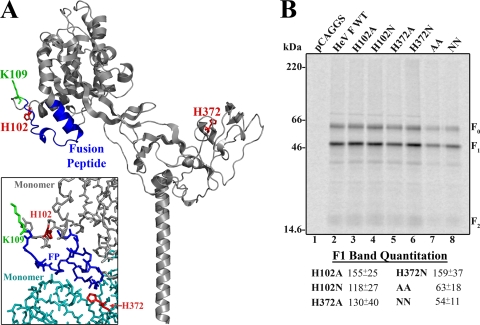
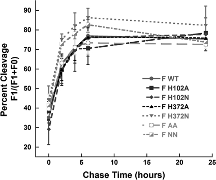
Cathepsin L cleavage of F does not require specific residues upstream of or at the cleavage site (K109) itself (5, 16), and the mechanism by which cathepsin L recognizes and specifically cleaves F is unclear. Modeling of the Hendra virus F amino acid sequence onto the prefusion structure of parainfluenza virus 5 (PIV5) (34) indicates that two of the three ectodomain histidine residues (H102 and H372) are positioned near the cathepsin L cleavage site following residue K109 (Fig. 1 A). In the monomer, H372 is located distally from K109, yet trimerization places H372 from one monomer directly beneath the FP and cleavage site of the neighboring monomer (Fig. 1A, inset). We hypothesized that protonation of histidine residues could cause local conformational changes, potentially modulating cathepsin L cleavage, though these hypothesized conformational changes would not be due to direct modulation of K109 interactions since the predicted distances from K109 to either H102 or H372 are 12 Å and 27 Å, respectively (α-carbon to α-carbon distances). To test the role of H102 and H372 in cathepsin L cleavage, each was mutated individually or together to alanine (A) or asparagine (N), which has a side chain volume similar to that of histidine. Surface expression levels and cleavage of wild-type (WT) and mutant F proteins were examined by cell surface biotinylation as previously described (8). All mutant F proteins were surface expressed and cleaved, though levels of the H102A/H372A (AA) and H102N/H372N (NN) proteins were decreased compared to those of the wild type (Fig. 1B). To examine cleavage kinetics, Vero cells transiently transfected with wild-type or mutant pCAGGS-Hendra virus F were metabolically labeled for 30 min and chased for 0 to 24 h. Band density corresponding to F0 and F1 was quantitated, and percent cleavage was defined as the density of F1/(F1+F0). Cleavage kinetics of all Hendra virus F mutants were not significantly different from wild-type levels (Fig. 2). These data suggest that protonation of histidines in the region of the cleavage site is not involved in cathepsin L processing of Hendra virus F.
FIG. 1.
Structural modeling and surface expression of Hendra virus F H102 and H372 mutants. (A) Homology model of the Hendra virus F monomer based on the crystal structure of PIV5 F, shown as a ribbon diagram (image generated using PyMOL; Delano Scientific [www.pymol.org]): red, H102 and H372; blue, fusion peptide (FP); green, P1 cleavage site residue K109. The locations of H102 and H372 in the trimeric protein are shown in the box insert using the same color scheme except with an additional monomer shown in teal. (B) Surface expression of transiently transfected wild-type and Hendra virus F mutants in Vero cells following metabolic labeling (3 h). Surface proteins were biotinylated prior to immunoprecipitation, and the total and surface populations were separated by streptavidin pull-down. Proteins were analyzed via 15% SDS-PAGE and visualized using autoradiography. F1 band quantitation via densitometry is shown normalized to wild-type levels plus or minus one standard deviation. The surface expression levels represent the averages of data from three independent experiments, with one representative gel shown.
FIG. 2.
Total protein cleavage time points for WT and mutant F protein. Vero cells transiently transfected with wild-type or mutant Hendra virus F were metabolically labeled (3 h) and chased for the indicated times. The total protein population was immunoprecipitated and analyzed by 15% SDS-PAGE and autoradiography. Band intensity was quantitated using the ImageQuant 5.2 software program (GE Healthcare, Piscataway, NJ), and percent cleavage was defined as the intensity of F1/(F1+F0). Error bars are plus or minus one standard deviation and represent the average of data from three independent experiments.
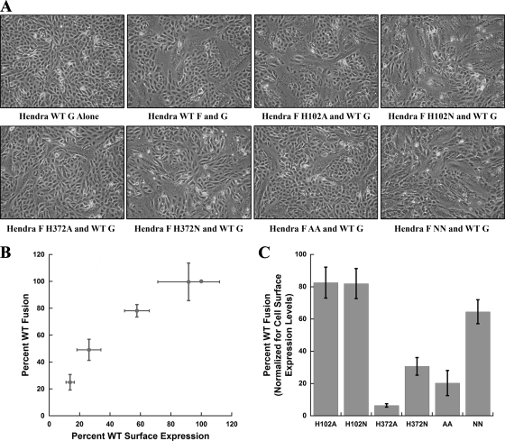
Since the mutants were expressed on the cell surface in the mature, cleaved form, fusion was examined using a syncytium assay (Fig. 3 A). Mutations at H102 did not significantly alter syncytium formation, but large reductions in fusion were observed for F proteins containing an H372 mutation. To quantitatively analyze fusion, a reporter gene assay was utilized. Since the single mutations all resulted in increased surface density (Fig. 1B) while decreases were observed for the double mutants, analysis of the effects of surface density on WT Hendra virus F fusion was first performed. Previous work with other class I viral fusion proteins, including PIV5 F and influenza virus hemagglutinin (HA), has shown that surface density correlates with the final extent of fusion over a range of densities (6). Aguilar et al. (1) demonstrated that increasing the amount of NiV G and NiV F DNA transfected results in an increase in syncytium formation. However, a direct correlation between F surface expression and fusion has not been previously reported for henipaviruses. To assess this, Vero cells were transfected with various amounts of wild-type pCAGGS-Hendra virus F and biotinylated as described previously (8); reporter gene analyses using the same DNA amounts were performed alongside the biotinylation experiments. Increased surface densities clearly led to increases in fusion, though the correlation was not completely linear (Fig. 3B). These data were then utilized to generate a percent WT fusion level for each mutant normalized for the observed cell surface expression levels. Mutations at H102 did not significantly alter fusion (Fig. 3C). However, cell-cell fusion was dramatically reduced (10% to 20% of wild-type values) with the H372A and AA mutant F proteins. A partial restoration in fusion was seen for the H372N and NN mutants, suggesting that side chain volume plays a role; however, fusion levels were only 30% to 60% of those of the wild type (Fig. 3C). While histidine residues proximal to the influenza virus HA fusion peptide have been shown to regulate low-pH triggering, Hendra virus F-mediated fusion occurs at neutral pH, and incubation at low pH has no effect on fusogenicity (A. Chang and R. E. Dutch, unpublished results). These data indicate that mutations at H372 result in a hypofusogenic protein, suggesting that side chain packing within this region may strongly modulate F protein triggering, potentially by altering protein stability.
FIG. 3.
Syncytium assay, reporter gene analysis, and correlation of wild-type and mutant F protein surface expression versus fusion activity. (A) Representative syncytium images from three independent experiments for wild-type and mutant Hendra virus F proteins. Vero cells transiently transfected with wild-type pCAGGS-Hendra virus G and wild-type or mutant pCAGGS-Hendra virus F were kept at 37°C for 24 to 48 h posttransfection, and photographs were taken using a Nikon digital camera mounted atop a Nikon TS100 microscope with a 10× objective. (B) Correlation between surface expression and fusogenicity for wild-type Hendra virus F. Vero cells transiently transfected with various amounts of wild-type Hendra virus F DNA were biotinylated, and reporter gene analysis was performed simultaneously using the same DNA quantities. (C) Reporter gene analysis of equal μg of wild-type or mutant Hendra virus F in pCAGGS normalized to average cell surface expression levels. Vero cells transiently transfected with wild-type Hendra virus G, wild-type or mutant Hendra virus F, and T7 luciferase were overlaid with BSR cells 18 h posttransfection, lysed, and assayed for luciferase activity (n = 5 to 8; ±95% confidence interval [CI]).
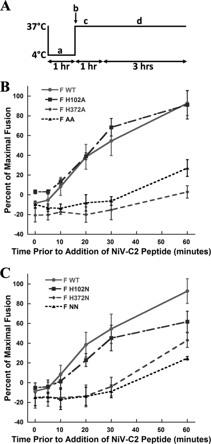
The hypofusogenic phenotype of the H372 mutants could be explained by changes in the stability of the prefusion form following cleavage, resulting in altered fusion kinetics. To examine fusion kinetics, sensitivity over time to peptides which block formation of the postfusion six-helix bundle was examined (NiV C2; corresponding to HRB of the highly homologous Nipah virus F protein; generously provided by Chris Broder [Uniformed Services University of the Health Sciences]). One hundred nM NiV C2 has been shown to inhibit Henipavirus F-mediated cell-cell fusion (4). Similar peptides inhibit many other class I fusion proteins (11-13, 23, 33, 35-36). Vero cells transfected with wild-type pCAGGS-Hendra virus F and G and T7 luciferase were overlaid with target BSR cells on ice for 1 h. Prewarmed Dulbecco's modified Eagle medium (DMEM) was added to initiate fusion, and at the indicated time points, DMEM containing 100 nM NiV C2 or 100 nM NiV SC (scrambled control peptide) was added. Cells were kept at 37°C for 3 h, and luciferase activity was assayed (Fig. 4 A). Cell-cell fusion kinetics for the wild-type Hendra virus F protein showed a steep increase in membrane fusion events from the 5- to 20-min time points (Fig. 4B, solid line). Approximately 50% of cell-cell fusion events became insensitive to the addition of NiV C2 by 30 min, with the majority of membrane fusion events complete by 60 min (Fig. 4B, solid line). Fusion kinetics for the H102A and H102N proteins closely resembled wild-type kinetics, consistent with overall fusion levels (Fig. 4B and C). In contrast, cell-cell fusion was dramatically slowed for F proteins containing mutations at H372. For all H372 mutants, no fusion was observed during the first 30 min, in stark contrast to results for the wild type. After 30 min, fusion was observed for the H372N and NN proteins, which reach 20 to 40% of maximal fusion within 60 min (Fig. 4C). The small amount of fusion observed for the H372A protein occurred long after fusion was complete for the wild-type protein. Combined, these data demonstrate that mutation of H372 to either alanine or asparagine decreases the initial rate of membrane fusion potentially by increasing the energetic barrier for Hendra virus F triggering.
FIG. 4.
Cell-cell reporter gene fusion kinetics for wild-type and mutant Hendra virus F proteins. (A) Diagram of the experimental setup: a, binding of BSR cells; b, addition of DMEM, NiV-SC peptide, or NiV-C2 peptides (0-min time point); c, addition of NiV-C2 at indicated time points; d, continued incubation of cells. (B and C) Cell-cell fusion kinetics for wild-type and mutant F proteins. Reporter gene analysis was performed as described above following binding of BSR cells at 4°C, addition of either DMEM, NiV-SC, or NiV-C2 at the indicated times, and continued incubation for 3 h at 37°C. Percent maximal fusion is defined as the amount of fusion which occurred during a given time point as a fraction of membrane fusion for a given construct over the duration of the experiment (3 h) in the absence of any peptide. Error bars are 95% confidence intervals (n = 3 to 6).
While most paramyxovirus F proteins, including the Hendra virus F protein, are thought to be triggered by specific interactions with a homotypic attachment protein (reviewed in reference 28), mutations within paramyxovirus F proteins which alter stability of the prefusion form (8, 15, 21-22, 27) can also strongly modulate triggering. H372 is modeled to be near a conserved domain, termed CBF1, in the Hendra virus F prefusion structure. Studies suggest that CBF1, which is structurally composed of β-sheets, is important for F protein folding (9), likely playing a critical role in stabilization of the fusion peptide following proteolytic cleavage, with the CBF1 domain from one monomer interacting with the fusion peptide from the neighboring monomer. Mutations in CBF1 in Hendra virus F resulted in folding defects which could not be rescued at decreased temperatures. Given the proximity of H372 to CBF1 in Hendra virus F, changes in side chain packing could stabilize the fusion peptide following cleavage and thus decrease the ability of the protein to trigger efficiently. In the model structure, H372 is predicted to be surrounded by polar and nonpolar residues (within 5 Å of the side chain), including I425, N423, Q342, F376, and two FP residues, A125 and T129. Extension out to 10 Å reveals that H372 is also near four additional FP residues (A126, I128, T129, and V132), suggesting that substitution of H372 with a smaller residue (H372A) could alter the packing depth of the more C-terminal portion of the FP following cleavage.
Studies from other systems also implicate the fusion peptide and surrounding residues as regulators of F-promoted membrane fusion (14, 24, 26, 29). The paramyxovirus fusion peptide is an important regulator of triggering, since conserved glycine residues within the FP have been shown to play a role in regulation and activation of F (26). Numerous mutations within the fusion peptide pocket of H5N1 influenza virus HA were shown to regulate the pH needed for HA activation (25), with only one mutation causing significant changes to HA expression or cleavage. Similar experiments using the H3 subtype of influenza virus HA also demonstrated changes in pH requirements upon mutation of certain fusion peptide-proximal residues (29). While influenza virus HA requires low pH for fusion promotion, the data presented here show that regulation of interactions with and around the fusion peptide is also critically important for triggering and fusion promotion of neutral-pH fusing systems. Thus, the decrease in the rate of triggering observed for the H372A mutant is consistent with a model in which residues surrounding the fusion peptide can act to regulate F-mediated fusion promotion independently of large changes in protein expression or cleavage.
Our data, therefore, strongly indicate that side chain packing near the fusion peptide (Fig. 1A, inset) is a strong modulator of Hendra virus F triggering, with a dramatic slowing in the rate of six-helix bundle formation observed when H372 is replaced with residues with smaller side chain volumes (Fig. 4). Modulation of side chain packing proximal to the FP could change the positioning of paramyxovirus FPs, thus altering the kinetics and efficiency of later conformational changes. Mutation of H372 may well stabilize interactions of the FP with the ectodomain following cleavage and thus affect triggering by substantially increasing the energy needed to project the FP toward the target cell membrane. Together, these data suggest a model by which packing around the fusion peptide affects both the rate and extent of F triggering.
Acknowledgments
We thank Christopher Broder (Uniformed Services University) for generously providing the inhibitory peptides, Lin-fa Wang (Australian Animal Health Laboratory) for providing the Hendra virus F and G plasmids, and Rachel Schowalter (NCI, NIH) for help in generating some of the Hendra virus F mutant constructs. We also acknowledge members of the Dutch lab for critically reviewing the manuscript.
This work was supported by NIAID/NIH grant R01AI051517 and NIH grant U54 AI057157 from the Southeastern Regional Center of Excellence for Emerging Infections and Biodefense.
Footnotes
Published ahead of print on 11 August 2010.
REFERENCES
- 1.Aguilar, H. C., K. A. Matreyek, C. M. Filone, S. T. Hashimi, E. L. Levroney, O. A. Negrete, A. Bertolotti-Ciarlet, D. Y. Choi, I. McHardy, J. A. Fulcher, S. V. Su, M. C. Wolf, L. Kohatsu, L. G. Baum, and B. Lee. 2006. N-Glycans on Nipah virus fusion protein protect against neutralization but reduce membrane fusion and viral entry. J. Virol. 80:4878-4889. [DOI] [PMC free article] [PubMed] [Google Scholar]
- 2.Baker, K. A., R. E. Dutch, R. A. Lamb, and T. S. Jardetzky. 1999. Structural basis for paramyxovirus-mediated membrane fusion. Mol. Cell 3:309-319. [DOI] [PubMed] [Google Scholar]
- 3.Bossart, K. N., L. F. Wang, B. T. Eaton, and C. C. Broder. 2001. Functional expression and membrane fusion tropism of the envelope glycoproteins of Hendra virus. Virology 290:121-135. [DOI] [PubMed] [Google Scholar]
- 4.Bossart, K. N., L. F. Wang, M. N. Flora, K. B. Chua, S. K. Lam, B. T. Eaton, and C. C. Broder. 2002. Membrane fusion tropism and heterotypic functional activities of the Nipah virus and Hendra virus envelope glycoproteins. J. Virol. 76:11186-11198. [DOI] [PMC free article] [PubMed] [Google Scholar]
- 5.Craft, W. W., Jr., and R. E. Dutch. 2005. Sequence motif upstream of the Hendra virus fusion protein cleavage site is not sufficient to promote efficient proteolytic processing. Virology 341:130-140. [DOI] [PubMed] [Google Scholar]
- 6.Dutch, R. E., S. B. Joshi, and R. A. Lamb. 1998. Membrane fusion promoted by increasing surface densities of the paramyxovirus F and HN proteins: comparison of fusion reactions mediated by simian virus 5 F, human parainfluenza virus type 3 F, and influenza virus HA. J. Virol. 72:7745-7753. [DOI] [PMC free article] [PubMed] [Google Scholar]
- 7.Eaton, B. T., C. C. Broder, D. Middleton, and L. F. Wang. 2006. Hendra and Nipah viruses: different and dangerous. Nat. Rev. Microbiol. 4:23-35. [DOI] [PMC free article] [PubMed] [Google Scholar]
- 8.Gardner, A. E., and R. E. Dutch. 2007. A conserved region in the F(2) subunit of paramyxovirus fusion proteins is involved in fusion regulation. J. Virol. 81:8303-8314. [DOI] [PMC free article] [PubMed] [Google Scholar]
- 9.Gardner, A. E., K. L. Martin, and R. E. Dutch. 2007. A conserved region between the heptad repeats of paramyxovirus fusion proteins is critical for proper F protein folding. Biochemistry 46:5094-5105. [DOI] [PMC free article] [PubMed] [Google Scholar]
- 10.Harcourt, B. H., A. Tamin, T. G. Ksiazek, P. E. Rollin, L. J. Anderson, W. J. Bellini, and P. A. Rota. 2000. Molecular characterization of Nipah virus, a newly emergent paramxyovirus. Virology 271:334-349. [DOI] [PubMed] [Google Scholar]
- 11.Joshi, S. B., R. E. Dutch, and R. A. Lamb. 1998. A core trimer of the paramyxovirus fusion protein: parallels to influenza virus hemagglutinin and HIV-1 gp41. Virology 248:20-34. [DOI] [PubMed] [Google Scholar]
- 12.Lamb, R. A., S. B. Joshi, and R. E. Dutch. 1999. The paramyxovirus fusion protein forms an extremely stable core trimer: structural parallels to influenza virus haemagglutinin and HIV-1 gp41. Mol. Membr. Biol. 16:11-19. [DOI] [PubMed] [Google Scholar]
- 13.Lambert, D. M., S. Barney, A. L. Lambert, K. Guthrie, R. Medinas, D. E. Davis, T. Bucy, J. Erickson, G. Merutka, and S. R. Pettaway, Jr. 1996. Peptides from conserved regions of paramyxovirus fusion (F) proteins are potent inhibitors of viral fusion. Proc. Natl. Acad. Sci. U. S. A. 93:2186-2191. [DOI] [PMC free article] [PubMed] [Google Scholar]
- 14.Lee, J. K., A. Prussia, T. Paal, L. K. White, J. P. Snyder, and R. K. Plemper. 2008. Functional interaction between paramyxovirus fusion and attachment proteins. J. Biol. Chem. 283:16561-16572. [DOI] [PMC free article] [PubMed] [Google Scholar]
- 15.Luque, L. E., and C. J. Russell. 2007. Spring-loaded heptad repeat residues regulate the expression and activation of paramyxovirus fusion protein. J. Virol. 81:3130-3141. [DOI] [PMC free article] [PubMed] [Google Scholar]
- 16.Moll, M., S. Diederich, H. D. Klenk, M. Czub, and A. Maisner. 2004. Ubiquitous activation of the Nipah virus fusion protein does not require a basic amino acid at the cleavage site. J. Virol. 78:9705-9712. [DOI] [PMC free article] [PubMed] [Google Scholar]
- 17.Murray, K., P. Selleck, P. Hooper, A. Hyatt, A. Gould, L. Gleeson, H. Westbury, L. Hiley, L. Selvey, B. Rodwell, et al. 1995. A morbillivirus that caused fatal disease in horses and humans. Science 268:94-97. [DOI] [PubMed] [Google Scholar]
- 18.O'Sullivan, J. D., A. M. Allworth, D. L. Paterson, T. M. Snow, R. Boots, L. J. Gleeson, A. R. Gould, A. D. Hyatt, and J. Bradfield. 1997. Fatal encephalitis due to novel paramyxovirus transmitted from horses. Lancet 349:93-95. [DOI] [PubMed] [Google Scholar]
- 19.Pager, C. T., W. W. Craft, Jr., J. Patch, and R. E. Dutch. 2006. A mature and fusogenic form of the Nipah virus fusion protein requires proteolytic processing by cathepsin L. Virology 346:251-257. [DOI] [PMC free article] [PubMed] [Google Scholar]
- 20.Pager, C. T., and R. E. Dutch. 2005. Cathepsin L is involved in proteolytic processing of the Hendra virus fusion protein. J. Virol. 79:12714-12720. [DOI] [PMC free article] [PubMed] [Google Scholar]
- 21.Paterson, R. G., C. J. Russell, and R. A. Lamb. 2000. Fusion protein of the paramyxovirus SV5: destabilizing and stabilizing mutants of fusion activation. Virology 270:17-30. [DOI] [PubMed] [Google Scholar]
- 22.Plattet, P., J. P. M. Langedijk, L. Zipperle, M. Vandevelde, C. Örvell, and A. Zurbriggen. 2009. Conserved leucine residue in the head region of morbillivirus fusion protein regulates the large conformational change during fusion activity. Biochemistry 48:9112-9121. [DOI] [PubMed] [Google Scholar]
- 23.Rapaport, D., M. Ovadia, and Y. Shai. 1995. A synthetic peptide corresponding to a conserved heptad repeat domain is a potent inhibitor of Sendai virus-cell fusion: an emerging similarity with functional domains of other viruses. EMBO J. 14:5524-5531. [DOI] [PMC free article] [PubMed] [Google Scholar]
- 24.Rawling, J., B. Garcia-Barreno, and J. A. Melero. 2008. Insertion of the two cleavage sites of the respiratory syncytial virus fusion protein in Sendai virus fusion protein leads to enhanced cell-cell fusion and a decreased dependency on the HN attachment protein for activity. J. Virol. 82:5986-5998. [DOI] [PMC free article] [PubMed] [Google Scholar]
- 25.Reed, M. L., H.-L. Yen, R. M. DuBois, O. A. Bridges, R. Salomon, R. G. Webster, and C. J. Russell. 2009. Amino acid residues in the fusion peptide pocket regulate the pH of activation of the H5N1 influenza virus hemagglutinin protein. J. Virol. 83:3568-3580. [DOI] [PMC free article] [PubMed] [Google Scholar]
- 26.Russell, C. J., T. S. Jardetzky, and R. A. Lamb. 2004. Conserved glycine residues in the fusion peptide of the paramyxovirus fusion protein regulate activation of the native state. J. Virol. 78:13727-13742. [DOI] [PMC free article] [PubMed] [Google Scholar]
- 27.Seth, S., A. L. Goodman, and R. W. Compans. 2004. Mutations in multiple domains activate paramyxovirus F protein-induced fusion. J. Virol. 78:8513-8523. [DOI] [PMC free article] [PubMed] [Google Scholar]
- 28.Smith, E. C., A. Popa, A. Chang, C. Masante, and R. E. Dutch. 2009. Viral entry mechanisms: the increasing diversity of paramyxovirus entry. FEBS J. 276:7217-7227. [DOI] [PMC free article] [PubMed] [Google Scholar]
- 29.Thoennes, S., Z.-N. Li, B.-J. Lee, W. A. Langley, J. J. Skehel, R. J. Russell, and D. A. Steinhauer. 2008. Analysis of residues near the fusion peptide in the influenza hemagglutinin structure for roles in triggering membrane fusion. Virology 370:403-414. [DOI] [PMC free article] [PubMed] [Google Scholar]
- 30.Wang, L.-F., B. H. Harcourt, M. Yu, A. Tamin, P. A. Rota, W. J. Bellini, and B. T. Eaton. 2001. Molecular biology of Hendra and Nipah viruses. Microbes Infect. 3:279-287. [DOI] [PubMed] [Google Scholar]
- 31.Wang, L. F., M. Yu, E. Hansson, L. I. Pritchard, B. Shiell, W. P. Michalski, and B. T. Eaton. 2000. The exceptionally large genome of Hendra virus: support for creation of a new genus within the family Paramyxoviridae. J. Virol. 74:9972-9979. [DOI] [PMC free article] [PubMed] [Google Scholar]
- 32.White, J. M., S. E. Delos, M. Brecher, and K. Schornberg. 2008. Structures and mechanisms of viral membrane fusion proteins: multiple variations on a common theme. Crit. Rev. Biochem. Mol. Biol. 43:189-219. [DOI] [PMC free article] [PubMed] [Google Scholar]
- 33.Wild, T. F., and R. Buckland. 1997. Inhibition of measles virus infection and fusion with peptides corresponding to the leucine zipper region of the fusion protein. J. Gen. Virol. 78:107-111. [DOI] [PubMed] [Google Scholar]
- 34.Yin, H. S., X. Wen, R. G. Paterson, R. A. Lamb, and T. S. Jardetzky. 2006. Structure of the parainfluenza virus 5 F protein in its metastable, prefusion conformation. Nature 439:38-44. [DOI] [PMC free article] [PubMed] [Google Scholar]
- 35.Young, J. K., D. Li, M. C. Abramowitz, and T. G. Morrison. 1999. Interaction of peptides with sequences from the Newcastle disease virus fusion protein heptad repeat regions. J. Virol. 73:5945-5956. [DOI] [PMC free article] [PubMed] [Google Scholar]
- 36.Zhao, X., M. Singh, V. N. Malashkevich, and P. S. Kim. 2000. Structural characterization of the human respiratory syncytial virus fusion protein core. Proc. Natl. Acad. Sci. U. S. A. 97:14172-14177. [DOI] [PMC free article] [PubMed] [Google Scholar]


