A 63-year-old man presented with acute left sylvian artery ischemic stroke. His medical history included hypertension, diabetes and pericardiocentesis for pericardial tamponade after a gunshot wound to the chest during the Salvadoran Civil War 26 years previously.
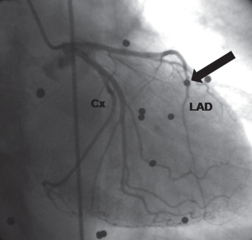
Echocardiography performed to investigate the etiology of stroke revealed an extensive left ventricular apical aneurysm (AA). Subsequent 64-multislice computed tomographic coronary angiography and standard coronary angiography were performed. A 30° right anterior oblique caudal view of the left coronary circulation (Figure 1) showed one bullet (arrow) in the mid left anterior descending artery with distal diminished flow. There was no sign of atherosclerotic disease that could have explained the AA. Multiple bullets were also found throughout the mediastinum.
Figure 1).
Cx Circumflex artery; LAD Left anterior descending artery
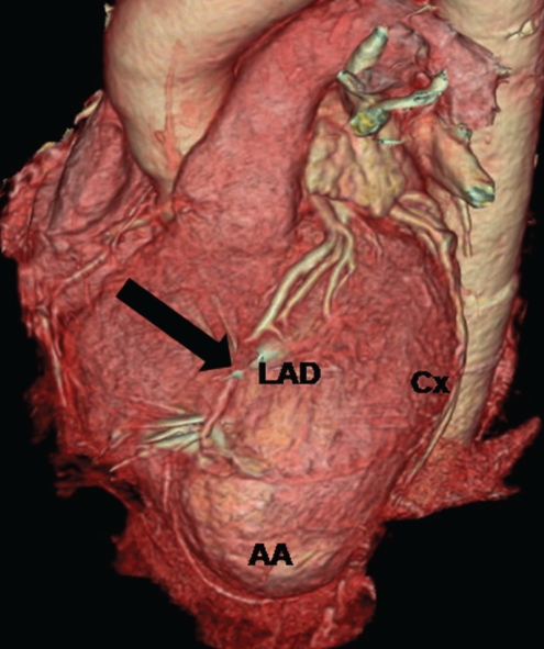
A surface-rendered three-dimensional reconstruction of the heart and coronary arteries (Figure 2) confirmed the intracoronary localization of the bullet with a filling artifact (arrow) due to shadowing from the bullet’s metallic composition.
Figure 2).
AA Apical aneurism; Cx Circumflex artery; LAD Left anterior descending artery
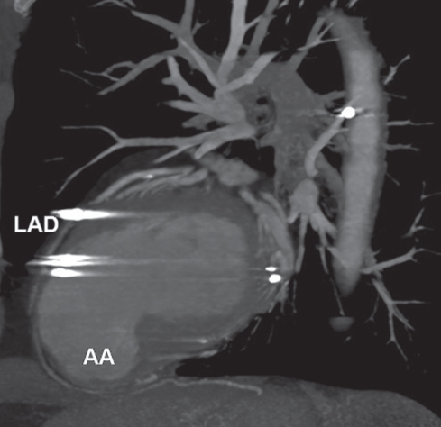
Mid left anterior descending artery bullet localization caused artery obstruction with secondary myocardial infarction and subsequent development of the AA. This AA, which is shown on a multiplanar reconstruction (Figure 3), led to an embolic stroke years later. Bullet extraction was not attempted. The patient was discharged on oral anticoagulation with warfarin.
Figure 3).
AA Apical aneurism; LAD Left anterior descending artery
Valvular injuries, fistulas, bullet embolus, constrictive pericarditis, myocardial infarction and aneurysm are all possible late complications after a penetrating cardiac injury (1). Clinicians should be aware of these potential complications.
REFERENCE
- 1.Evans J, Gray LA, Rayner A. Principles for the management of penetrating cardiac wounds. Am Surg. 1979;189:777–84. doi: 10.1097/00000658-197906000-00015. [DOI] [PMC free article] [PubMed] [Google Scholar]


