Abstract
Primary cardiac paraganglioma (pheochromocytoma) is very rare, constituting only 1% of cardiac tumours. A case of a 44-year-old woman presenting with angina chest pain and a tumour with dual blood supply from both the right and left coronary arteries is reported.
Keywords: Angina, Cardiac paraganglioma, Dual coronary blood supply
Abstract
Le paragangliome cardiaque primitif (phéochromocytome) est très rare, puisqu’il représente seulement 1 % des tumeurs cardiaques. Est présenté le cas d’une femme de 44 ans ayant consulté pour des douleurs thoraciques angineuses et une tumeur à double irrigation par les artères coronaires droite et gauche.
CASE PRESENTATION
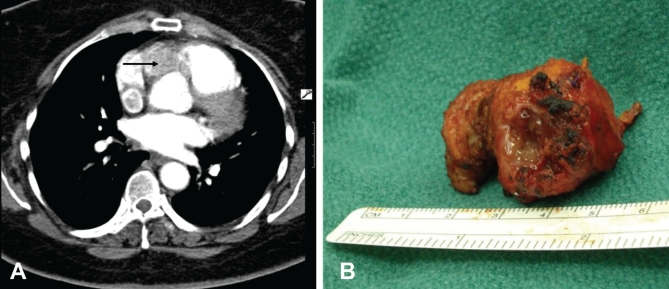
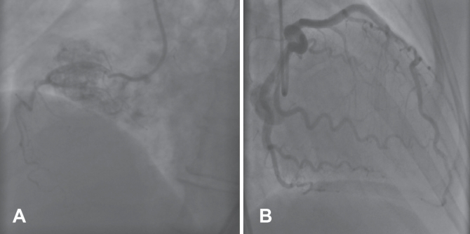
A 44-year-old woman with diabetes mellitus, hypercholesterolemia and a three-year history of hypertension, presented to the King Faisal Specialist Hospital and Research Centre (Jeddah, Saudi Arabia) with a history of recurrent chest pain consistent with angina. The physical examination was unremarkable except for obesity with a body mass index of 36 kg/m2. A transthoracic echocardiogram revealed an incidental finding of a homogeneous mass anterior to the aorta and right coronary artery. Left and right ventricular function was normal with no valvular pathology identified. Multislice computed tomography of the chest confirmed a 4 cm × 3 cm soft tissue mass overlying the anterior surface of the aortic root, encroaching on the right coronary ostium and extending through the right ventricular wall (Figure 1A). Computed tomography scans of the abdomen and pelvis were unremarkable. At cardiac catheterization, a ‘tumour blush’ with blood supply derived from both right and left coronary arteries was identified, but there was no evidence of occlusive coronary artery disease (Figures 2A and 2B).
Figure 1).
A Computed tomography scan of the chest demonstrating a mass overlying the aortic root and extending through the right ventricular muscle fibres (arrow). B Resected cardiac paraganglioma
Figure 2).
Coronary angiogram showing right coronary artery tumour blush (A) and left circumflex coronary arteries (B)
At operation, a dark red tumour was identified. The tumour was found to overlay the right atrioventricular groove and right coronary artery; it arose from the aortic root, encased the right coronary ostium and extended through the right ventricle. Attempts to dissect the tumour from the heart resulted in episodes of hypertension (blood pressure of 220/110 mmHg). The concentration of plasma noradrenaline during tumour manipulation exceeded 25,000 pg/mL (normal range 70 pg/mL to 750 pg/mL), and the concentration of plasma dopamine was 487 pg/mL (normal range less than 30 pg/mL). Once cardiopulmonary bypass was established, manipulation of the tumour had no effect on blood pressure. The tumour was successfully removed and the right coronary artery was revascularized using a saphenous vein graft.
The postoperative course was uneventful with complete resolution of hypertension, and the patient was discharged home eight days postoperatively. The resected tumour measured 4 cm × 3 cm × 1.8 cm, and was solid and yellowish in colour (Figure 1B). The histological examination revealed slight pleomorphic cells with abundant granular cytoplasm. The tumour cells were arranged in solid nests with minimal stroma and many blood vessels. Sustentacular cells were positive for S-100. The tumour cells were strongly positive for chromogranin and synaptophysin. These features were consistent with paraganglioma. During follow-up, the patient had normal blood pressure and her iodine-131-metaiodobenzylguanidine scintigraphy showed no evidence of local recurrence or distant metastasis.
DISCUSSION
The term paraganglionic tissue was first described by Kohn (1) in 1900. It originates from primitive neural crest and is classified histologically as chromaffin (catecholamine secreting) and nonchromaffin (noncatecholamine secreting). Tumours arising from paraganglionic tissue (paragangliomas) of the chromaffin type are called pheochromocytoma and those of nonchromaffin are called chemodectomas. Cardiac paragangliomas are extremely rare and may arise from visceral autonomic paraganglia (atrium or interatrial septum) or branchiomeric paraganglia (branchial arch, coronary, pulmonary or aortopulmonary) (2–4). The most common site for tumour development is the left atrium, possibly explained by the close proximity of paraganglionic cells to the left atrium. Cardiac pheochromocytomas are extremely vascular and their blood supply is exclusively derived from coronary circulation as demonstrated in our patient.
The clinical features of cardiac pheochromocytoma may be subtle: headache, sweating, flushing and palpitation may be absent, but hypertension is almost always present. However, these tumours can cause angina without the presence of occlusive coronary artery disease. We speculate that angina could be related to episodes of hypertension, coronary spasm or an element of coronary steal competing with myocardial blood supply.
CONCLUSION
Primary cardiac paragangliomas may present with dual blood supply from coronary circulation. The multimodality imaging studies in assessing cardiac tumour are important in planning a surgical strategy. Excision of cardiac pheochromocytoma can be safely performed using cardiopulmonary bypass with or without some form of coronary artery bypass grafting. Long-term follow-up is warranted for tumour recurrence, blood pressure response and patency of coronary bypass grafting.
REFERENCES
- 1.Kohn A. Uben den Bau Und doe Entwicklung der Sug. Carotiddruse. Arch fur Mikr Anat. 1900;56:81–148. [Google Scholar]
- 2.Orringer MB, Sisson JC, Glazer G, et al. Surgical treatment of cardiac pheochromocytoma. J Thorac Cardiovasc Surg. 1985;89:753–7. [PubMed] [Google Scholar]
- 3.Fitzgerald PJ, Ports TA, Cheitlin MD, Magilligan DJ, Tyrrell JB. Intracardiac pheochromocytoma with dual coronary blood supply: Case report and review of the literature. Cardiovasc Surg. 1995;3:557–61. doi: 10.1016/0967-2109(95)94459-a. [DOI] [PubMed] [Google Scholar]
- 4.Jeevanandam V, Oz MC, Shapiro B, Barr MI, Marboe C, Rose EA. Surgical management of cardiac pheochromocytoma resection versus transplantation. Ann Surg. 1995;221:415–9. doi: 10.1097/00000658-199504000-00013. [DOI] [PMC free article] [PubMed] [Google Scholar]


