Abstract
Although the general cytotoxicity of selenite is well established, the mechanism by which this compound crosses cellular membranes is still unknown. Here, we show that in Saccharomyces cerevisiae, the transport system used opportunistically by selenite depends on the phosphate concentration in the growth medium. Both the high and low affinity phosphate transporters are involved in selenite uptake. When cells are grown at low Pi concentrations, the high affinity phosphate transporter Pho84p is the major contributor to selenite uptake. When phosphate is abundant, selenite is internalized through the low affinity Pi transporters (Pho87p, Pho90p, and Pho91p). Accordingly, inactivation of the high affinity phosphate transporter Pho84p results in increased resistance to selenite and reduced uptake in low Pi medium, whereas deletion of SPL2, a negative regulator of low affinity phosphate uptake, results in exacerbated sensitivity to selenite. Measurements of the kinetic parameters for selenite and phosphate uptake demonstrate that there is a competition between phosphate and selenite ions for both Pi transport systems. In addition, our results indicate that Pho84p is very selective for phosphate as compared with selenite, whereas the low affinity transporters discriminate less efficiently between the two ions. The properties of phosphate and selenite transport enable us to propose an explanation to the paradoxical increase of selenite toxicity when phosphate concentration in the growth medium is raised above 1 mm.
Keywords: Membrane Trafficking, Selenium, Transcription Regulation, Transport Metals, Yeast, Phosphate Transport, Saccharomyces cerevisiae
Introduction
Although toxic at high concentrations, selenium is required in many cells, because it is translationally incorporated as selenocysteine into selenoproteins that perform specific and essential functions (1). Cells must ensure selenium uptake to sustain this metabolism. However, little is known about selenium transport. Because selenium and sulfur are chalcogen elements that have many chemical properties in common, selenium shares metabolic pathways with sulfur. Accordingly, selenate was shown to be taken up by the yeast Saccharomyces cerevisiae sulfate permeases (2). Similarly, in plants, selenate is taken up by roots via the high affinity sulfate transporters (3).
On the other hand, specific selenite transporters have never been identified so far. The sulfate ABC transporter of Escherichia coli mediates selenite uptake in addition to that of selenate (4). However, repression of the expression of that transporter does not quench selenite uptake completely, indicating that an alternative entry pathway exists for selenite (5). In S. cerevisiae, which does not possess selenoproteins, an energy-dependent uptake of selenite, distinct from that of selenate, was reported. Characterization of the kinetics of selenite uptake suggested the existence of two transport systems: a high affinity system at low selenite concentration and a low affinity system at higher concentration (6). Recently, a study of selenite uptake by wheat (Triticum aestivum) roots showed it to be an active process competitively inhibited by phosphate, suggesting a role for the plant phosphate transporters in selenite uptake (7). Interestingly, in S. cerevisiae, a correlation between resistance to the toxicity of selenite and the expression of a high affinity phosphate transporter has been evidenced (8).
In this study, we asked whether the selenite and phosphate oxyanions, which have similar sizes and charges at pH 6, share the same pathways to enter S. cerevisiae cells. Phosphate is an essential nutrient required for numerous biological processes such as biosynthesis of nucleic acids and phospholipids. In S. cerevisiae, the inorganic phosphate (Pi) acquisition system is composed of five transporters (9). Three of them (Pho87p, Pho90p, and Pho91p) are constitutively transcribed and take up phosphate with low affinity (10). The high affinity transport system, composed of Pho84p and Pho89p, is transcriptionally up-regulated by the phosphate signal transduction (PHO)2 pathway in response to Pi starvation (11–13). This well characterized regulatory pathway requires the transcription factor Pho4p, whose nuclear or cytoplasmic localization depends on the cyclin/cyclin-dependent kinase complex Pho80p-Pho85p and the cyclin-dependent kinase inhibitor Pho81p. When phosphate is limiting, the cyclin-dependent kinase inhibitor Pho81p inactivates the Pho80p-Pho85p complex, leading to accumulation of unphosphorylated Pho4p in the nucleus and subsequent activation of phosphate-responsive genes (14–17). When phosphate is abundant, Pho4p is phosphorylated by the Pho80p-Pho85p complex and exported to the cytoplasm by the receptor Msn5p, where it becomes unable to activate transcription. One of the genes up-regulated by the PHO pathway is SPL2, a negative regulator of the low affinity phosphate transporters (18). Inhibition of the low affinity phosphate transport is likely to occur through a physical interaction with Spl2 because both Pho87p and Pho90p have an SPX (SYG1, pho81, XPR1) domain that has been shown to bind the regulatory protein Spl2p (19). Thus, although the low affinity phosphate transporters are not transcriptionally regulated in response to external phosphate availability, their transport activity is inhibited post-transcriptionally following phosphate starvation. Overall, the above regulatory mechanism results in cells that use either the high affinity or the low affinity transport systems, depending on phosphate availability.
In this study, we show that selenite toxicity is dependent on phosphate concentrations in the growth medium. Selenite uptake measurements allow us to establish that both the high affinity phosphate transporter Pho84p as well as the low affinity carriers mediate selenite uptake in S. cerevisiae and that phosphate and selenite compete for uptake by both these systems. Primacy of one transport system on the other depends on the phosphate concentration conditions used to grow the cells.
EXPERIMENTAL PROCEDURES
Strains and Media
The S. cerevisiae strains used in this study are derived from strain BY4742 (MATa his3Δ1 leu2Δ lys2Δ0 ura3Δ0). The parental and all the single mutants were purchased from Euroscarf. The double and triple mutant strains were constructed by replacing the entire reading frame of the reference gene with a PCR-generated marker cassette containing either the URA3kl or the LEU2 genes (20). All of the disruptions were verified by PCR analysis. The constructed strains are as follows: Δpho87-Δpho90 (BY4742 pho87::KanMX4; pho90::URA3kl), Δpho87-Δpho91 (BY4742 pho87::KanMX4; pho91::URA3kl), Δpho90-Δpho91 (BY4742 pho90::KanMX4;pho91::URA3kl), Δpho84-Δspl2 (BY4742 pho84::KanMX4; spl2::URA3kl), and Δpho87-Δpho90-Δpho91 (BY4742 pho87::KanMX4; pho90::URA3kl; pho91::LEU2). Plasmid pRS426 and its derivatives expressing PHO84 (pPHO84) or PHO87 (pPHO87) from the ADH1 promoter are described in Ref. 9. YPD medium contained 1% yeast extract (Difco), 1% Bacto-tryptone (Difco), and 2% glucose. Standard synthetic dextrose (SD) minimal medium contained 0.67% yeast nitrogen base (Difco), 2% glucose, and 50 mg/liter of histidine, leucine, lysine, and uracil and was buffered at pH 6.0 by the addition of 50 mm MES-NaOH. This medium contained 7.3 mm phosphate. Phosphate-depleted SD medium was prepared as described (21). Then 2% glucose, 50 mg/liter of histidine, leucine, lysine, and uracil, and 50 mm MES were added to the filtered phosphate-depleted medium, and the pH was adjusted to 6.0 with HCl. The medium was filtered through a 0.2-μm filter unit (Nalgene). This medium contained less than 50 μm phosphate (21) and did not support growth of the parental strain. Phosphate was added to this medium at the indicated concentrations from a 100 mm potassium phosphate pH 6.0 solution.
Toxicity Assays
The strains were always pregrown overnight at 30 °C in SD medium containing the amount of phosphate used in the following experiments with the exception of strains containing the Δpho4 or Δpho84 deletions, which do not grow well in low phosphate medium. These strains were pregrown at 0.6 or 0.8 mm phosphate prior to dilution to the desired phosphate concentration and incubated further for at least 4 h.
For growth inhibition, the cells were inoculated in phosphate-defined medium at an OD650 of 0.12 and left to grow at 30 °C for 1 h. Then 5 mm Na2SeO3 (Sigma) was added to half of the cultures, and growth was monitored by following the OD650 as a function of time. For viability assays, the cells were inoculated in phosphate-defined medium to obtain an OD650 of 0.025 and left to grow at 30 °C. When the OD650 reached 0.1, sodium selenite, selenate, or selenide was added to 1 ml of the cultures at the desired final concentration. After 1 h at 30 °C under agitation, the samples were diluted 1000-fold in water. To monitor cell viability, 200-μl aliquots of this dilution were plated in duplicate onto YPD agar plates. The plates were left to grow for 2 days at 30 °C prior to scoring.
Phosphate Uptake
The strains were pregrown overnight as indicated above. The cells were inoculated in phosphate-defined medium to obtain an OD650 of 0.4 and left to grow at 30 °C. When the OD650 reached 1.5, the cells were harvested by centrifugation and washed with SD medium without phosphate. The cells were resuspended in SD medium without phosphate at a density of 1 OD650/ml and incubated at 30 °C for 5 min prior to the addition of [32P]Pi (PerkinElmer Life Science). Uptake measurements were initiated by the addition of 0.1 ml of [32P]Pi (final concentrations from 5 to 7500 μm; specific activity between 103 and 106 dpm·nmol−1) to 0.9 ml of cell suspension. The assays were terminated at 3 and 6 min by the addition of 1 ml of ice-cold 0.5 m phosphate, pH 6.0. Cell suspensions were then filtered using 0.45-μm nitrocellulose filters (Schleicher & Schuell) and washed twice with 3 ml of 0.25 m phosphate, pH 6.0. The radioactivity retained on the filter was measured by liquid scintillation counting. The rates of transport are given in pmol/min and per OD650. The Km and Vmax values were derived from iterative nonlinear fits of the theoretical Michaelis equation to the experimental values, using the Levenberg-Marquardt algorithm as described previously (22). When experimental values could not be fitted to a single hyperbola, a partition method was employed, as recommended (23). Km1 and Vmax 1 values were derived from the experimental data obtained for phosphate concentrations ranging from 5 to 50 μm. The theoretical contribution of the first uptake system was subtracted from the subsequent experimental values (100–5000 μm). The resulting data were used to determine Km and Vmax values for the second uptake system.
Selenite Uptake
The strains were pregrown overnight as indicated above. The cells were inoculated in phosphate-defined medium to obtain an OD650 of 0.4 and left to grow at 30 °C. When the OD650 reached 1.5, the cells were harvested by centrifugation and washed with SD medium without phosphate. The cells were resuspended in SD medium without phosphate at a density of 5 OD650/ml and incubated at 30 °C for 10 min prior to the addition of Na2SeO3. For determination of the kinetic parameters for selenite uptake, selenite concentrations were in the range 1–20 mm. At time intervals, 10 ml of cell suspension were removed from the incubation mixture, and the reaction was stopped by the addition of 1 ml of ice-cold 1 m phosphate, pH 6.0. The samples were centrifuged (5000 × g for 5 min), washed twice with 10 ml of water, and lyophilized.
Selenium content of the cells was determined by inductively coupled plasma mass spectrometry (ICP/MS) on a Thermo Fisher PQ Excell quadripole spectrometer in the collision cell mode. The mass of each yeast sample was estimated by the difference of weight between the filled and empty microtubes (microbalance Sartorius ME 36S, range 30 g/1 μg). The yeast samples were digested with 3 ml of HNO3 (67–69%; Plasmapur SGS) and 1 ml of H2O2 (30%; Suprapur Merck) in a closed vessel microwave oven (Ethos 900; Thermo Fisher). The residues were then diluted with 10 ml of pure water. The ICP/MS mass spectrometer was calibrated (range, 0–200 μg/liter) against a reagent blank solution, and several selenium standard solutions were obtained after dilutions from a concentrated certified standard (selenium, 999 ± 5 mg/liter; Certipur Merck). The data were recorded for the four selenium isotopes 76Se, 78Se, 80Se, and 82Se. Selenium concentrations calculated from each isotope were analogous. The results are the means of data obtained on each isotope and are expressed in μg of selenium·g−1 (dry weight). Conversion in pmol·OD650−1 was made using a mean atomic mass of 79 and by considering that 1 g of yeast (dry weight) corresponds to 6200 OD650.
RESULTS
Effect of Phosphate on Selenite Toxicity
The toxicity of selenite in yeast is well documented, although the mechanisms of toxicity are less well understood (24, 25). Selenite uptake is the first step in selenium metabolism that ultimately leads to toxicity. To investigate the possibility that selenite enters yeast cells via the orthophosphate transport system, the influence of the concentration of phosphate in the growth medium on the toxicity of sodium selenite was analyzed. We compared the effect of the addition of 5 mm of sodium selenite on the growth of yeast cells (strain BY4742) cultivated in the presence of increasing concentrations of potassium phosphate (SD with 0.1, 0.2, 0.4, or 0.8 mm Pi or standard high Pi SD (7.3 mm Pi)) (Fig. 1A). In the absence of selenite, whatever the concentration of phosphate, growth rates were identical (t½ = 135 min at 30 °C). We observed, however, that in the medium supplemented with 0.1 mm phosphate, upon reaching the late exponential phase, phosphate depletion became limiting for cell growth. In the presence of 5 mm selenite, growth rates and plateau values were clearly dependent on phosphate concentrations. Up to 0.8 mm Pi, the cells were much more affected by selenite toxicity in the medium containing the lowest phosphate concentration. Surprisingly, at high phosphate concentration, selenite toxicity increased.
FIGURE 1.
Selenite toxicity is dependent on phosphate concentration. A, cells (strain BY4742) were grown at 30 °C in phosphate-depleted SD medium supplemented with phosphate as follows: 0.1 mm (□, ■), 0.2 mm (△, ▴), 0.4 mm (○, ●), 0.8 mm (⧫, ◊), and 7.3 mm (×, *). The cultures were divided in two, and 5 mm Na2SeO3 was added to one sample (filled symbols). Cell growth was monitored by measuring the OD650 at various times. B, cells (strain BY4742) were incubated in SD medium supplemented with the indicated phosphate concentration. When the OD650 reached 0.1, Na2SeO3 was added to the cultures (0 mm, black bars; 5 mm, gray bars; 10 mm, white bars). After 1 h of incubation at 30 °C, the samples were diluted and plated onto YPD-agar. Cell viability was determined after 2 days of growth at 30 °C. The results are expressed as percentages of survival compared with control samples incubated in the absence of selenite. The error bars represent the means and ranges of two independent experiments.
To determine whether this inhibition of growth was due to increased selenite lethality, cell viability was assayed after a short term exposure to selenite. The cells, grown exponentially in medium containing various concentrations of phosphate, were incubated for 1 h in the same medium with 5 or 10 mm selenite, diluted, and plated on rich medium plates (high phosphate). The survival rates were determined by counting colonies after 2 days growth at 30 °C (Fig. 1B). In a medium containing 0.1 mm Pi, exposure to 5 mm selenite reduced cell survival by nearly 90%. In contrast, at 0.4 mm Pi, more than 80% of the cells were resistant to 5 mm selenite. When the phosphate concentration was raised further, viability of the cells decreased to reach ∼50% in high phosphate medium. These results show that selenite not only inhibits the growth of yeast cells but also induces mortality.
In a previous paper, we showed that extracellular reduction of selenite into hydrogen selenide (HSe−) led to increased cellular accumulation and toxicity of selenium (26). To determine whether sodium selenide toxicity was dependent on phosphate concentration, we measured cell survival after a 5-min exposure to 40 μm Na2Se in medium containing increasing concentrations of Pi. This resulted in ∼50% mortality of the cells, independently of phosphate concentration, suggesting that these two forms of inorganic selenium (selenite and selenide) are internalized by different pathways, as suggested previously (27). Because our sample of sodium selenite might contain traces of selenate, sodium selenate toxicity was also assayed. Exposure to 5 mm selenate resulted in low mortality (<20%), irrespective of phosphate concentration. Thus, the toxicity of selenite cannot originate from trace amounts of selenate contaminating the selenite solution.
Selenite Toxicity in Mutants of the PHO Pathway
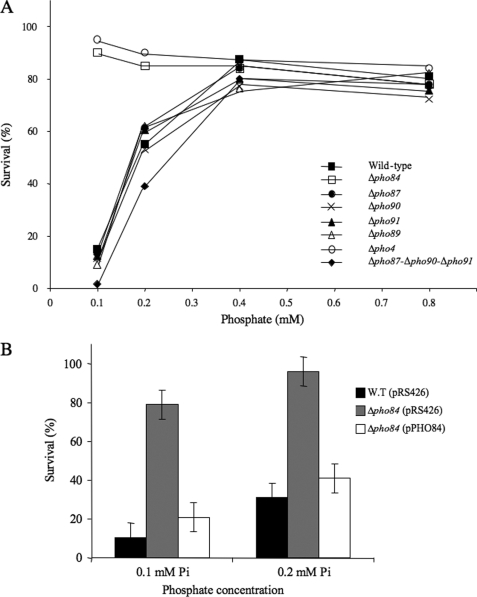
In a first set of experiments, the toxicity of selenite was analyzed in Pi-limited medium. Viability assays were performed in strains disrupted for one of the phosphate transporter genes (PHO84, PHO87, PHO89, PHO90, and PHO91). The cells were pregrown with increasing Pi concentrations, exposed to 5 mm selenite, plated, and scored to assess cell survival (Fig. 2A). Whatever the concentration of phosphate in the range 0.1–0.8 mm, each of the single mutants in the low affinity carriers (Δpho87, Δpho90, and Δpho91) displayed curves similar to that of the parental strain, with high selenite toxicity at low Pi concentrations. However, a triple mutant Δpho87-Δpho90-Δpho91 was slightly more sensitive to selenite than the wild-type strain. At 0.1 mm Pi, survival was 1.4% for the triple mutant as compared with 15% for wild type. In contrast, the Δpho84 strain was very resistant to selenite toxicity at low Pi concentrations. The Δpho4 strain, in which PHO84 is not expressed, was also very resistant to selenite toxicity at low Pi concentrations. As shown in Fig. 2B, expression, in the Δpho84 strain, of PHO84 from the constitutive ADH1 promoter restored selenite toxicity values close to those observed in the wild type. These results suggest that Pho84p plays a major role in the toxicity of selenite, at least up to 0.4–0.5 mm phosphate.
FIGURE 2.
Selenite toxicity in mutants of the phosphate transport pathway. A, various strains, as indicated in the figure, were grown at 30 °C in SD medium supplemented with the indicated phosphate concentration. When the OD650 reached 0.1, 5 mm Na2SeO3 was added to the cultures. After 1 h of incubation at 30 °C, the samples were diluted and plated onto YPD-agar. Cell viability was determined after 2 days growth at 30 °C. The results are expressed as percentages of survival compared with control samples incubated in the absence of selenite. The values are the means of at least three independent experiments. Standard deviations between these experiments were lower than 15%. B, cells expressing either the control plasmid pRS426 or pPHO84 expressing PHO84 from the ADH1 promoter were grown and treated as in A. The error bars represent the means and ranges of two independent experiments.
In the case of a Δpho89 strain, the survival curve was similar to that of the wild type. This indicates that, under our experimental conditions, this transporter is not involved in selenite toxicity. However, both the transcription and the activity of Pho89p are strongly pH-dependent, with an optimum pH of >8.0 (13). Although no effect of the inactivation of its gene was observed, a potential role for Pho89p in selenite toxicity cannot be excluded but must be studied in different conditions. For this reason, this high affinity transporter was not considered in the remainder of our study.
Selenite Uptake Is Reduced in a Δpho84 Mutant
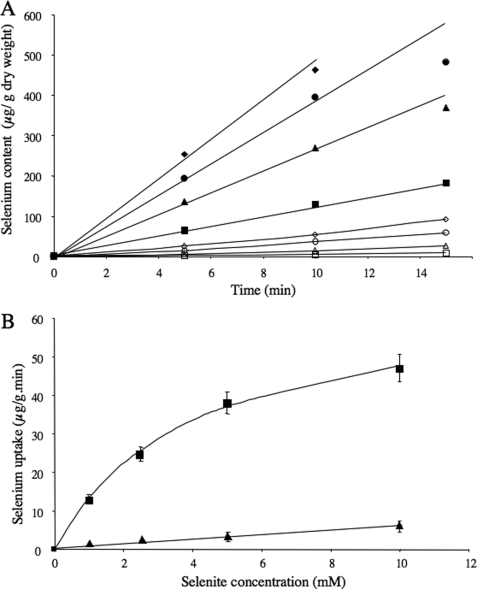
The uptake of selenite by BY4742 yeast cells was measured using cells grown in Pi-limited conditions (0.3 mm). The cells were washed and resuspended in SD medium without Pi. The cell suspensions were incubated at 30 °C in the presence of various concentrations of selenite (1–10 mm), and aliquots were removed at various times to measure the amount of total incorporated selenium (Fig. 3A). Cellular accumulation of selenium was linear over a period of at least 10 min. The rate of selenite uptake, as a function of selenite concentration, could be adequately described by the Michaelis-Menten equation, allowing us to derive a Km of 4 ± 1 mm and a Vmax of 67 ± 4 μg of selenium·g−1·min−1 (or 136 pmol of selenium·OD−1·min−1) (Fig. 3B). The rate of selenite uptake, at the selenite concentration of 5 mm, was 76 pmol of selenium·OD−1·min−1 (Table 1). The uptake of 5 mm selenite, by the Δpho84 mutant assayed in the same conditions, was only 11 pmol of selenium·OD−1·min−1, indicating that deletion of PHO84 drastically reduced selenite uptake. These results show that, in Pi-limited conditions, transport of selenite is ensured mostly by Pho84p. In addition, the kinetics of selenite uptake were measured in the presence of 0.5 mm Pi in the uptake medium (Fig. 3B and Table 1). Whatever the selenite concentration, accumulation of selenium was reduced at least 10 times, showing that selenite uptake by Pho84p is inhibited by phosphate.
FIGURE 3.
Uptake of selenium by BY4742 cells grown in SD medium supplemented with 0.3 mm Pi. A, cells were harvested by centrifugation, washed, and resuspended in SD medium without phosphate. Uptake of 1 mm (□, ■), 2.5 mm (▴, △), 5 mm (○, ●), and 10 mm (⧫, ◊) Na2SeO3 was measured as described under “Experimental Procedures,” in the absence (filled symbols) or the presence (open symbols) of 0.5 mm potassium phosphate. The samples were analyzed for their total selenium content. B, rates of selenite uptake in the absence of phosphate (■), determined from A, were fitted to the Michaelis-Menten equation as described under “Experimental Procedures.” In the presence of 0.5 mm phosphate (▴), the rate of selenite uptake increased roughly linearly with the selenite concentration.
TABLE 1.
Rates of selenite uptake and inhibition by phosphate of wild-type and Pi transporter-defective strains in the presence of 5 mm selenite
The selenite uptake measurements were performed as described under “Experimental Procedures.” The errors are standard deviations. ND, not determined.
| Pi concentration in the growth medium | Strain |
V |
|
|---|---|---|---|
| 0 mm Pia | 0.5 mm Pia | ||
| pmol of SeO32−·min−1·OD650−1 | |||
| 0.3 mm | BY4742 | 76 ± 10 | 6.4 ± 1 |
| 7.3 mm | BY4742 | 27 ± 4 | 24 ± 4 |
| 0.3 mm | Δpho84 | 11 ± 4 | ND |
| 7.3 mm | Δpho84 | 20 ± 4 | ND |
| 0.3 mm | Δpho84-Δspl2 | 21 ± 4 | ND |
| 7.3 mm | Δpho84-Δspl2 | 21 ± 3 | ND |
| 7.3 mm | Δpho87 | 31 ± 5 | 14 ± 4 |
| 7.3 mm | Δpho87 (pPHO87) | 30 ± 4 | 26 ± 3 |
| 7.3 mm | Δpho85 | 87 ± 10 | 11 ± 2 |
a Concentration of phosphate in the uptake medium.
Inhibition of Phosphate Uptake by Selenite in Cells Grown in Low Pi Conditions
Because the addition of phosphate in the assay reduced selenium accumulation, phosphate uptake was expected to be inhibited by selenite. Therefore, [32P]Pi transport kinetic parameters were measured in the presence of increasing concentrations of selenite (Table 2).
TABLE 2.
Phosphate uptake parameters and selenite inhibition constants of wild-type and Pi transporter-defective strains
The uptake experiments were performed as described under “Experimental Procedures” on two independently grown cultures, apart from BY4742 grown in 0.3 mm Pi (four independent experiments), BY4742 grown in 7.3 mm Pi, and Δspl2 grown in 0.3 mm Pi (three independent experiments). Seven different concentrations of phosphate were used to determine the kinetic parameters. The Pi concentrations were in the range 5–500 μm for high affinity uptake and 0.1–7.5 mm for low affinity uptake measurements. To determine the phosphate uptake parameters in the Δspl2 strain, 10 concentrations of phosphate were used (5–5000 μm). Selenite inhibition constants were determined from the measurements of the apparent Km for phosphate in the presence of 2.5, 5, and 10 mm selenite. The Ki was deduced from the slope of the plot of Kmapp as a function of selenite concentration. The errors are standard deviations.
| Pi concentration | Strain | Vmax | Km | Ki |
|---|---|---|---|---|
| pmol Pi·min−1·OD650−1 | μm | mm | ||
| 0.3 mm | BY4742 | 860 ± 80 | 20 ± 4 | 4.6 ± 1.7 |
| 0.3 mm | Δpho87-Δpho90-Δpho91 | 2000 ± 300 | 25 ± 3 | 5.5 ± 1.8 |
| 0.3 mm | Δspl2 | Vmax 1 600 ± 140 | Km 1 19 ± 6 | |
| Vmax 2 1700 ± 400 | Km 2 4000 ± 1300 | |||
| 0.3 mm | Δpho84 | 700 ± 150 | 5000 ± 1600 | |
| 0.3 mm | Δpho84-Δspl2 | 1400 ± 300 | 5900 ± 1800 | |
| 7.3 mm | BY4742 | 1600 ± 400 | 4300 ± 1600 | 9.0 ± 2.0 |
| 7.3 mm | Δpho87 | 440 ± 50 | 25 ± 6 | |
| 7.3 mm | Δpho84 | 1700 ± 200 | 6000 ± 2000 | 9.5 ± 2.0 |
| 7.3 mm | Δpho84-Δspl2 | 2000 ± 500 | 5800 ± 2000 |
First, we analyzed phosphate uptake in conditions identical to those previously used to measure selenite uptake (strain BY4742, 0.3 mm Pi). The rate of phosphate accumulation at concentrations ranging from 5 to 500 μm could be fitted to a single Michaelis-Menten equation, giving a Vmax of 860 pmol of Pi incorporated per min/OD650 and a Km of 20 μm. The Km for Pi uptake is in the micromolar range, suggesting that only high affinity phosphate transport is operative at this phosphate concentration. To support this idea, we compared the rate of phosphate uptake in the wild-type strain and the Δpho84 mutant grown in 0.3 mm Pi, using a phosphate concentration of 0.3 mm in the assay. Rates of 840 and 43 pmol of Pi·OD−1·min−1 were determined, respectively. These results indicate that in the growth conditions used above, Pho84p is the major contributor to phosphate uptake. Kinetic studies were also performed in the presence of 1, 2.5, 5, and 10 mm selenite. The results show that selenite competitively inhibits phosphate uptake by BY4742 cells with a Ki of 4.6 mm (Table 2). It is noteworthy that this value is close to the Km determined for selenite uptake. This is in agreement with the conclusion that the same transporter, Pho84p, is responsible for the major part of the uptake of both phosphate and selenite. In a previous paper (8), the authors found that selenite did not compete with phosphate transport. However, they did not go beyond a 10-fold molar excess of selenite, which may not have been enough to observe an effect, because of the large difference of Km for phosphate or selenite transport.
We also measured the Ki of selenite toward phosphate uptake in the Δpho87-Δpho90-Δpho91 strain. Experimental conditions were identical to those used above with the wild-type strain. As expected, the Km for phosphate (25 μm) and the Ki for selenite (5.5 mm) were similar to those determined with the BY4742 strain, which confirms that the selenite transport kinetic values determined above correspond to Pho84p-mediated uptake.
Selenite Toxicity in High Phosphate Medium
Transport of selenite by Pho84p cannot explain the increase in selenite toxicity observed with cells grown in high Pi conditions, because Pho84p is not significantly expressed in these conditions (11, 18). In agreement with this idea, when assayed for cell survival after exposure to 5 or 10 mm selenite, the single mutants in the high affinity transporter genes (PHO84 and PHO89), as well as the Δpho4 and Δspl2 strains, grown in high phosphate medium, displayed selenite survival values comparable with those of the wild-type strain (Fig. 4). In contrast, single mutant strains in the low affinity Pi transporters, as well as the double and triple mutants, were more resistant to selenite. An increased resistance of the Δpho87, Δpho90, and Δpho91 mutants has been previously reported by Pinson et al. (8). Several studies have shown that mutations in the low affinity phosphate transporters resulted in derepression of the PHO-regulated genes (8, 10, 28), leading to increased transcription of PHO84 and to inactivation of the low affinity transporters through derepression of SPL2. Thus, in these mutants, Pho84p is responsible for most of the phosphate uptake, whatever the phosphate conditions.
FIGURE 4.
Selenite toxicity in high Pi medium. The cells were incubated in SD medium. When the OD650 reached 0.1, Na2SeO3 was added to the cultures (0 mm, black bars; 5 mm, gray bars; 10 mm, white bars). After 1 h of incubation at 30 °C, the samples were diluted and plated onto YPD-agar. Cell viability was determined after 2 days growth at 30 °C. The results are expressed as percentages of survival compared with control samples incubated in the absence of selenite. The error bars represent the means and ranges of two independent experiments. W.T, wild type.
To confirm the induction of the PHO pathway upon the deletion of a single low affinity transporter gene, we compared kinetic parameters for phosphate uptake in wild-type cells grown in high Pi medium and in a Δpho87 strain (Table 2). Values corresponding to low affinity Pi transport were observed for the wild-type strain (Km = 4.3 mm, Vmax = 1600 pmol of Pi·OD−1·min−1). This Km was higher than that usually associated with the low affinity Pi transporters (4 mm instead of 0.2–0.8 mm). This difference might be related to the pH conditions of our assay, because Pi transport is usually assayed at pH 4.5 instead of pH 6.0. As expected, deletion of PHO87 led to a Km in the micromolar range showing that, in this mutant, only high affinity phosphate transporters are functional. Another circumstance in which the PHO regulon is constitutively expressed is in the Δpho85 mutant. In such a strain, inactivation of the Pho85p kinase leads to accumulation of unphosphorylated Pho4p in the nucleus and thus to constitutive activation of the PHO pathway. We found that this strain was also very resistant to selenite toxicity at high Pi concentrations (Fig. 4). Finally, overexpression of plasmid-encoded PHO84, in the Δpho87 mutant, did not alter selenite survival values, whereas expression of PHO87 reduced them to wild-type levels.
Altogether, these results suggest that, when phosphate is abundant in the medium, strains expressing the low affinity transporters (wild type, Δpho4, Δpho89, Δpho84, Δspl2, and Δpho87 (pPHO87)) are more sensitive to selenite toxicity than strains using mostly Pho84p to uptake phosphate (Δpho85, Δpho87, Δpho90, Δpho91, and Δpho87 (pPHO84)). One possible explanation is that suppression of low affinity transport influences intracellular selenite metabolism, leading to lower H2Se production and, therefore, lower toxicity. Because down- or up-regulation of the sulfate assimilation pathway might interfere with selenium metabolism, we asked whether this pathway was altered in the deletion mutants. For this purpose, the strains were grown on Pb(II)/sulfate plates (29). Wild-type and single deletion mutants in the low affinity Pi transport system displayed equivalent coloring (result not shown), suggesting that these mutations do not interfere with the metabolism of sulfate.
A more likely hypothesis is that low affinity Pi transporters are able to uptake selenite and that, in high Pi medium, they are more efficient than Pho84p in importing this ion. The following experiments were performed to assess this idea.
Selenite Toxicity in a Δspl2 Strain
To establish that the low affinity transporters are involved in the uptake of selenite, we used a Δspl2 strain grown in Pi-limited medium. In this strain, low affinity transport is active whatever the phosphate concentration. First, we determined the contribution of each transport system to total phosphate uptake in the Δspl2 mutant. As expected from previous studies (18), deletion of SPL2 resulted in activation of the low affinity transporters (Table 2). The observed values of [32P]Pi incorporated could not be fitted with a single hyperbola but were correctly fitted with a double hyperbola curve. The results show that the high affinity system (Km1 = 19 μm) uptakes phosphate with a Vmax 1 of 600 pmol of Pi·OD−1·min−1. Activation of the low affinity transporters allowed us to determine a Km2 of 4 mm and a Vmax 2 of 1700 pmol of Pi·OD−1·min−1 for low affinity Pi transport.
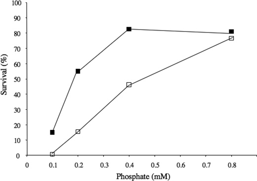
Then we assayed selenite toxicity in the Δspl2 strain. As shown in Fig. 5, inactivation of SPL2 resulted in increased selenite sensitivity, as compared with the wild-type cells. At 0.1 mm Pi, survival of the Δspl2 cells exposed to 5 mm selenite was less than 1%. These results show that deletion of SPL2 leads to the activation of the low affinity transport system with a concomitant increase in selenite toxicity.
FIGURE 5.
Sensitivity to selenite toxicity of a Δspl2 strain grown in low Pi medium. Strains BY4742 (■) and Δspl2 (□) were grown at 30 °C in SD medium supplemented with the indicated phosphate concentrations. When the OD650 reached 0.1, 5 mm Na2SeO3 was added to the cultures. After 1 h of incubation at 30 °C, the samples were diluted and plated onto YPD-agar. Cell viability was determined after 2 days of growth at 30 °C. The results are expressed as percentages of survival compared with control samples incubated in the absence of selenite. The values are the means of at least three independent experiments. Standard deviations between these experiments were lower than 15%.
Selenite Uptake by the Low Affinity Transport System
To demonstrate that the low affinity phosphate transporters can import selenite, we compared the initial rates of selenite uptake at a fixed selenite concentration of 5 mm in Δpho84 and Δpho84-Δspl2 cells grown in low Pi medium (Table 1). Phosphate uptake was also determined in the same conditions (Table 2). In the strain that expresses Spl2p, activation of the PHO pathway led to down-regulation of the low affinity transporters. As a consequence, the Vmax of Pi uptake was twice higher in the Δpho84-Δspl2 strain than in the Δpho84 strain. This higher activity of the low affinity transporters was paralleled by a 2-fold increase of selenite uptake, establishing that the low affinity transporters are able to carry selenite inside cells.
Selenite uptake by the low affinity transporters was also measured in cells grown in high Pi medium. In these conditions, the wild-type, Δpho84, and Δpho84-Δspl2 strains exhibited phosphate uptake rate values very similar to that measured with the Δpho84-Δspl2 strain grown in low Pi medium (Table 2). The rates of selenite uptake by the three strains grown in high Pi medium were also very similar to that measured with the Δpho84-Δspl2 strain grown in low Pi medium (Table 1).
Finally, we determined the kinetic parameters for selenite uptake in the wild-type strain grown in high Pi conditions. A Km of 7.7 ± 3 mm and a Vmax of 40 ± 8 μg of selenium·g−1·min−1 (or 81 pmol of selenium·OD−1·min−1) were determined. Measurement of phosphate uptake in the presence of various concentrations of phosphate and selenite indicated that selenite competitively inhibited phosphate uptake, with a Ki value close to 9 mm (Table 2). This Ki value is comparable with the Km for selenite transport by low affinity carriers, as measured in selenite uptake experiments. This result confirms that the low affinity Pi transport system is competent for selenite transport.
Phosphate Inhibition of Selenite Uptake
The experimental kinetic values determined above allow us to calculate a ratio between the Km values for selenite and phosphate of ∼250 for Pho84p and ∼2 for the low affinity transporters. Therefore, we anticipated that, in high Pi medium, phosphate would inhibit selenite uptake much more efficiently in strains expressing Pho84p than in strains expressing the low affinity system. To verify this prediction, we compared the rates of selenite uptake in the presence of 0.5 mm Pi in the assay of Δpho87 and Δpho85 cells to those of strains expressing low affinity transporters (wild type and Δpho87 (pPHO87)). The data reported in Table 1 show that, indeed, selenite uptake was severely inhibited by phosphate in Pho84p-expressing strains, whereas little inhibition was observed in strains expressing low affinity Pi transporters. These results indicate that, when extracellular Pi is abundant, selenite is taken up more efficiently by the latter transporters than by Pho84p.
DISCUSSION
Active selenite transport by S. cerevisiae has already been reported (6), but no transporter had been identified so far. In this study, we demonstrate that the high affinity phosphate transporter Pho84p, as well as the low affinity carriers, are able to uptake sodium selenite in S. cerevisiae. The transport system used opportunistically by selenite depends on the phosphate concentration in the medium. At low Pi concentrations (up to 0.4 mm), Pho84p is the major contributor to selenite uptake. When phosphate is abundant, the role of Pho84p becomes negligible, and selenite is internalized through one or all of the low affinity Pi transporters. Other toxic metalloids are taken up adventitiously by the existing transport system. For instance, the phosphate transport system is also known to take up arsenate in both prokaryotes and eukaryotes (30, 31). Another example is that of the sulfate transporters of yeast and plants that have low selectivity for sulfate versus analogous selenate or chromate (2, 3, 32).
Selenite uptake measurements in cells expressing either the high affinity Pi transporter Pho84p or the low affinity Pho87p, Pho90p, and Pho91p indicate that both systems are able to transport selenite inside the cells. Comparison of the Vmax/Km of the transporters for Pi or for selenium (Table 3) shows that Pho84p is slightly more efficient than the low affinity carriers for selenite transport. However, Pho84p has a much higher affinity for Pi than for selenium. Thus, the high affinity transporter is very selective for Pi, whereas the low affinity system is much less discriminating.
TABLE 3.
Efficiency and selectivity of phosphate transporters for phosphate and selenite uptakes
The values are deduced from the data in Table II for Pi uptake and from values given in the text for selenite uptake.
| Pi transporter expressed | Strain and growth conditions | Vmax/Km for Pi | Vmax/Km for selenite | Selectivitya |
|---|---|---|---|---|
| pmol OD−1·min−1·mm−1 | pmol OD−1·min−1·mm−1 | |||
| Pho84p | BY4742 and 0.3 mm Pi | 43000 | 34 | 1260 |
| Low affinity | BY4742 and 7.3 mm Pi | 370 | 10.5 | 35 |
a Selectivity is defined as the ratio between the Vmax/Km for Pi uptake and that for selenium uptake.
Selenite toxicity results correlate well with selenium uptake measurements, indicating that mortality of S. cerevisiae cells is directly dependent on the amount of internalized selenium by the phosphate transport system. In the wild-type strain grown in very low Pi conditions (0.1 mm), selenite toxicity is high. When the phosphate concentration in the medium is increased up to 0.4 mm Pi, selenite toxicity is reduced. This effect is easily accounted for by phosphate inhibition of the Pho84p-mediated selenite uptake. When phosphate concentration in the culture medium is further increased, the transport of phosphate (and of selenite) is progressively taken over by the low affinity carriers. Because these carriers are less specific, the advantage of phosphate over selenite (in term of Vmax/Km) is reduced, and selenite uptake/toxicity increases. This mechanism implies that, at very high phosphate concentrations, selenite resistance should improve again. This effect was, indeed, observed previously (8). In the Δpho84 strain grown in low Pi medium, resistance to selenite toxicity can be attributed to the concomitant inactivation of PHO84 and down-regulation of low affinity transport that affects both phosphate and selenite transport (Tables 1 and 2) and thus results in low selenite (and phosphate) uptake.
The triple mutant Δpho87-Δpho90-Δpho91 was more sensitive to selenite than the wild-type strain in Pi-limited medium. In agreement with previous studies (10), we observed that in Pi-limited medium the triple-disruptant strain exhibited a 2-fold higher Vmax of Pi uptake than the wild-type strain (Table 2). It was shown that the higher activity in this strain was accompanied by enhanced transcription of the PHO84 gene (10). Increased activity of Pho84p, in phosphate and selenite transport, explains the higher sensitivity of this strain to selenite as compared with the wild-type cells. Another strain that is very sensitive to selenite at low Pi concentrations is the Δspl2 strain. In this case, loss of the regulation by Spl2p results in activation of low affinity transporters. In this strain, both the high and low affinity Pi transport systems are active in Pi-limited conditions, resulting in higher selenite uptake and toxicity.
On the contrary, in high Pi medium, each of the single, as well as the double and the triple low affinity Pi transporter mutants, were more resistant to selenite than the wild-type strain or mutants of the high affinity transport system. Increased selenite resistance of the low affinity phosphate transporter mutants has been observed previously (8). The higher resistance to selenite of such mutants was ascribed to an overexpression of Pho84p. However, the mechanism by which expression of Pho84p led to selenite resistance remained unknown. The results obtained in this study provide a good explanation for this behavior. Phosphate uptake measurements in the single low affinity Pi transporter mutant Δpho87 confirm that, in high Pi conditions, Pho84p is overexpressed, and the low affinity transport system is concomitantly down-regulated. Additionally, we show that inhibition by phosphate of Pho84p-mediated selenite transport is much more effective than that of the low affinity transport system. As an example, with a simple calculation using the selenite and phosphate kinetic constants determined here, we find that in the presence of 5 mm selenite, the addition of 7.3 mm Pi (as in the high Pi medium) inhibits by more than 150-fold the uptake of selenite by Pho84p. In the same conditions, the uptake of selenite by the low affinity transporters is reduced by only 50%. Therefore, reduced uptake of selenite by cells expressing Pho84p, as compared with cells using the low affinity Pi transporters, is responsible for the paradoxical resistance of these strains to selenite, in high Pi conditions.
Competitive inhibition of SeO32− uptake by phosphate has long been documented in various plant species (7, 33), in the green alga Chlamydomonas reinhardtii (34), and in the yeast Candida albicans (35). Thus, selenite uptake by the phosphate transport pathway could be a general mechanism, at least in plants, algae, and fungi.
Acknowledgments
We gratefully acknowledge Sébastien Sannac and Caroline Oster for valuable support in ICP/MS measurements. We thank Erin O'Shea for providing the plasmids pRS426, pPHO84, and pPHO87.
Footnotes
- PHO
- phosphate signal transduction
- SD
- synthetic dextrose
- MES
- 4-morpholineethanesulfonic acid
- ICP/MS
- inductively coupled plasma mass spectrometry.
REFERENCES
- 1.Rayman M. P. (2000) Lancet 356, 233–241 [DOI] [PubMed] [Google Scholar]
- 2.Cherest H., Davidian J. C., Thomas D., Benes V., Ansorge W., Surdin-Kerjan Y. (1997) Genetics 145, 627–635 [DOI] [PMC free article] [PubMed] [Google Scholar]
- 3.Sors T. G., Ellis D. R., Salt D. E. (2005) Photosynth. Res. 86, 373–389 [DOI] [PubMed] [Google Scholar]
- 4.Lindblow-Kull C., Kull F. J., Shrift A. (1985) J. Bacteriol. 163, 1267–1269 [DOI] [PMC free article] [PubMed] [Google Scholar]
- 5.Turner R. J., Weiner J. H., Taylor D. E. (1998) Biometals 11, 223–227 [DOI] [PubMed] [Google Scholar]
- 6.Gharieb M. M., Gadd G. M. (2004) Mycol. Res. 108, 1415–1422 [DOI] [PubMed] [Google Scholar]
- 7.Li H. F., McGrath S. P., Zhao F. J. (2008) New Phytol. 178, 92–102 [DOI] [PubMed] [Google Scholar]
- 8.Pinson B., Merle M., Franconi J. M., Daignan-Fornier B. (2004) J. Biol. Chem. 279, 35273–35280 [DOI] [PubMed] [Google Scholar]
- 9.Wykoff D. D., O'Shea E. K. (2001) Genetics 159, 1491–1499 [DOI] [PMC free article] [PubMed] [Google Scholar]
- 10.Auesukaree C., Homma T., Kaneko Y., Harashima S. (2003) Biochem. Biophys. Res. Commun. 306, 843–850 [DOI] [PubMed] [Google Scholar]
- 11.Bun-Ya M., Nishimura M., Harashima S., Oshima Y. (1991) Mol. Cell. Biol. 11, 3229–3238 [DOI] [PMC free article] [PubMed] [Google Scholar]
- 12.Martinez P., Zvyagilskaya R., Allard P., Persson B. L. (1998) J. Bacteriol. 180, 2253–2256 [DOI] [PMC free article] [PubMed] [Google Scholar]
- 13.Zvyagilskaya R. A., Lundh F., Samyn D., Pattison-Granberg J., Mouillon J. M., Popova Y., Thevelein J. M., Persson B. L. (2008) FEMS Yeast Res. 8, 685–696 [DOI] [PubMed] [Google Scholar]
- 14.Oshima Y. (1997) Genes Genet. Syst. 72, 323–334 [DOI] [PubMed] [Google Scholar]
- 15.Ogawa N., DeRisi J., Brown P. O. (2000) Mol. Biol. Cell 11, 4309–4321 [DOI] [PMC free article] [PubMed] [Google Scholar]
- 16.Persson B. L., Lagerstedt J. O., Pratt J. R., Pattison-Granberg J., Lundh K., Shokrollahzadeh S., Lundh F. (2003) Curr. Genet. 43, 225–244 [DOI] [PubMed] [Google Scholar]
- 17.Persson B. L., Petersson J., Fristedt U., Weinander R., Berhe A., Pattison J. (1999) Biochim. Biophys. Acta 1422, 255–272 [DOI] [PubMed] [Google Scholar]
- 18.Wykoff D. D., Rizvi A. H., Raser J. M., Margolin B., O'Shea E. K. (2007) Mol. Cell 27, 1005–1013 [DOI] [PMC free article] [PubMed] [Google Scholar]
- 19.Hürlimann H. C., Pinson B., Stadler-Waibel M., Zeeman S. C., Freimoser F. M. (2009) EMBO Rep. 10, 1003–1008 [DOI] [PMC free article] [PubMed] [Google Scholar]
- 20.Baudin A., Ozier-Kalogeropoulos O., Denouel A., Lacroute F., Cullin C. (1993) Nucleic Acids Res. 21, 3329–3330 [DOI] [PMC free article] [PubMed] [Google Scholar]
- 21.Bisson L. F., Thorner J. (1982) Genetics 102, 341–359 [DOI] [PMC free article] [PubMed] [Google Scholar]
- 22.Dardel F. (1994) Comput. Appl. Biosci. 10, 273–275 [DOI] [PubMed] [Google Scholar]
- 23.Burns D. J., Tucker S. A. (1977) Eur. J. Biochem. 81, 45–52 [DOI] [PubMed] [Google Scholar]
- 24.Letavayová L., Vlasáková D., Spallholz J. E., Brozmanová J., Chovanec M. (2008) Mutat. Res. 638, 1–10 [DOI] [PubMed] [Google Scholar]
- 25.Pinson B., Sagot I., Daignan-Fornier B. (2000) Mol. Microbiol. 36, 679–687 [DOI] [PubMed] [Google Scholar]
- 26.Tarze A., Dauplais M., Grigoras I., Lazard M., Ha-Duong N. T., Barbier F., Blanquet S., Plateau P. (2007) J. Biol. Chem. 282, 8759–8767 [DOI] [PubMed] [Google Scholar]
- 27.Ganyc D., Self W. T. (2008) FEBS Lett. 582, 299–304 [DOI] [PMC free article] [PubMed] [Google Scholar]
- 28.Hürlimann H. C., Stadler-Waibel M., Werner T. P., Freimoser F. M. (2007) Mol. Biol. Cell 18, 4438–4445 [DOI] [PMC free article] [PubMed] [Google Scholar]
- 29.Cost G. J., Boeke J. D. (1996) Yeast 12, 939–941 [DOI] [PubMed] [Google Scholar]
- 30.Rothstein A. (1963) J. Gen. Physiol. 46, 1075–1085 [DOI] [PMC free article] [PubMed] [Google Scholar]
- 31.Rosen B. P., Liu Z. (2009) Environ. Int. 35, 512–515 [DOI] [PMC free article] [PubMed] [Google Scholar]
- 32.Pereira Y., Lagniel G., Godat E., Baudouin-Cornu P., Junot C., Labarre J. (2008) Toxicol. Sci. 106, 400–412 [DOI] [PubMed] [Google Scholar]
- 33.Hopper J. L., Parker D. R. (1999) Plant Soil 210, 199–207 [Google Scholar]
- 34.Riedel G. F., Sanders J. G. (1996) Environ. Toxicol. Chem. 15, 1577–1583 [Google Scholar]
- 35.Falcone G., Nickerson W. J. (1963) J. Bacteriol. 85, 754–762 [DOI] [PMC free article] [PubMed] [Google Scholar]


