

“A Letter to my Younger Colleagues” is a series of essays written by selected senior Canadian paediatricians, who were named as outstanding mentors by a prominent group of their younger peers. I hope you enjoy and treasure the rich pearls of wisdom that each author offers, based on a lifetime of professional practice and personal reflections.
Andrew Lynk MD
Assistant Editor, Paediatrics & Child Health
One of my greatest pleasures (and challenges) during my career was participating in weekly professor’s rounds. In that setting, a child and a parent(s) were chosen by the residents because the diagnosis was not apparent or the investigations, based on the differential diagnosis, were at a dead end. With previous approval, the parent and child were invited to attend the rounds. The history and physical examination was reviewed by a resident, followed by my evaluation with the patient and the parents. Together with the resident staff, we explored various possibilities without the benefit of laboratory or radiological data.
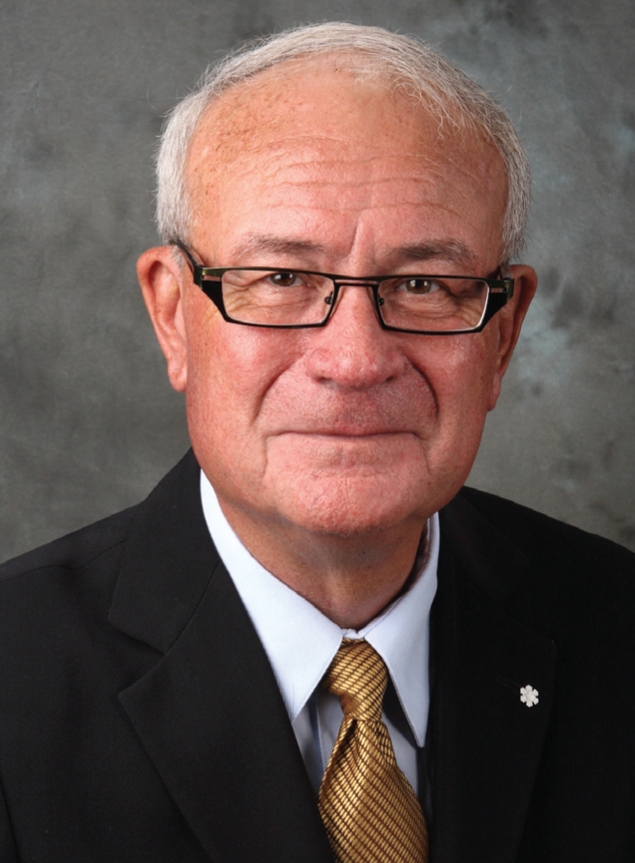
Dr Robert HA Haslam
One of my outstanding professors during my residency training was the father of paediatric neurology, Dr Frank Ford. Dr Ford firmly believed that if there was uncertainty of the diagnosis following a careful and thorough history and physical examination, it was unlikely that laboratory tests would be useful. Although my interaction with Dr Ford preceded the discovery of computed tomography and magnetic resonance imaging, he taught me how important a repeat history and physical examination is when unravelling the diagnosis in a difficult case. I believe that it remains the most important ‘test’ that we perform on our patients, in spite of the wide array of investigations and procedures that are currently available.
I will illustrate the value of repeating a history and physical examination when a diagnosis is not forthcoming, by citing a few examples in which, in fact, the diagnosis became evident during the course of professor’s rounds, with important input from the resident staff.
A family from Africa had recently immigrated to Canada. Several months later, their two-year-old child became irritable and anorectic; weight loss was significant. She became febrile, developed chronic diarrhea and became malnourished. A physical examination showed generalized lymphadenopathy, and the tip of her spleen was palpable. On review of the family history, we learned that the family lived in an area where AIDS was endemic, and that several family members had died from HIV. The parents did not inform the immigration authorities that one of them had acquired the disease fearing that they may be denied entry into Canada. The diagnosis of HIV was confirmed in their daughter, and the patient and her parents were followed by the infectious disease team.
A two-and-a-half-year-old boy with a cerebral palsy diagnosis was followed at another centre. He was the second child of unrelated parents. The pregnancy was planned, and the mother’s health was normal during the full-term gestation. The labour was normal, and the child’s birth weight was 3.5 kg. The Apgar score was 9 at 1 min, and 9 at 5 min. The infant breastfed normally and was discharged following a normal examination. The child’s early developmental milestones were normal in the language and social spheres, but the gross motor skills were delayed in that he did not sit up until after his first birthday and pulled to a stand with difficulty approximately six months later. A multidisciplinary team made the cerebral palsy diagnosis and referred him to a physical and occupational therapist for ongoing therapy. Because of concern by the therapists that the child had made no gross or fine motor gains in the following nine months, he was referred for additional evaluation. During professor’s rounds, it was clear that the mother never believed that her child was normal in gross motor development and, in fact, was concerned that he had lost skills during the past few months because he was no longer able to stand unassisted and was still not walking. She also noted that he experienced constant urinary dribbling and, on questioning, had never observed him to have an erection. The examination confirmed that the patient’s language skills were advanced. He was very attentive and interactive. The general physical examination was normal. The child’s tone was reduced in all extremities. His sitting posture was stooped, and he was unable to pull to a stand or bear weight on his legs. The cranial nerve examination was normal, including the optic nerves and surrounding retina. The deep tendon reflexes were absent in the upper extremities and markedly increased in the legs. The plantar responses were extensor bilaterally. A sensory examination, including light touch and pain, suggested a loss of sensation above the mid-cervical area. Imaging studies showed a tumour in the C5-C6 area and, at operation, an astrocytoma was partially removed.
A three-month-old infant was referred from another province for a magnetic resonance imaging scan of the head. The pregnancy and delivery history were normal. The Apgar scores were 7 at 1 min, and 8 at 5 min. The infant was moved to the intensive care nursery on the day of birth because of difficulty in maintaining a normal body temperature. The newborn screening tests for hypothyroidism and phenylketonuria were reported to be normal. Following discharge, the infant fed poorly and had significant constipation. The parent’s placed the child between them at night to manage ongoing hypothermia. Several emergency admissions to hospital were necessary because of bradycardia. On one occasion, the patient experienced a brief cardiac arrest that was treated successfully. A comprehensive series of metabolic and imaging studies were normal. Magnetic resonance imaging was not available; thus, the infant was subsequently transferred by air ambulance to our hospital for the study. On admission, when examined during teaching rounds, the diagnosis was obvious. The child had mottled skin, a large umbilical hernia and a characteristic hoarse cry. Laboratory studies confirmed the diagnosis of hypothyroidism. The patient was immediately started on thyroid replacement therapy and, within 10 days, showed marked improvement in alertness, body tone and temperature, and appetite. It was later determined that a technician working in the provincial laboratory fudged the results of the newborn thyroid screen, so he/she could take an early lunch. The referring paediatrician never considered hypothyroidism as a possibility because of the normal screening test. The infant did not undergo a magnetic resonance imaging scan! Follow-up showed that the patient had moderate cognitive delay and required educational assistance. The family successfully sued the provincial government to provide funding for lifelong support of their daughter.
The actual cases summarized above highlight several critical points encountered in the assessment and management of ‘difficult’ clinical problems. A repeat family history can be extremely helpful as documented in the first case. When an inborn error of metabolism or metabolic disorder is under consideration, remember that families do not like to spontaneously talk about family members who have been institutionalized with a cognitive or physical problem, or infants and children who have died suddenly. Never underestimate the concerns of a parent. As noted in the second case, the mother was very concerned that her son had a progressive disorder, in spite of reassurance from several physicians that he only had a ‘mild case of cerebral palsy’. Each patient demonstrated the importance of a careful reexamination. Finally, in cases in which the history and physical examination is definitely pointing to a definitive diagnosis, repeat the laboratory test that seems to be getting in the way of the correct diagnosis.
BIOGRAPHICAL NOTE: ROBERT HA HASLAM
Dr Haslam grew up in Saskatoon and attended the University of Saskatchewan (Saskatoon, Saskatchewan), receiving his medical degree in 1960. Following a rotating internship, he briefly practiced family medicine in rural Saskatchewan.
In 1962, Dr Haslam was accepted into the Johns Hopkins paediatric training program (Maryland, USA). During his fifth year, he was chosen by Professor Robert Cooke to be the Chief Resident, overseeing the educational and clinical experiences for the medical students and residents. In 1967, he began a fellowship (1967–1970) in child neurology at the University of Kentucky (Kentucky, USA) with the late Professor David B Clark, a world renowned academic child neurologist.
His first academic position was as Director of the John F Kennedy Institute (Maryland, USA), a facility affiliated with Johns Hopkins Hospital. The Institute fosters teaching, education, clinical care and research in infants and children with developmental disabilities in a multidisciplinary setting. While there, Dr Haslam assisted with the introduction of child neurology into the developmental disabilities paediatric training program (1970–1975). Then, he became Head of Pediatrics (1975–1986) at the University of Calgary and The Alberta Children’s Hospital (Calgary, Alberta), assisting in the development of a vibrant academic paediatric program. Following this, he moved to Toronto, Ontario, to become Professor and Chairman of the Department of Paediatrics, University of Toronto and Paediatrician-in-Chief at The Hospital for Sick Children (1986–1996). During this challenging but rewarding time, he cochaired the undergraduate curriculum renewal committee, assisted in the decentralization of paediatrics in metropolitan Toronto, established Canada’s first academic alternative funding program, and initiated the fostering of a clinician scientist research training program. After returning to Calgary, he served as a mentor (2000–2008) to child neurology residents in the outpatient longitudinal clinic at the Alberta Children’s Hospital. During Dr Haslam’s career, he volunteered on many boards and foundations, and has conducted research programs focused on the developmentally disabled child. He had been honoured by receiving the Ross Award (2002), The AAP Capute Award (2003) and the Order of Canada (CM) (2007).