Abstract
Measles virus (MV) is the causative agent for acute measles and subacute sclerosing panencephalitis (SSPE). Although numerous mutations have been found in the MV genome of SSPE strains, the mutations responsible for the neurovirulence have not been determined. We previously reported that the SSPE Osaka-2 strain but not the wild-type strains of MV induced acute encephalopathy when they were inoculated intracerebrally into 3-week-old hamsters. The recombinant MV system was adapted for the current study to identify the gene(s) responsible for neurovirulence in our hamster model. Recombinant viruses that contained envelope-associated genes from the Osaka-2 strain were generated on the IC323 wild-type MV background. The recombinant virus containing the M gene alone did not induce neurological disease, whereas the H gene partially contributed to neurovirulence. In sharp contrast, the recombinant virus containing the F gene alone induced lethal encephalopathy. This phenotype was related to the ability of the F protein to induce syncytium formation in Vero cells. Further study indicated that a single T461I substitution in the F protein was sufficient to transform the nonneuropathogenic wild-type MV into a lethal virus for hamsters.
Measles virus (MV) is a member of the Morbillivirus genus in the Paramyxoviridae family, and its genome is a nonsegmented single-stranded RNA of negative polarity. The MV genome contains N, P, M, F, H, and L genes. The genome is covered with the nucleocapsid (N) protein, which is transcribed from the N gene. The P gene encodes the phospho-protein (P), which forms the replicase complex with the large (L) polymerase protein encoded by the L gene. In addition to encoding the P protein, the P gene encodes accessory C and V proteins, which counteract antiviral host defense. The envelope of the virion consists of the two transmembrane glycoproteins, fusion (F) and hemagglutinin (H), which are transcribed from the F and H genes, respectively. The M gene encodes the matrix (M) protein, which associates with nucleocapsids and with the cytoplasmic regions of the F and H proteins (25). The signaling lymphocyte activation molecule (SLAM, also designated CD150) is the primary receptor for wild-type MV (22, 27, 69), and specific amino acid changes (N481Y or S546G) in the MV H protein present in some MV strains are required for interaction between the H protein and CD46 (23, 26, 39, 42, 46, 56, 62).
MV is the causative agent of measles and, on very rare occasions, causes subacute sclerosing panencephalitis (SSPE). SSPE is a fatal degenerative disease caused by persistent MV infection of the central nervous system (59). On rare occasions, MV has been isolated from brain cells of patients with SSPE by cocultivation with cell lines susceptible to MV (48). Genetic analyses revealed that these viruses derived from SSPE patients (SSPE strains) contain numerous mutations, and the existence of characteristic or frequently found mutations common to SSPE strains was suggested (3, 14). The M gene of SSPE strains seems particularly vulnerable to mutation, and its expression is restricted. In many SSPE strains, an A-to-G-biased hypermutation occurred in the genome and destroyed the M protein-coding frames. In some cases, translation of the M protein is complicated by a transcriptional defect that leads to an almost exclusive synthesis of dicistronic P-M mRNA (4, 12, 13, 61). Elucidation of the mechanism of the aberrant read-through transcription at the P-M gene junction revealed that a single deletion or mutation at the P gene end was responsible (5). Another characteristic change of the structural protein found in SSPE strains is located in the F protein (8, 16, 47, 57). A mutation in the termination codon of the F protein in some strains resulted in an elongated cytoplasmic domain. Another type of mutation created a premature termination codon in the F protein-coding frame and resulted in a shortened cytoplasmic domain. In some strains, including the SSPE Osaka-2 strain, one mutation did not change the length of the cytoplasmic domain, but multiple mutations in the reading frame caused nonconservative amino acid substitutions. Some of the mutations developed in this domain can be explained by the biased hypermutation that is more frequently and extensively found in the M gene, as mentioned above (15, 16). However, the effects of these hypermutations as well as of other sporadic mutations in the genome of SSPE strains have been poorly understood.
The ability to induce syncytia in Vero cells is one of the characteristic features of SSPE strains in vitro. Triggered by the binding of the H protein to the cellular receptor, the F protein plays a central role in virus-cell and cell-to-cell fusion. We previously demonstrated that the F protein of three SSPE strains (Osaka-1, Osaka-2, and Osaka-3 strains) induced syncytia in Vero cells when coexpressed with the H protein from any MV strain, including the wild-type MVs (6). In addition, the region responsible for the enhanced fusion was further narrowed to the extracellular domain of the F protein. Whether the enhanced fusion activity is related to the neurovirulence is still unclear.
MVs generally do not induce neurological disease in experimental small animals. Brain-adapted strains can replicate in newborn animals, but neurovirulence is reduced as the animal ages (1, 7, 9, 24). Transgenic mice expressing human CD46 or SLAM were established and observed for MV infection (19, 37, 45, 50, 52, 60). In a transgenic line which expresses SLAM ubiquitously (60), infection of the central nervous system was examined, and this revealed that MV could induce an acute neurological syndrome in mice less than 3 weeks old. In contrast, SSPE strains show strong neurovirulence and can induce lethal neurological disease in immunocompetent, genetically unaltered animals (2, 31, 32, 33, 64, 70). We previously reported that the SSPE Osaka-2 strain induced acute encephalopathy in 3-week-old hamsters several days after intracerebral inoculation (29). Therefore, it is important to analyze the differential roles of the mutations found in the genome of SSPE strains to understand the molecular mechanism of MV persistence in the brain and the pathogenesis of SSPE. Cathomen et al. described the role of mutations found in SSPE strains by adapting the recombinant MV system (10). In a similar system, Patterson et al. also described the role of the mutation in the M gene (52). These results indicated that expression of the defective M protein or defects in the cytoplasmic tail of the envelope glycoproteins resulted in the attenuation of MV neurovirulence in mice, but enhancing factors responsible for MV neurovirulence have not been determined. Because genetically unaltered young adult hamsters are highly susceptible to infection with SSPE strains, show clear symptoms, and tolerate infection for 2 or 3 days and because, practically, a higher titer of virus stock can be inoculated into brain, we have been using hamsters for the analyses of MV neurovirulence. The recombinant MV system (53, 58, 65) was adapted for the current study to identify the gene(s) responsible for neurovirulence in our hamster model.
We found that mutations in the F and H protein-coding genes of the SSPE Osaka-2 strain were responsible for neurovirulence. Further investigation demonstrated that a single amino acid substitution in the F protein transformed the nonneuropathogenic wild-type MV IC323 strain into a lethal virus similar to the SSPE Osaka-2 strain in hamsters.
MATERIALS AND METHODS
Cells and viruses.
CHO cells expressing CD46 (CHO/CD46) cells (a gift of Y. Murakami, Department of Pharmaceutical Sciences, Hokkaido University) (30) were cultured in RPMI 1640 medium (Nissui Pharmaceutical Co., Tokyo, Japan) supplemented with 5% heat-inactivated fetal calf serum (FCS) and 0.7 mg of hygromycin (Nacalai Tesque, Tokyo, Japan) per ml. CHO/SLAM and Vero/SLAM cells (gifts of Y. Yanagi, Department of Virology, Graduate School of Medical Sciences, Kyushu University) (51, 69) were cultured in Dulbecco's modified Eagle's medium (DMEM; Nissui Pharmaceutical) supplemented with 10% FCS and 0.5 mg of G418 (Nacalai Tesque) per ml. B95a cells (34) were grown in RPMI 1640 medium supplemented with 10% FCS. Vero, 293T, CHO, IMR-32, SK-N-SH, A172, and U-251 cells (63) were cultured in DMEM supplemented with 10% FCS.
Isolation of sibling viruses (Osaka-2/FrV and Osaka-2/FrB) of the SSPE Osaka-2 strain was described previously (48). These sibling viruses were neurovirulent when they were injected into hamster brains (29).
Plasmids.
Plasmids were cloned in the mammalian expression vector pME18S, which contained genes encoding the entire F or H protein region, as described previously (6). Plasmids encoding chimeric F proteins, OSA2-ext and OSA2-cyt, were previously described as Osa2/Toy and Mas/Osa2, respectively (6). The nucleotide sequences of the M, F, and H genes of the Osaka-2 strain were reported previously (4, 23, 47). Plasmids encoding mutant F proteins were constructed by a PCR-based method with synthetic primer pairs, and the inserts were confirmed by sequencing. Plasmids encoding the mutant MV genome were based on p(+)MV323, which encodes the antigenomic full-length cDNA of the wild-type IC-B strain (65). For construction of plasmids to prepare recombinant viruses containing the M gene, SalI and BstEII sites located in the 3′ noncoding regions of the P and M genes, respectively, were used for cloning. Because the BstEII site is found in the coding region but not in the 3′ noncoding region of the M gene of the Osaka-2 strain, synthetic primers were used to destroy the BstEII site in the coding region (5′-3997CCTTCAACCTGCTAGTGACC4016-3′ and 5′-4016GGTCACTAGCAGGTTGAAGG3997-3′) and to create a BstEII site in the 3′ noncoding region (5′-4820GCGGTTGGGTCACCTCGACCGC4799-3′). To construct plasmids for preparation of recombinant viruses containing the F and H genes, BsmBI and SpeI sites and SpeI and PacI sites were used for cloning, respectively.
Transient expression of F and H glycoproteins and indirect immunofluorescence microscopy.
Cells grown on Sonic seal slide wells (Nalge Nunc International, Rochester, NY) were transfected with 0.5 μg of F-containing plasmids and 0.5 μg of H-containing plasmids per well using Lipofectamine LTX transfection reagent (Invitrogen, Carlsbad, CA) according to the manufacturer's protocol.
Transfected cells were incubated at 35°C for 24 h. The cells were washed once with phosphate-buffered saline (pH 7.4), air dried, and fixed with a 1:1 acetone-methanol mixture at room temperature for 2 min. Monoclonal antibody to the H protein was used for the immunofluorescent staining as described previously (6).
Preparation of recombinant MV that expresses proteins derived from SSPE.
Recombinant MVs were generated from cDNAs by using CHO/SLAM cells and the vaccinia virus carrying T7 RNA polymerase, vTF7-3, according to a previously described procedure (66). To obtain cell-free virus stock, infected B95a cells were treated with cytochalasin D (CD; Sigma, St. Louis, MO) as previously described (28), and the resulting virus-like particles (CD-VLP) were stored at −85°C. Infectivity titers of the virus stock were determined by determining the number of PFU in B95a cells or in other cell lines such as Vero or Vero/SLAM cells.
Virus inoculation into and recovery from hamsters and histopathological examination.
Three-week-old female Syrian golden hamsters (SLC-Japan, Shizuoka, Japan) were gently anesthetized with ether, and 50 μl of properly diluted CD-VLP stock was inoculated into the right hemisphere of the brain. Selected hamsters from each group were sacrificed, and the brains were removed and subjected to either virus rescue or histopathological examination. Virus was recovered from brain cells by cocultivation with B95a cells. Total cellular RNA was extracted from the recovered virus-infected cells or directly from the brain according to a previously described method (4). The RNA was reverse transcribed, and the regions containing the M, F, or H gene were amplified by PCR and sequenced. Removed whole brains were fixed with 10% formalin (Wako Pure Chemicals, Osaka, Japan). Sections were prepared and stained with hematoxylin and eosin and evaluated histopathologically. All animal experiments were performed according to the Guide for Animal Experimentation, Osaka City University, in the infected-animal room at the biosafety level 2 Laboratory Center of the Medical School.
RESULTS
The M gene of the SSPE Osaka-2 strain is not a determinant of neurovirulence.
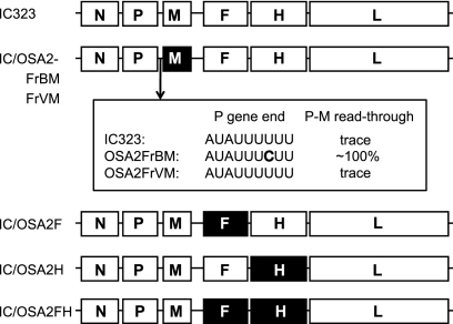
Many mutations are found in the MV genome from SSPE strains, and the M gene is the most affected. To test the consequences of M gene mutation on neurovirulence in our hamster model, we generated recombinant viruses in which the M gene of the IC323 strain was replaced with that of the SSPE Osaka-2 strain (Fig. 1). Because the difference in the P gene end sequences of the two sibling viruses (Osaka-2/FrB and Osaka-2/FrV) of the strain (29) affected transcription of the M gene (5), the P gene end region of each sibling virus was included in the constructs. The resulting recombinant IC/OSA2FrBM virus produces almost exclusively dicistronic P-M mRNA, whereas the IC/OSA2FrVM virus produces monocistronic P and M mRNAs.
FIG. 1.
Schematic diagram of the genome of the recombinant MVs. The protein-coding regions (N, P, M, F, H, and L) of the IC323 strain are shown as open boxes. The M, F, and H protein-coding regions derived from the Osaka-2 strain are shown as filled boxes. The sequence of the P gene end of the two sibling viruses (OSA2FrBM and OSA2FrVM) of the Osaka-2 strain and the extents of the transcriptional read-through at the P-M gene junction are indicated.
The recombinant viruses were propagated on B95a cells, and CD-VLP were prepared and stored as described in Materials and Methods. The CD-VLP stock was titrated on B95a cells (Fig. 2 A). The titers of the recombinant viruses containing the M gene derived from both sibling viruses were similar when viruses were titrated in B95a cells. The CD-VLP stock was also titrated on Vero/SLAM cells, and the titers were higher than in B95a cells and comparable between the two viruses. In contrast, no detectable cytopathic effects such as syncytium formation were observed in CD-VLP-infected Vero cells.
FIG. 2.
(A) Plaque titration of the CD-VLP of recombinant MV containing the M gene of either OSA2FrV (IC/OSA2FrVM; black bars) or OSA2FrB (IC/OSA2FrBM; gray bars). Three different kinds of cell lines were used for the titration. The means ± standard deviations for triplicate samples are shown. (B and C) Growth kinetics of the recombinant MVs containing the M gene. B95a cells were infected with each recombinant MV at a multiplicity of infection of 0.01 PFU/cell. At indicated time points, culture fluids were collected, an equal volume of medium was added, and cells were frozen and thawed once, clarified by centrifugation, and then titrated. Filled circles, IC323; open triangles, IC/OSA2FrBM; open circles, IC/OSA2FrVM. Data shown are for infectious cell-free viruses released into the culture medium (B) and cell-associated viruses prepared by the freezing and thawing of infected cells (C).
Infectious cell-free virus production was assayed by B95a cell culture (Fig. 2B and C). Wild-type IC323 virus produced significant amounts of cell-free virus particles, whereas the IC/OSA2FrBM and IC/OSA2FrVM viruses produced minimum amounts of both the extracellular and intracellular cell-free virus particles. This suggested that the defective expression of the Osaka-2 M gene affected virus particle production, probably during the budding process, which was demonstrated by another nonproductive SSPE strain (52).
The recombinant IC/OSA2FrBM and IC/OSA2FrVM viruses were inoculated intracerebrally into 3-week-old hamsters, and neurovirulence was evaluated (Table 1). No hamster developed any neurological signs during the course of observation. In the same manner, no hamster that was inoculated with the IC323 virus (850 PFU/brain or 11,500 PFU/brain) showed any symptoms during the course of observation.
TABLE 1.
Inocula of recombinant viruses containing the M gene of the SSPE Osaka-2 strain and neurovirulence in hamsters
| Virus | Expt no. | Titer (PFU) by cell typea |
Incidence of diseaseb | |
|---|---|---|---|---|
| Vero | B95a | |||
| IC323 | 1 | <0.5 | 850 | 0/6 |
| 2 | <0.5 | 11,500 | 0/4 | |
| OSA2FrVM | 1 | <0.5 | 3,150 | 0/5 |
| OSA2FrBM | 1 | <0.5 | 2,250 | 0/5 |
Titer (PFU) of the inoculated virus (50 μl/brain) was determined either on Vero cells or on B95a cells.
Number of diseased hamsters/number of inoculated hamsters. Hamsters showed no symptoms of disease onset after intracerebral inoculation until the time of death or sacrifice at the terminal stage, with one exception. Hamster 216 died 16 days after intracerebral inoculation without any neurological symptoms except for continuous weight loss, and the death was considered unrelated to the recombinant virus infection.
Recombinant MV containing the F or H gene of the SSPE Osaka-2 strain was defective in infectious cell-free virus production.
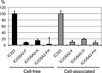
Genes coding for the two envelope glycoproteins are also frequently mutated in most SSPE strains though the extent of the mutation occurring in the F and H genes is less significant than that occurring in the M gene. To ascertain the effects of the mutations developed in the F and H genes on the functions of their proteins, we prepared recombinant viruses containing either the F or H gene or both genes of the SSPE Osaka-2/FrV strain (Fig. 1). The recombinant viruses were prepared by a method similar to that described above for the viruses containing the M gene. B95a cells were infected with the CD-VLP stock, and the culture medium was harvested at 72 h p.i. and titrated to quantify infectious cell-free virus production (Fig. 3). The production level of virus carrying the F or H gene (IC/OSA2F and IC/OSA2H, respectively) was 8.3% ± 2.4% or 14.7% ± 4.6%, respectively, of that of the parental IC323 virus. Cell-free virus production by the IC/OSA2FH virus, the F and H double mutant, was further restricted and decreased to 3.3% ± 20.6% of that produced by the IC323 virus. The yield of intracellular viruses prepared by freezing and thawing of infected cells was also low (Fig. 3). The results indicated that the amino acid substitutions developed in the Osaka-2 F and H proteins affected virus particle production.
FIG. 3.
Infectious cell-free virus production by the recombinant MVs. B95a cells were infected with each recombinant MV at a multiplicity of infection of 0.01 PFU/cell. At 72 h p.i., the virus released into the culture fluid (cell-free) or the cell-associated virus prepared by the freezing and thawing of infected cells (cell-associated) was titrated on B95a cells. The virus titer in cells infected with the IC323 strain was set to 100%. Bars indicate the means with standard deviations for triplicate samples.
Recombinant MV containing the F gene of the SSPE Osaka-2 strain infects cell lines of neural origin.
To determine whether the recombinant viruses containing the F or H gene of the Osaka-2 strain have different cell tropisms, a series of cell lines of different origins were used, including cell lines of neural origin, IMR-32 and SK-N-SH from human neuroblastoma, A172 from human oligodendroglioma, and U-251 from human astrocytoma (Table 2). As expected, all the viruses infected SLAM-positive cell lines such as B95a and CHO/SLAM cells and induced syncytia, whereas no virus infected CD46-positive CHO/CD46 cells. Recombinant viruses containing the F gene of the Osaka-2 strain (IC/OSA2FH and IC/OSA2F) infected Vero, IMR-32, and SK-N-SH cell lines and induced syncytia. A172 cells were also positive, but U-251 cells were negative by the immunofluorescence test. In contrast, recombinant viruses containing the H gene of the Osaka-2 strain (IC/OSA2H) had a similar cell tropism to the wild-type IC323 virus. These results suggest that the F protein of the Osaka-2 strain played a key role in the tropism and syncytium formation of some types of cells of neural origin.
TABLE 2.
Susceptibility of various cell lines to recombinant MV
| Cell line | Detection of virusa |
|||||||
|---|---|---|---|---|---|---|---|---|
| IC323 |
IC/OSA2FH |
IC/OSA2F |
IC/OSA2H |
|||||
| Syncytia | IFA | Syncytia | IFA | Syncytia | IFA | Syncytia | IFA | |
| B95a | + | + | + | + | + | + | + | + |
| Vero | − | + | + | + | + | + | − | − |
| IMR-32 | − | − | + | + | + | + | − | − |
| SK-N-SH | − | + | + | + | + | + | − | − |
| A172 | − | − | − | + | − | + | − | − |
| U-251 | − | − | − | − | − | − | − | − |
| CHO/CD46 | − | − | − | − | − | − | − | − |
| CHO/SLAM | + | + | + | + | + | + | + | + |
Determined by the presence (+) or absence (−) of syncytium formation and of immunofluorescent staining. IFA, immunofluorescence assay.
The F and H genes of the SSPE Osaka-2 strain are determinants of neurovirulence.
To test the neurovirulence of the recombinant viruses, IC323-based viruses containing either the F or H gene or both genes of the SSPE Osaka-2 strain (IC/OSA2F, IC/OSA2H, or IC/OSA2FH, respectively) were inoculated into hamster brains (Table 3). All the hamsters inoculated with the IC/OSA2FH virus (4,250 PFU/brain as assayed by B95a cells) developed neurological signs such as hyperactivity and seizures 3 or 4 days postinfection (p.i.). The symptoms observed in the hamsters were similar to those of animals inoculated with the parental Osaka-2 strain. All of these hamsters died or became moribund within 2 days after the onset of symptoms. The hamsters inoculated with the IC/OSA2F virus (2,450 PFU/brain as assayed by B95a cells) also showed neurological signs within 4 days p.i. and died or became moribund within 3 days after the onset. Smaller amounts of the IC/OSA2F virus (245 or 25 PFU/brain as assayed by B95a cells) induced the disease in three out of four hamsters, but one or two of the hamsters recovered.
TABLE 3.
Inocula of recombinant viruses containing the F or H gene of the SSPE Osaka-2 strain and neurovirulence in hamsters
| Virus | Expt no. | Titer (PFU) by cell typea |
No. of days to onset of disease (survival time [days])b | Incidence of diseasec | |
|---|---|---|---|---|---|
| Vero | B95a | ||||
| IC/OSA2FH | 1 | 25 | 4,250 | 3 (2), 3 (2), 3 (2*), 4 (1*), 4 (2*), 4 (2*) | 6/6 |
| IC/OSA2F | 1 | 150 | 2,450 | 3 (2*), 3 (2*), 3 (3), 3 (3*), 3 (3*), 4 (2) | 6/6 |
| 2 | 150 | 2,450 | 3 (3), 3 (3*) | 2/2 | |
| 15 | 245 | 5 (2), 5 (2*), 20 (−), − (−) | 3/4 | ||
| 1.5 | 25 | 5 (121), 6 (−), 7 (3), − (−) | 3/4 | ||
| IC/OSA2H | 1 | <0.5 | 750 | 9 (8*), 26 (1*), − (−), − (−), − (−),− (−) | 2/6 |
| 2 | <0.5 | 2,900 | 7 (4*), 11 (3*), − (−), − (−) | 2/4 | |
| <0.05 | 290 | 15 (3), − (−), − (−), − (−) | 1/4 | ||
| <0.005 | 29 | 23 (3), − (−), − (−), − (−) | 1/4 | ||
| IC323 | 1 | 2,000 | − (−), − (−), − (−), − (−), − (−), − (−) | 0/6 | |
| IC/F:OSA2F | 1 | 122 | 2,000 | 3 (3*), 3 (3*) | 2/2 |
| IC/F:OSA2ext | 1 | 12 | 2,000 | 3 (1), 3 (2), 3 (2), 4 (2) | 4/4 |
| IC/F:OSA2cyt | 1 | 0.16 | 2,000 | − (−), − (−), − (−), − (−) | 0/4 |
| 2 | 2.0 | 25,000 | − (−), − (−), − (−), − (−)d | 0/4 | |
| IC/F:L197I | 1 | 0.1 | 2,000 | − (−), − (−), − (−), − (−) | 0/4 |
| IC/F:Y442D | 1 | 5 | 2,000 | 4 (−), 4 (−), 4 (−), 5 (41), 5 (−),− (−) | 5/6 |
| IC/F:T461I | 1 | 110 | 2,000 | 4 (2*), 4 (4), 4 (5), 4 (−), 5 (1*), 5 (4) | 6/6 |
| IC/F:E478G | 1 | <0.01 | 2,000 | − (−), − (−), − (−), − (−) | 0/4 |
The titer of the inoculated virus (50 μl/brain) was determined in either Vero cells or B95a cells.
Number of days from intracerebral inoculation to the onset of the disease is shown (−, no symptoms). In parentheses, the number of days from onset of disease to death or sacrifice at the terminal stage, indicated by an asterisk, is shown (−, animal did not die).
Number of diseased hamsters/number of inoculated hamsters.
Hamster 248 died 21 days after intracerebral inoculation without any neurological symptoms, and the virus was not recovered; the death was considered unrelated to the recombinant virus infection.
The hamsters inoculated with the IC/OSA2H virus (2,900 PFU/brain as assayed by B95a cells) also showed neurological signs, and two of the four hamsters died; the minimum amount of the IC/OSA2H virus that could kill a hamster was 29 PFU/brain. However, the incidence of neurovirulence was much lower than that of the hamsters inoculated with the IC/OSA2F virus, and the incubation period was longer and varied.
Recombinant MVs containing the envelope genes of the SSPE Osaka-2 strain affected pyramidal cells in fields CA1 through CA3 of the hippocampus.
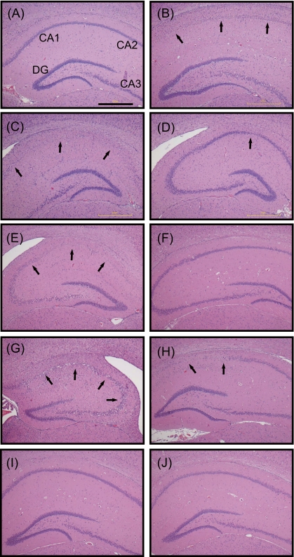
Some of the hamsters inoculated with the recombinant MVs were sacrificed on the verge of death or at the endpoint of the observation, and hematoxylin and eosin-stained sections of the brains were prepared (Fig. 4 ). Hamsters inoculated with the IC323 virus had no lesions throughout the brain (Fig. 4A), whereas severe lesions were found in fields CA1 through CA3 of the hippocampi of hamsters inoculated with the IC/OSA2FH (Fig. 4B) or IC/OSA2F (Fig. 4C) virus. The regions were equally affected in both hemispheres, and the pyramidal cells showed necrosis. The hamsters inoculated with IC/OSA2H (Fig. 4D) that developed neurological signs and were sacrificed showed minimal lesions with necrotic neurons in the CA1 field of the hippocampus. These histopathological observations and the incidence of neurovirulence indicate the relative importance of the F gene for the effective spread in the brain.
FIG. 4.
Histopathological findings of the hippocampus. Neuronal loss and degenerated neurons were observed with different extents of severity in fields CA1 through CA3 of hamsters inoculated with the IC/OSA2FH, IC/OSA2F, IC/OSA2H, IC/F:OSA2F-ext, IC/F:T461I, and IC/F:Y442D (B to E, G, and H), whereas no lesion was detected in the brain of the hamsters inoculated with the wild-type IC323, IC/F:OSA2-cyt, IC/F:L197I, or IC/F:E478G virus (A, F, I, and J). In hamsters inoculated with the IC/OSA2H virus (D), neuronal damage was restricted to the CA1 field of the hippocampus. Proliferation and activation of astroglia and microglia were also noted in the affected areas. Inoculated viruses were the following: IC323 (hamster 262; showed no symptoms and was sacrificed at 70 days p.i.) (A); IC/OSA2FH (hamster 290; showed symptoms at 4 days p.i. and was sacrificed at 6 days p.i.) (B); IC/OSA2F (hamster 176; showed symptoms at 3 days p.i. and was sacrificed at 6 days p.i.) (C); IC/OSA2H (hamster 187; showed symptoms at 7 days p.i. and was sacrificed at 12 days p.i.) (D); IC/F:OSA2-ext (hamster 237; showed symptoms at 3 days p.i. and died at 5 days p.i.) (E); IC/F:OSA2-cyt (hamster 245; showed no symptoms and was sacrificed at 280 days p.i.) (F); IC/F:T461I (hamster 275; showed symptoms at 5 days p.i. and was sacrificed at 6 days p.i.) (G); IC/F:Y442D (hamster 268; showed symptoms at 4 days p.i. and was sacrificed at 168 days p.i.) (H); IC/F:L197I (hamster 258; showed no symptoms and was sacrificed at 70 days p.i.) (I); IC/F:E478G (hamster 278; showed no symptoms and was sacrificed at 70 days p.i.) (J). CA1, CA2, CA3, and DG (dentate gyrus) fields are indicated in panel A. Scale bar, 500 μm. Panels A, B, and G to J show the hippocampus area of the left hemisphere, and panels C to F show the hippocampus area of the right hemisphere of the brain. The arrows indicate the abnormal appearance of fields CA1 through CA3 due to the necrosis of pyramidal cells.
The extracellular domain of the F protein is responsible for neurovirulence in hamsters.
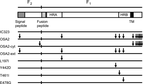
We previously demonstrated syncytium formation in Vero cells when the F protein from the SSPE strain was coexpressed with the H protein from any MV strain (6). The region of the F protein responsible for syncytium formation was assigned principally to the extracellular domain though the cytoplasmic domain also played a role to some extent in the enhanced fusogenic activity. To investigate the differential effects of the amino acid substitutions in each domain in the infected cells, additional recombinant viruses were prepared. Recombinant viruses (IC/F:OSA2-ext and IC/F:OSA2-cyt) were recovered (Fig. 5) from full-length plasmids containing a chimeric F gene either of the extracellular or cytoplasmic domain, respectively, of the Osaka-2 strain. The IC/F:OSA2-ext virus infected and induced syncytia in Vero cells at a rate similar to that of the IC/OSA2F virus, whereas the IC/F:OSA2-cyt virus did not infect Vero cells (data not shown). Both the IC/F:OSA2-ext and IC/F:OSA2-cyt viruses produced infectious cell-free virus at approximately 5% of the IC323 virus level (data not shown).
FIG. 5.
Schematic diagram of the MV F protein and constructs. HRA and HRB, two highly conserved heptad repeat domains, are shown; regions of signal peptide and fusion peptide are indicated as shaded boxes. TM, transmembrane domain (black box). Arrows indicate amino acids different from the those of the IC323 strain. For the F and H coexpression experiments by transfection, the fragments were inserted into a eukaryotic expression vector, pME18S. For the recombinant MV constructions, plasmids were prepared by replacing the whole F gene fragment from the basic plasmid containing the IC323 viral genome.
The IC/F:OSA2-ext and IC/F:OSA2-cyt viruses were inoculated into hamster brains at a titer of 2,000 PFU per brain (Table 3). Hamsters inoculated with the IC/F:OSA2-ext virus developed neurological signs such as general seizures 3 or 4 days p.i. and died 1 or 2 days after the onset of seizures, whereas hamsters inoculated with the IC/F:OSA2-cyt virus showed no symptoms. In a further study, four hamsters that were inoculated with the larger amount of the IC/F:OSA2-cyt virus (25,000 PFU per brain) did not develop the disease. Therefore, the extracellular domain of the F protein of the Osaka-2 strain is responsible for neurovirulence in hamsters.
A single amino acid substitution in the extracellular domain of the F protein is responsible for enhanced fusogenicity in Vero cells and neurovirulence in hamsters.
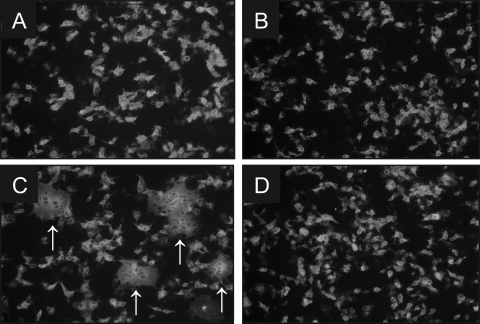
Because I10V, an isoleucine-to-valine substitution at amino acid 10 in the N-terminal region of the F protein, is included in the signal peptide that is deleted after synthesis, only four amino acids, L197I, Y442D, T461I, and E478G, are different between the IC323 and the Osaka-2 strains in the extracellular domain of the F protein. We then prepared four additional mutant plasmids that expressed the F protein with a single amino acid substitution. The resulting F plasmids were individually coexpressed with the IC323 H protein in B95a or Vero cells, and the ability to induce syncytium formation (Fig. 6) was examined. As expected, all four mutants induced syncytia in B95a cells within 24 h posttransfection, indicating that these F mutant proteins were functionally expressed on the cell surface (data not shown). On the other hand, only the T461I mutant efficiently induced syncytia in Vero cells at 24 h posttransfection (Fig. 6C). The Y442D mutant also induced syncytia, but it took 72 h or more to make small ones (data not shown).
FIG. 6.
Syncytium formation induced by a different kind of mutant F protein. Vero cells were cotransfected with plasmid DNA encoding the H and F proteins of MV. The H gene was derived from the IC323 strain while the F gene was from one of the mutant F genes. At 24 h p.i., cells were fixed and stained with a monoclonal antibody to the H protein and fluorescein isothiocyanate-conjugated anti-mouse antibody. (A) IC/F:L197I. (B) IC/F:Y442D. (C) IC/F:T461I. (D) IC/F:E478G. Arrows in panel C indicate syncytia formed in cells cotransfected with the F gene containing the T461I substitution. No syncytia were found in cells cotransfected with other F genes.
Four recombinant viruses containing the F gene that expressed the F protein with a single amino acid substitution found in the extracellular domain of the Osaka-2 F protein were recovered (Fig. 5) and inoculated into hamster brain. Hamsters inoculated with the virus containing the T461I F protein mutation (IC/F:T461I) developed neurological signs 4 or 5 days p.i. and died 4 or 5 days after the onset of signs (Table 3). Hamsters inoculated with the IC/F:Y442D virus also developed neurological signs 4 or 5 days p.i. but recovered and survived for at least 24 weeks. Two other mutant viruses (IC/F:L197I and IC/F:E478G virus) did not produce any neurological signs.
Some of the dead or dying hamsters were subjected to histopathological examination (Fig. 4). As found in the brains of hamsters inoculated with the IC/OSA2FH or IC/OSA2F virus, severe lesions were found around the CA1 field of the hippocampus of the brains from the hamsters inoculated with the IC/F:OSA2-ext or IC/F:T461I virus (Fig. 4E and G). Old lesions with neuronal loss and astrogliosis were documented in the CA1 field of the hippocampus of the brain from hamsters inoculated with the IC/F:Y442D virus (Fig. 4H). In contrast, no lesion was found in the brains of hamsters inoculated with the IC/F:OSA2-cyt, IC/F:L197I, or IC/F:E478G virus (Fig. 4F, I, and J). Therefore, the severity of the lesions correlated with neurovirulence.
DISCUSSION
SSPE strains of MV contain numerous mutations, especially in the M, F, and H genes, which result in structural alterations of the proteins encoded by these genes. It is important to ascertain whether these changes are simple accumulations during long-term persistence of the virus or changes necessary for effective virus spread in the brain. Because SLAM, the primary entry receptor for wild-type MV, is not expressed on neural cells and because basically SSPE strains do not use CD46 as an alternative receptor (28, 63), it is reasonable to speculate that changes or adaptations that use an unidentified receptor are introduced in the envelope proteins. The recent development of MV rescue system technology allowed us to identify the molecular determinant(s) of MV responsible for the phenotypic change in vitro and for neurovirulence. Cathomen et al. first described the role of mutations found in SSPE strains by adapting the recombinant MV system (10, 11). In a similar system, Patterson et al. also described the role of the mutation in the M gene (52). However, these experiments used a recombinant MV system based on the Edmonston strain (10, 11, 52) and CD46 transgenic mice (10, 52), and hence CD46 would be used as the receptor for the infection. Therefore, we generated recombinant viruses containing SSPE genes from a vector plasmid based on the wild-type MV IC323 strain (65). In this study, we analyzed the functional alterations of the envelope-associated proteins both in vitro and in vivo.
As previously reported (10, 52), recombinant MVs in which the M gene was deleted or replaced by the hypermutated M gene from SSPE strains were infectious but showed reduced productivity of the extracellular cell-free virion. We also generated infectious recombinant viruses containing the M gene of the Osaka-2 strain, and the virus-infected cells showed a nonproductive nature. Although the hypermutation of the M gene resulting in the defective expression of the M protein is the most characteristic feature in the genome of many SSPE strains, including the Osaka-2 strain, replacement of the M gene alone did not confer a neurovirulent phenotype in hamsters. Rather, it is possible to consider the M gene mutation as a contributing factor to attenuation. It should be noted that a matrix-less MV lost acute pathogenicity but penetrated more deeply into the brain parenchyma than the Edmonston MV in mice expressing the human receptor CD46 and defective for the interferon type I system (10); and the recombinant MV containing the biased hypermutated M gene of the Biken strain, from which the defective M protein was produced, induced prolonged infection occurring as long as 30 to 50 days after that caused by MV (52). We do not exclude the possibility that the defect of the M protein in the Osaka-2 strain makes some contribution to its persistence in the brain.
Experiments using the transient coexpression of the F and H proteins demonstrated that the extracellular domain of the F protein was responsible for efficient syncytium formation in SLAM-negative Vero cells as well as in cell lines expressing SLAM or CD46 (6). The region responsible for the enhanced fusogenicity was further narrowed to the level of a single amino acid in the current study. Substitution of a single amino acid (T461I) was sufficient for enhancement of fusion activity. This residue is located in the heptad repeat B (HRB) domain, which is adjacent to the transmembrane domain (20, 35). The HRB domain is thought to be involved in the conformational change during the fusion process to make a six-helix bundle structure with the heptad repeat A (HRA) domain, which is adjacent to the fusion peptide (reviewed in reference 36). Although fusogenic activity was lower than that caused by the T461I substitution, another amino acid change, Y442D, also induced syncytia in Vero cells. Doyle et al. (17) reported the alteration of fusogenic activity of mutant F proteins with amino acid substitutions at positions 94, 367, and 462. Okada et al. (49) also described the enhanced fusogenicity resulting from a single G464W substitution in the F protein, which was developed in a Vero cell-adapted wild-type T11 strain. These results suggested that the conformational change of the F protein due to an amino acid substitution at a different site could result in a similar enhancement of fusion activity. We previously described the effects of amino acid substitutions in the F protein from three SSPE strains (Osaka-1, Osaka-2, and Osaka-3) on syncytium formation in Vero cells. These three SSPE strains replicate and induce syncytia in Vero cells, and the SSPE F protein was responsible for syncytium formation. The T461I substitution was found in the F protein of the Osaka-3 strain as well as in the Osaka-2 strain. However, substitutions found in the F protein of the Osaka-1 strain do not overlap with those in the Osaka-2 strain. It is possible that other substitution(s) or combinations could induce similar conformational changes in the protein and could result in the same phenotype, i.e., enhanced fusion. We are currently preparing recombinant viruses containing the F and H genes from the Osaka-1 strain to verify the idea suggested by the results obtained from the analysis of the Osaka-2 strain.
A series of recombinant viruses containing mutations in the F gene were generated on the IC323 wild-type strain background. Expression of the F gene with a single T461I substitution in the F protein (IC/F:T461I virus) was sufficient to induce lethal encephalopathy in hamsters. Expression of the F gene with a Y442D substitution in the F protein (IC/F:Y442D virus) also induced severe neurological symptoms though these hamsters recovered quickly from the disease. Considering that the Y442D substitution induced syncytium formation less efficiently than the T461I substitution in the F and H cotransfection experiments, there is a good correlation between the extent of the fusion activity in vitro and the severity of neurovirulence in hamsters. As previously described (10, 11), MV bearing an F protein with a shortened cytoplasmic tail induced enhanced cell fusion. The virus lost acute pathogenicity but penetrated more deeply into the brain parenchyma than the Edmonston MV in the CD46-transgenic, interferon type I-deficient mice. The result indicated that defects in the cytoplasmic tail of the F protein resulted in attenuation of MV neurovirulence in mice. The role of the F gene in neurovirulence of other viruses has been reported. The F gene of rodent brain-adapted mumps virus was a major determinant of neurovirulence in neonatal Lewis rats (40). Lemon et al. showed that expression of the F gene alone of the neurovirulent strain was sufficient to induce significant levels of hydrocephalus in the animals. However, there was no correlation between fusogenicity and neurovirulence, and the mechanism whereby the mumps virus F protein modulates neurovirulence remains unknown. The direct correlation of enhanced fusogenicity and neurovirulence of the Osaka-2 strain will be evaluated by similar experiments with the Osaka-1 strain, the F protein of which has different substitutions, as discussed above.
In addition to the F protein, the H protein contributed to neurovirulence to some extent. Some (30 to 50%) of the hamsters inoculated with the recombinant virus expressing the H protein of the Osaka-2 strain (IC/OSA2H virus) developed the same neurological symptoms as those inoculated with the virus containing the Osaka-2 F gene (IC/OSA2F virus). The incidence was much lower in hamsters inoculated with the IC/OSA2H virus than in those inoculated with the IC/OSA2F virus, and the onset was delayed. This minor but definite contribution of the H gene to neurovirulence was masked by the major contribution of the F gene in the hamsters inoculated with the IC/OSA2FH virus. Further study will be needed in order to differentiate the role of the H protein in neurovirulence. Duprex et al. described the role of the H gene of the CAM/RB strain, a rodent brain-adapted MV, in neurovirulence in newborn mice (18). Further study revealed that the combination of two amino acid substitutions (R195G and N200S) in the stem 2 region of MV H protein determined neurovirulence (44). However, these two amino acid substitutions are unique to the CAM/RB strain and are not found in SSPE strains. Therefore, the role and mechanism of the Osaka-2 H protein in neurovirulence could be different. It should be noted that the recombinant virus containing the CAM/RB-H gene replicated in the brains of mice to a lesser extent than the parental CAM/RB virus, indicating the requirement of other genes for full neurovirulence. This is consistent with our result of minimum neurovirulence in the recombinant virus expressing the Osaka-2 H protein in hamsters. Further investigation is required to address the question of which amino acid substitution(s) is responsible for neurovirulence.
The mechanism of the restricted production of infectious cell-free virus by the SSPE F- and H-containing virus is largely unknown. It is possible that the interaction between the F and H proteins with the M protein is restricted because of the alteration of the cytoplasmic domain of the envelope glycoproteins. Cathomen et al. suggested the association of M with the cytoplasmic tails of the glycoproteins might negatively influence their fusion efficiency (11). However, the restricted production of infectious cell-free virus by the mutant F protein that contained only the extracellular domain of the Osaka-2 strain cannot be explained by this mechanism. Our preliminary data indicated that even a single substitution in the extracellular domain could greatly alter cell-free virus production (unpublished observations). The F protein is highly conserved, and only one or two amino acid differences are found among wild-type MV strains. This implies the elimination of viruses containing a mutant F protein that affects the production of infectious virions. It is possible that such mutations found in SSPE strains are allowed or selected in a specific environment such as the brain.
The mechanism of the spread of MV in the brain is poorly understood. A hypothesis of transsynaptic spread of MV in neurons without interaction between the H protein and the host cellular receptor was proposed (38). Further investigation suggested that neurokinin-1 (NK-1), a member of the neurotachykinin family of G protein-coupled neurotransmitter receptors, served as a receptor for the MV F protein (43). Because the fusion-inhibitory peptide (FIP; z-d-Phe-l-Phe-Gly) is known to inhibit fusion by paramyxovirus (54, 55) and because the tripeptide sequence is identical to the active site of substance P, a ligand for NK-1, it is possible that the SSPE F protein is more effective for microfusion at the synaptic cleft. However, it is equally possible that the virus spreads by microfusion via a specific interaction between the H protein and an unidentified host cellular receptor at the synapse. It should be noted that syncytium formation was observed in the CA1 field of one hamster brain that was inoculated with the Osaka-2 strain (29). In a study of MV spread in neurons using rat hippocampal slice culture (21), MV showed a retrograde spread from CA1 to CA3 pyramidal cells. This observation is consistent with the result we observed in the hippocampal region, which showed that the CA1 field was the most affected and that the CA2 and CA3 fields were also affected in severe cases. The apparent target region of the recombinant virus containing the SSPE H gene was also the CA1. Because binding of the H protein with the host cellular receptor is usually required for the induction of a conformational change in the F protein, the differential role of the H protein in virus spread in the brain should be clarified. To understand this, substitutions appearing in the SSPE H protein should be evaluated individually in relation to the interaction with the yet unidentified receptor(s) on Vero cells (5), epithelial cells (41, 67, 68), and neural cells, as well as the known receptors SLAM and CD46.
In conclusion, our findings indicate that the F gene of the Osaka-2 strain of MV is a major determinant of neurovirulence, regardless of whether a specific interaction between the H protein and the host cellular receptor is involved. A single amino acid substitution in the F protein is sufficient for neurovirulence. Studies of recombinant viruses containing the F gene with mutations found in different SSPE strains are necessary to understand the relationship between the increased fusogenicity and neurovirulence. In addition, identification of an unidentified alternative receptor in Vero and neural cells will provide new insights into the mechanism of MV spread in the brain and the pathogenesis of SSPE and open a way to develop a novel therapy.
Acknowledgments
We thank Y. Murakami and Y. Yanagi for providing cell lines and B. Moss for vaccinia virus vTF7-3. We also thank E. Nishiguchi and R. Tanaka for technical assistance and the staff of the Central Laboratory of Osaka City University Medical School for technical support.
This work was supported by a Grant-in-Aid for Scientific Research (number 19591216) from the Ministry of Education, Science, Sports, and Culture of Japan.
Footnotes
Published ahead of print on 18 August 2010.
REFERENCES
- 1.Albrecht, P., and H. P. Schumacher. 1971. Neurotropic properties of measles virus in hamsters and mice. J. Infect. Dis. 124:86-93. [DOI] [PubMed] [Google Scholar]
- 2.Albrecht, P., T. Burnstein, M. J. Klutch, H. T. Hicks, and F. A. Ennis. 1977. Subacute sclerosing panencephalitis: experimental infection in primates. Science 195:64-66. [DOI] [PubMed] [Google Scholar]
- 3.Ayata, M., A. Hirano, and T. C. Wong. 1989. Structural defect linked to nonrandom mutations in the matrix gene of Biken strain subacute sclerosing panencephalitis virus defined by cDNA cloning and expression of chimeric genes. J. Virol. 63:1162-1173. [DOI] [PMC free article] [PubMed] [Google Scholar]
- 4.Ayata, M., K. Hayashi, T. Seto, R. Murata, and H. Ogura. 1998. The matrix gene expression of subacute sclerosing panencephalitis (SSPE) virus (Osaka-1 strain): a comparison of two sibling viruses isolated from different lobes of an SSPE brain. Microbiol. Immunol. 42:773-780. [DOI] [PubMed] [Google Scholar]
- 5.Ayata, M., K. Komase, M. Shingai, I. Matsunaga, Y. Katayama, and T. C. Wong. 2002. Mutations affecting transcriptional termination in the P gene end of subacute sclerosing panencephalitis viruses. J. Virol. 76:13062-13068. [DOI] [PMC free article] [PubMed] [Google Scholar]
- 6.Ayata, M., M. Shingai, X. Ning, M. Matsumoto, T. Seya, S. Otani, T. Seto, S. Ohgimoto, and H. Ogura. 2007. Effect of the alterations in the fusion protein of measles virus isolated from brains of patients with subacute sclerosing panencephalitis on syncytium formation. Virus Res. 130:260-268. [DOI] [PubMed] [Google Scholar]
- 7.Baringer, J. R., and J. F. Griffith. 1970. Experimental measles virus encephalitis. A light, phase, fluorescence, and electron microscopic study. Lab. Invest. 23:335-346. [PubMed] [Google Scholar]
- 8.Billeter, M. A., R. Cattaneo, P. Spielhofer, K. Kaelin, M. Huber, A. Schmid, K. Baczko, and V. ter Meulen. 1994. Generation and properties of measles virus mutations typically associated with subacute sclerosing panencephalitis. Ann. N. Y. Acad. Sci. 724:367-377. [DOI] [PubMed] [Google Scholar]
- 9.Byington, D. P., and K. P. Johnson. 1972. Experimental subacute sclerosing panencephalitis in the hamster: correlation of age with chronic inclusion-cell encephalitis. J. Infect. Dis. 126:18-26. [DOI] [PubMed] [Google Scholar]
- 10.Cathomen, T., B. Mrkic, D. Spehner, R. Drillien, R. Naef, J. Pavlovic, A. Aguzzi, M. A. Billeter, and R. Cattaneo. 1998. A matrix-less measles virus is infectious and elicits extensive cell fusion: consequences for propagation in the brain. EMBO J. 17:3899-3908. [DOI] [PMC free article] [PubMed] [Google Scholar]
- 11.Cathomen, T., H. Y. Naim, and R. Cattaneo. 1998. Measles viruses with altered envelope protein cytoplasmic tails gain cell fusion competence. J. Virol. 72:1224-1234. [DOI] [PMC free article] [PubMed] [Google Scholar]
- 12.Cattaneo, R., A. Schmid, G. Rebmann, K. Baczko, V. ter Meulen, W. J. Bellini, S. Rozenblatt, and M. A. Billeter. 1986. Accumulated measles virus mutations in a case of subacute sclerosing panencephalitis: interrupted matrix protein reading frame and transcription alteration. Virology 154:97-107. [DOI] [PubMed] [Google Scholar]
- 13.Cattaneo, R., G. Rebmann, A. Schmid, K. Baczko, V. ter Meulen, and M. A. Billeter. 1987. Altered transcription of a defective measles virus genome derived from a diseased human brain. EMBO J. 6:681-688. [DOI] [PMC free article] [PubMed] [Google Scholar]
- 14.Cattaneo, R., A. Schmid, M. A. Billeter, R. D. Sheppard, and S. A. Udem. 1988. Multiple viral mutations rather than host factors cause defective measles virus gene expression in a subacute sclerosing panencephalitis cell line. J. Virol. 62:1388-1397. [DOI] [PMC free article] [PubMed] [Google Scholar]
- 15.Cattaneo, R., A. Schmid, D. Eschle, K. Baczko, V. ter Meulen, and M. A. Billeter. 1988. Biased hypermutation and other genetic changes in defective measles viruses in human brain infections. Cell 55:255-265. [DOI] [PMC free article] [PubMed] [Google Scholar]
- 16.Cattaneo, R., A. Schmid, P. Spielhofer, K. Kaelin, K. Baczko, V. ter Meulen, J. Pardowits, S. Flanagan, B. K. Rima, S. A. Udem, and M. A. Billeter. 1989. Mutated and hypermutated genes of persistent measles viruses which caused lethal human brain diseases. Virology 173:415-425. [DOI] [PubMed] [Google Scholar]
- 17.Doyle, J., A. Prussia, L. K. White, A. Sun, D. C. Liotta, J. P. Snyder, R. W. Compans, and R. K. Plemper. 2006. Two domains that control prefusion stability and transport competence of the measles virus fusion protein. J. Virol. 80:1524-1536. [DOI] [PMC free article] [PubMed] [Google Scholar]
- 18.Duprex, W. P., I. Duffy, S. McQuaid, L. Hamill, S. L. Cosby, M. A. Billeter, J. Schneider-Schaulies, V. ter Meulen, and B. K. Rima. 1999. The H gene of rodent brain-adapted measles virus confers neurovirulence to the Edmonston vaccine strain. J. Virol. 73:6916-6922. [DOI] [PMC free article] [PubMed] [Google Scholar]
- 19.Duprex, W. P., S. McQuaid, B. Roscic-Mrkic, R. Cattaneo, C. McCallister, and B. K. Rima. 2000. In vitro and in vivo infection of neural cells by a recombinant measles virus expressing enhanced green fluorescent protein. J. Virol. 74:7972-7979. [DOI] [PMC free article] [PubMed] [Google Scholar]
- 20.Eckert, D. M., and P. S. Kim. 2001. Mechanisms of viral membrane fusion and its inhibition. Annu. Rev. Biochem. 70:777-810. [DOI] [PubMed] [Google Scholar]
- 21.Ehrengruber, M. U., E. Ehler, M. A. Billeter, and H. Y. Naim. 2002. Measles virus spreads in rat hippocampal neurons by cell-to-cell contact and in a polarized fashion. J. Virol. 76:5720-5728. [DOI] [PMC free article] [PubMed] [Google Scholar]
- 22.Erlenhoefer, C., W. J. Wurzer, S. Löffler, S. Schneider-Schaulies, V. ter Meulen, and J. Schneider-Schaulies. 2001. CD150 (SLAM) is a receptor for measles virus but is not involved in viral contact-mediated proliferation inhibition. J. Virol. 75:4499-4505. [DOI] [PMC free article] [PubMed] [Google Scholar]
- 23.Furukawa, K., M. Ayata, M. Kimura, T. Seto, I. Matsunaga, R. Murata, T. Yamano, and H. Ogura. 2001. Hemadsorption expressed by cloned H genes from subacute sclerosing panencephalitis (SSPE) viruses and their possible progenitor measles viruses isolated in Osaka, Japan. Microbiol. Immunol. 45:59-68. [DOI] [PubMed] [Google Scholar]
- 24.Griffin, D. E., J. Mullinix, O. Narayan, and R. Johnson. 1974. Age dependence of viral expression: comparative pathogenesis of two rodent-adapted strains of measles virus in mice. Infect. Immun. 126:690-695. [DOI] [PMC free article] [PubMed] [Google Scholar]
- 25.Griffin, D. E. 2007. Measles virus, p. 1551-1585. In D. M. Knipe, P. M. Howley, D. E. Griffin, R. A. Lamb, M. A. Martin, B. Roizman, and S. E. Straus (ed.), Fields Virology, 5th ed. Lippincott Williams & Wilkins, Philadelphia, PA.
- 26.Hsu, E. C., F. Sarangi, C. Iorio, M. S. Sidhu, S. A. Udem, D. L. Dillehay, W. Xu, P. A. Rota, W. J. Bellini, and C. D. Richardson. 1998. A single amino acid change in the hemagglutinin protein of measles virus determines its ability to bind CD46 and reveals another receptor on marmoset B cells. J. Virol. 72:2905-2916. [DOI] [PMC free article] [PubMed] [Google Scholar]
- 27.Hsu, E. C., C. Iorio, F. Sarangi, A. A. Khine, and C. D. Richardson. 2001. CDw150(SLAM) is a receptor for a lymphotropic strain of measles virus and may account for the immunosuppressive properties of this virus. Virology 279:9-21. [DOI] [PubMed] [Google Scholar]
- 28.Ishida, H., M. Ayata, M. Shingai, I. Matsunaga, Y. Seto, Y. Katayama, N. Iritani, T. Seya, Y. Yanagi, O. Matsuoka, T. Yamano, and H. Ogura. 2004. Infection of different cell lines of neural origin with subacute sclerosing panencephalitis (SSPE) virus. Microbiol. Immunol. 48:277-287. [DOI] [PubMed] [Google Scholar]
- 29.Ito, N., M. Ayata, M. Shingai, K. Furukawa, T. Seto, I. Matsunaga, M. Muraoka, and H. Ogura. 2002. Comparison of the neuropathogenicity of two SSPE sibling viruses of the Osaka-2 strain isolated with Vero and B95a cells. J. Neurovirol. 8:6-13. [DOI] [PubMed] [Google Scholar]
- 30.Iwata, K., T. Seya, H. Ariga, and S. Nagasawa. 1994. Expression of a hybrid complement regulatory protein, membrane cofactor protein decay accelerating factor on Chinese hamster ovary: comparison of its regulatory effect with those of decay accelerating factor and membrane cofactor protein. J. Immunol. 152:3436-3444. [PubMed] [Google Scholar]
- 31.Johnson, K. P., and D. P. Byington. 1971. Subacute sclerosing panencephalitis (SSPE) agent in hamsters. I. Acute giant cell encephalitis in newborn animals. Exp. Mol. Pathol. 15:339-379. [DOI] [PubMed] [Google Scholar]
- 32.Johnson, K. P., and D. P. Byington. 1977. Subacute sclerosing panencephalitis: animal models, p. 511-515. In D. Schlessinger (ed.), Microbiology. American Society for Microbiology, Washington DC.
- 33.Katz, M., L. B. Rorke, W. S. Masland, G. B. Brodano, and H. Koprowski. 1970. Subacute sclerosing panencephalitis: isolation of a virus encephalitogenic for ferrets. J. Infect. Dis. 121:188-195. [DOI] [PubMed] [Google Scholar]
- 34.Kobune, F., H. Sakata, and A. Sugiura. 1990. Marmoset lymphoblastoid cells as a sensitive host for isolation of measles virus. J. Virol. 64:700-705. [DOI] [PMC free article] [PubMed] [Google Scholar]
- 35.Lamb, R. A. 1993. Paramyxovirus fusion: a hypothesis for changes. Virology 197:1-11. [DOI] [PubMed] [Google Scholar]
- 36.Lamb, R. A., and T. S. Jardetzky. 2007. Structural basis of viral invasion: lessons from paramyxovirus F. Curr. Opin. Struct. Biol. 17:427-436. [DOI] [PMC free article] [PubMed] [Google Scholar]
- 37.Lawrence, D. M., M. M. Vaughn, A. R. Belman, J. S. Cole, and G. F. Rall. 1999. Immune response-mediated protection of adult but not neonatal mice from neuron-restricted measles virus infection and central nervous system disease. J. Virol. 73:1795-1801. [DOI] [PMC free article] [PubMed] [Google Scholar]
- 38.Lawrence, D. M., C. E. Patterson, T. L. Gales, J. L. D'Orazio, M. M. Vaughn, and G. F. Rall. 2000. Measles virus spread between neurons requires cell contact but not CD46 expression, syncytium formation, or extracellular virus production. J. Virol. 74:1908-1918. [DOI] [PMC free article] [PubMed] [Google Scholar]
- 39.Lecouturier, V., J. Fayolle, M. Caballero, J. Carabana, M. L. Celma, R. Fernandez-Munoz, T. F. Wild, and R. Buckland. 1996. Identification of two amino acids in the hemagglutinin glycoprotein of measles virus (MV) that govern hemadsorption, HeLa cell fusion, and CD46 downregulation: phenotypic markers that differentiate vaccine and wild-type MV strains. J. Virol. 70:4200-4204. [DOI] [PMC free article] [PubMed] [Google Scholar]
- 40.Lemon, K., B. K. Rima, S. McQuaid, I. V. Allen, and W. P. Duprex. 2007. The F gene of rodent brain-adapted mumps virus is a major determinant of neurovirulence. J. Virol. 81:8293-8302. [DOI] [PMC free article] [PubMed] [Google Scholar]
- 41.Leonard, V. H. J., P. L. Sinn, G. Hodge, T. Miest, P. Devaux, N. Oezguen, W. Braun, P. B. McCray, Jr., M. B. McChesney, and R. Cattaneo. 2008. Measles virus blind to its epithelial cell receptor remains virulent in rhesus monkeys but cannot cross the airway epithelium and is not shed. J. Clin. Invest. 118:2448-2458. [DOI] [PMC free article] [PubMed] [Google Scholar]
- 42.Li, L., and Y. Qi. 2002. A novel amino acid position in hemagglutinin glycoprotein of measles virus is responsible for hemadsorption and CD46 binding. Arch. Virol. 147:775-786. [DOI] [PubMed] [Google Scholar]
- 43.Makhortova, N., P. Askovich, C. E. Patterson, L. A. Gechman, N. P. Gerard, and G. F. Rall. 2007. Neurokinin-1 enables measles virus trans-synaptic spread in neurons. Virology 362:235-244. [DOI] [PMC free article] [PubMed] [Google Scholar]
- 44.Moeller-Ehrlich, K., M. Ludlow, R. Beschorner, R. Meyermann, B. K. Rima, W. P. Duprex, S. Niewiesk, and J. Schneider-Schaulies. 2007. Two functionally linked amino acids in the stem 2 region of measles virus haemagglutinin determine infectivity and virulence in the rodent central nervous system. J. Gen. Virol. 88:3112-3120. [DOI] [PubMed] [Google Scholar]
- 45.Mrkic, B., J. Pavlovic, T. Rulicke, P. Volpe, C. J. Buchholz, D. Hourcade, J. P. Atkinson, A. Aguzzi, and R. Cattaneo. 1998. Measles virus spread and pathogenesis in genetically modified mice. J. Virol. 72:7420-7427. [DOI] [PMC free article] [PubMed] [Google Scholar]
- 46.Nielsen, L., M. Blixenkrone-Moller, M. Thylstrup, N. J. V. Hansen, and G. Bolt. 2001. Adaptation of wild-type measles virus to CD46 receptor usage. Arch. Virol. 146:197-208. [DOI] [PubMed] [Google Scholar]
- 47.Ning, X., M. Ayata, M. Kimura, K. Komase, K. Furukawa, T. Seto, N. Ito, M. Shingai, I. Matusnaga, T. Yamano, and H. Ogura. 2002. Alterations and diversity in the cytoplasmic tail of the fusion protein of subacute sclerosing panencephalitis virus strains isolated in Osaka, Japan. Virus Res. 86:123-131. [DOI] [PubMed] [Google Scholar]
- 48.Ogura, H., M. Ayata, K. Hayashi, T. Seto, O. Matsuoka, H. Hattori, K. Tanaka, K. Tanaka, Y. Takano, and R. Murata. 1997. Efficient isolation of subacute sclerosing panencephalitis virus from patient brains by reference to magnetic resonance and computed tomographic images. J. Neurovirol. 3:304-309. [DOI] [PubMed] [Google Scholar]
- 49.Okada, H., M. Itoh, K. Nagata, and K. Takeuchi. 2009. Previously unrecognized amino acid substitutions in the hemagglutinin and fusion proteins of measles virus modulate cell-cell fusion, hemadsorption, virus growth, and penetration rate. J. Virol. 83:8713-8721. [DOI] [PMC free article] [PubMed] [Google Scholar]
- 50.Oldstone, M. B., H. Lewicki, D. Thomas, A. Tishon, S. Dales, J. Patterson, M. Manchester, D. Homann, D. Naniche, and A. Holz. 1999. Measles virus infection in a transgenic model: virus-induced immunosuppression and central nervous system disease. Cell 98:629-640. [DOI] [PubMed] [Google Scholar]
- 51.Ono, N., H. Tatsuo, Y. Hidaka, T. Aoki, H. Minagawa, and Y. Yanagi. 2001. Measles viruses on throat swabs from measles patients use signaling lymphocytic activation molecule (CDw150) but not CD46 as a cellular receptor. J. Virol. 75:4399-4401. [DOI] [PMC free article] [PubMed] [Google Scholar]
- 52.Patterson, J. B., T. I. Cornu, J. Redwine, S. Dales, H. Lewicki, A. Holz, D. Thomas, M. A. Billeter, and M. B. Oldstone. 2001. Evidence that the hypermutated M protein of a subacute sclerosing panencephalitis measles virus actively contributes to the chronic progressive CNS disease. Virology 291:215-225. [DOI] [PubMed] [Google Scholar]
- 53.Radecke, F., P. Spielhofer, H. Schneider, K. Kaelin, M. Huber, C. Dotsch, G. Christiansen, and M. A. Billeter. 1995. Rescue of measles viruses from cloned DNA. EMBO J. 14:5773-5784. [DOI] [PMC free article] [PubMed] [Google Scholar]
- 54.Richardson, C. D., A. Scheid, and P. W. Choppin. 1980. Specific inhibition of paramyxovirus and myxovirus replication by oligopeptides with amino acid sequences similar to those at the N-terminal of the F1 or HA2 viral polypeptides. Virology 105:205-222. [DOI] [PubMed] [Google Scholar]
- 55.Richardson, C. D., and P. W. Choppin. 1983. Oligopeptides that specifically inhibit membrane fusion by paramyxoviruses: studies on the site of action. Virology 131:518-532. [DOI] [PubMed] [Google Scholar]
- 56.Rima, B. K., J. A. P. Earle, K. Baczko, V. ter Meulen, U. G. Liebert, C. Carstens, J. Carabana, M. Caballero, M. L. Celma, and R. Fernandez-Munoz. 1997. Sequence divergence of measles virus haemagglutinin during natural evolution and adaptation to cell culture. J. Gen. Virol. 78:97-106. [DOI] [PubMed] [Google Scholar]
- 57.Schmid, A., P. Spielhofer, R. Cattaneo, K. Baczko, V. ter Meulen, and M. A. Billeter. 1992. Subacute sclerosing panencephalitis is typically characterized by alterations in the fusion protein cytoplasmic domain of the persisting measles virus. Virology 188:910-915. [DOI] [PubMed] [Google Scholar]
- 58.Schneider, H., P. Spielhofer, K. Kaelin, C. Dötsch, F. Radecke, G. Sutter, and M. A. Billeter. 1997. Rescue of measles virus using a replication-deficient vaccinia-T7 vector. J. Virol. Methods 64:57-64. [DOI] [PubMed] [Google Scholar]
- 59.Schneider-Schaulies, J., V. ter Meulen, and S. Schneider-Schaulies. 2003. Measles infection of the central nervous system. J. Neurovirol. 9:247-252. [DOI] [PubMed] [Google Scholar]
- 60.Sellin, C. I., N. Davoust, V. Guillaume, D. Baas, M. F. Belin, R. Buckland, T. F. Wild, and B. Horvat. 2006. High pathogenicity of wild-type measles virus infection in CD150 (SLAM) transgenic mice. J. Virol. 80:6420-6429. [DOI] [PMC free article] [PubMed] [Google Scholar]
- 61.Seto, T., M. Ayata, K. Hayashi, K. Furukawa, R. Murata, and H. Ogura. 1999. Different transcriptional expression of the matrix gene of the two sibling viruses of the subacute sclerosing panencephalitis virus (Osaka-2 strain) isolated from a biopsy specimen of patient brain. J. Neurovirol. 5:151-160. [DOI] [PubMed] [Google Scholar]
- 62.Shibahara, K., H. Hotta, Y. Katayama, and M. Homma. 1994. Increased binding activity of measles virus to monkey red blood cells after long-term passage in Vero cell cultures. J. Gen. Virol. 75:3511-3516. [DOI] [PubMed] [Google Scholar]
- 63.Shingai, M., M. Ayata, H. Ishida, I. Matsunaga, Y. Katayama, T. Seya, H. Tatsuo, Y. Yanagi, and H. Ogura. 2003. Receptor use by vesicular stomatitis virus pseudotypes with glycoproteins of defective variants of measles virus isolated from brains of patients with subacute sclerosing panencephalitis. J. Gen. Virol. 84:2133-2143. [DOI] [PubMed] [Google Scholar]
- 64.Sugita, T., K. Shiraki, S. Ueda, N. Iwa, H. Shoji, M. Ayata, and S. Kato. 1984. Induction of acute myoclonic encephalopathy in hamsters by subacute sclerosing panencephalitis virus. J. Infect. Dis. 150:340-347. [DOI] [PubMed] [Google Scholar]
- 65.Takeda, M., K. Takeuchi, N. Miyajima, F. Kobune, Y. Ami, N. Nagata, Y. Suzaki, Y. Nagai, and M. Tashiro. 2000. Recovery of pathogenic measles virus from cloned cDNA. J. Virol. 74:6643-6647. [DOI] [PMC free article] [PubMed] [Google Scholar]
- 66.Takeda, M., S. Ohno, F. Seki, K. Hashimoto, N. Miyajima, K. Takeuchi, and Y. Yanagi. 2005. Efficient rescue of measles virus from cloned cDNA using SLAM-expressing Chinese hamster ovary cells. Virus Res. 108:161-165. [DOI] [PubMed] [Google Scholar]
- 67.Takeda, M., M. Tahara, T. Hashiguchi, T. A. Sato, F. Jinnouchi, S. Ueki, S. Ohno, and Y. Yanagi. 2007. A human lung carcinoma cell line supports efficient measles virus growth and syncytium formation via a SLAM- and CD46-independent mechanism. J. Virol. 81:12091-12096. [DOI] [PMC free article] [PubMed] [Google Scholar]
- 68.Takeuchi, K., N. Miyajima, N. Nagata, M. Takeda, and M. Tashiro. 2003. Wild-type measles virus induces large syncytium formation in primary human small airway epithelial cells by a SLAM(CD150)-independent mechanism. Virus Res. 94:11-16. [DOI] [PubMed] [Google Scholar]
- 69.Tatsuo, H., N. Ono, K. Tanaka, and Y. Yanagi. 2000. SLAM (CDw150) is a cellular receptor for measles virus. Nature 406:893-897. [DOI] [PubMed] [Google Scholar]
- 70.Thormar, H., K. Arnesen, and P. D. Mehta. 1977. Encephalitis in ferrets caused by a nonproductive strain of measles virus (D.R.) isolated from patient with subacute sclerosing panencephalitis. J. Infect. Dis. 136:229-238. [DOI] [PubMed] [Google Scholar]


