Abstract
Highly pathogenic avian influenza viruses (HPAIV) with reassorted NS segments from H5- and H7-type avian virus strains placed in the genetic background of the A/FPV/Rostock/34 HPAIV (FPV; H7N1) were generated by reverse genetics. Virological characterizations demonstrated that the growth kinetics of the reassortant viruses differed from that of wild-type (wt) FPV and depended on whether cells were of mammalian or avian origin. Surprisingly, molecular analysis revealed that the different reassortant NS segments were not only responsible for alterations in the antiviral host response but also affected viral genome replication and transcription as well as nuclear ribonucleoprotein (RNP) export. RNP reconstitution experiments demonstrated that the effects on accumulation levels of viral RNA species were dependent on the specific NS segment as well as on the genetic background of the RNA-dependent RNA polymerase (RdRp). Beta interferon (IFN-β) expression and the induction of apoptosis were found to be inversely correlated with the magnitude of viral growth, while the NS allele, virus subtype, and nonstructural protein NS1 expression levels showed no correlation. Thus, these results demonstrate that the origin of the NS segment can have a dramatic effect on the replication efficiency and host range of HPAIV. Overall, our data suggest that the propagation of NS reassortant influenza viruses is affected at multiple steps of the viral life cycle as a result of the different effects of the NS1 protein on multiple viral and host functions.
Since 1997, the emergence of a new H5N1 highly pathogenic avian influenza virus (HPAIV) has resulted in major losses to the poultry industry and caused over 460 human infections with approximately 60% mortality (12, 55). The virus first appeared in Asia but has now spread to many countries throughout Europe and Africa with the potential to cause a worldwide pandemic. H7-type HPAIV strains have caused outbreaks in Europe for many years and have resulted in some infections in humans, albeit with low mortality rates (18). With the rapid spread of H5N1 viruses to Europe, it is increasingly likely that H5-type viruses may reassort with H7-type HPAIV. We have previously shown that the NS segment of an H5N1 virus isolated in 1996 increased the replication and pathogenicity of an H7-type HPAIV (26) and were therefore interested in investigating the effect of other NS segments to determine how NS segments encoded by different H7 and H5 subtype viruses affect the virulence of an H7-type HPAIV. We were also interested in the effect of an NS segment from an H5N1 virus isolated after 1998, as it has previously been reported that the NS segment of such viruses cause enhanced replication in mammalian cells (52).
Phylogenetic analysis of influenza A virus NS segments shows that they can be classified into two groups: allele A and allele B (39). NS segments of human, swine, most equine, and some avian influenza viruses (IV) belong to allele A, while NS segments of one equine and other avian IV are grouped into allele B (24, 50). The only example of an allele B NS segment from a mammalian IV, A/Equine/Jilin/89, originated in China and caused severe disease and high mortality in horses. The virus appeared to have crossed to horses from an avian source, since all eight segments appeared to be avian-like (15). Although a previous study showed that an HPAIV (A/FPV/Rostock/34 [FPV; H7N1; allele A] virus) which received allele B NS segments resulted in attenuated variants in squirrel monkeys (51), we have shown that a reassortant of this strictly avian HPAIV carrying the allele B NS segment of A/Goose/Guangdong/1/96 (GD; H5N1) virus was more virulent than the wild-type H7N1 virus and can productively infect mice (26). A/Goose/Guangdong/1/96 was the source of the hemagglutinin (HA) and neuraminidase (NA) gene segments in the A/HongKong/483/97 (HK97) virus, which caused the first cluster of confirmed human cases of avian influenza infection in the world (4). The original GD virus itself has become established in geese and formed a distinct lineage in southern China (5, 21). Here, we aimed to examine the effect of another allele B NS segment of a virus isolated after 1998, A/Mallard/NL/12/2000 (Ma; H7N3) virus, as well as an allele A NS segment of a recent H5N1-type virus, A/Vietnam/1203/2004 (VN; H5N1) virus, on the replication and virulence of the H7N1 HPAIV strain FPV. VN is a human isolate from 2004 (21) and therefore emerged more recently than the GD H5N1 strain.
The NS segment encodes the nonstructural protein NS1, which is translated from unspliced mRNA, and the nuclear export protein NS2/NEP, which is translated from spliced mRNA transcripts. The NS1 protein has been shown to be a major pathogenicity factor. As such, it can impair host innate and adaptive immunity in a number of ways. It can bind to double-stranded RNA (dsRNA), thereby suppressing the activation of double-stranded RNA-activated protein kinase (PKR), a known stimulator of type I interferon (IFN) (3). Furthermore, it can inhibit retinoic acid-inducible gene I (RIG-I)-mediated induction of IFN by (i) binding to RIG-I and preventing it from binding to single-stranded RNA (ssRNA) bearing 5′ phosphates (32), (ii) forming an NS1/RIG-I-RNA complex (45), or (iii) interacting with the ubiquitin ligase tripartite motif 25 (TRIM25) and inhibiting TRIM25-mediated RIG-I caspase recruitment domain (CARD) ubiquitination (11). Another way NS1 impairs the production of IFN is by preventing the activation of transcription factors, such as activating transcription factor 2 (ATF-2)/c-Jun, NF-κB, and interferon regulatory factor 3 (IRF-3), IRF-5, and IRF-7, all of which stimulate IFN production (13, 19). By forming an inhibitory complex with NXF1/TAP, p15/NXT, Rae1/mrnp41, and E1B-AP5, which are important factors in the mRNA export machinery, NS1 decreases cellular mRNA transport in order to render cells highly permissive to IV replication (48). NS1 can also inhibit the 3′-end processing of cellular pre-mRNAs (including IFN pre-mRNA) through interaction with the cellular proteins CPSF30 (53) and PABII (6). A further NS1 activity is the direct blockade of the function of 2′-5′-oligoadenylate synthetase (OAS) (33) and PKR (3), important regulators of translation that can induce IFN production and the host apoptotic response.
Additionally, many studies have highlighted the importance of the interaction between the ribonucleoprotein (RNP) complex and NS1 for viral replication (10, 20, 28, 34). The NS1 protein was shown to interact with the RNP complex in vivo (28), and truncated NS1 affected production of viral RNA (vRNA) but not of cRNA and mRNA in infected Madin-Darby canine kidney (MDCK) cells, implicating NS1 in the regulation of replication (10). The influenza A virus NS1 protein was also found to regulate the time course of viral RNA synthesis during infection, as a mutant virus with two amino acid changes at positions 123 and 124 deregulated the normal time course of viral RNA synthesis (34). The influenza A virus polymerase is an integral component of the CPSF30-NS1A complex in infected cells (20), and the NS1 protein of influenza B virus is thought to form a complex with PKR and the RNP complex (8). The NS2/NEP protein interacts with the viral M1 protein and mediates the export of viral RNPs (vRNPs) from the nucleus to the cytoplasm (40) and has also been shown to play a role in the regulation of viral replication and transcription (46). However, a direct interaction between NS2/NEP and the viral polymerase complex has not been demonstrated.
It is therefore evident that both the NS1 and NS2/NEP proteins play important roles in viral pathogenicity and replication. However, it is still unclear how they act on the viral RNP complex and which mechanisms are involved. In this study, a series of recombinant A/FPV/Rostock/34 (FPV; H7N1) viruses with NS segments from FPV, A/Goose/Guangdong/1/96 (GD; H5N1) virus, A/Vietnam/1203/2004 (VN; H5N1) virus, and A/Mallard/NL/12/2000 (Ma; H7N3) virus were generated by reverse genetics. We show that NS segments from different H5- and H7-type HPAIV can alter the replication efficiency of FPV in mammalian and avian cells. Furthermore, we found that the effect of the NS segments on virus titers was not dependent on the NS allele. The results also indicate that the H5-type IV NS segment from a virus that emerged after 1998 did not increase virus replication of the H7-type FPV in mammalian cells. The exchange of NS segments did not only change the host antiviral response, including the type I interferon response and apoptosis, but also altered the production of viral RNA species and viral genome transport. In summary, the propagation of NS reassortant influenza viruses is affected at multiple steps of the viral life cycle as a result of the effects of the NS1 protein on multiple viral and host functions.
MATERIALS AND METHODS
Cells, virus infection, titration, and HA assay.
Human embryonic kidney (HEK) 293T cells (constitutively expressing the SV40 large T antigen), A549 cells (human alveolar epithelial cells), and MDCK cells were maintained in Dulbecco's modified Eagle's medium (DMEM) supplemented with 10% fetal calf serum (FCS) and antibiotics (100 U ml−1 penicillin and 0.1 mg ml−1 streptomycin) at 37°C. QT6 (quail fibroblast) cells were maintained in Ham's F-10 medium containing 1% l-glutamine, supplemented with 8% heat-inactivated FCS, 2% chicken serum, 2% tryptose phosphate broth, and streptomycin/penicillin. LMH (chicken hepatocellular carcinoma) cells were maintained in RPMI 1640 medium containing 1% l-glutamine supplemented with 8% heat-inactivated FCS, 2% chicken serum, and streptomycin/penicillin. The cells were incubated at 37°C with 5% CO2 and 95% humidity.
Confluent cells were washed with phosphate-buffered saline (PBS) and infected with the various viruses at the indicated multiplicity of infection in PBS-BA (PBS containing 0.2% bovine serum albumin [BSA], 1 mM MgCl2, 0.9 mM CaCl2, 100 U ml−1 penicillin, and 0.1 mg ml−1 streptomycin) for 1 h at room temperature (RT). The inoculum was aspirated, and the cells were incubated with DMEM-BA medium containing 0.2% BSA and antibiotics at 37°C.
Virus titer was determined via focus assay as described earlier (26). For the phenotype experiments, the diameters of focus plaques were measured, and the distribution curve was drawn with the statistics software Origin 8.0. The hemagglutination assay was performed according to standard procedures using 1.5% chicken red blood cells. The results represent the averages from three independent experiments.
Plasmid construction.
In order to clone the polymerase and NP genes of GD and FPV into pcDNA3.0 (FPV PB1, FPV PB22, and FPV PA; GD PB1, and GD PB2) and pcDNA3.1 (FPV NP; GD PA and GD NP) vectors, the PB2, PB1, and PA genes of FPV and the PB1 and PB2 genes of GD were PCR amplified from the corresponding polymerase I (Pol I)-driven plasmids using the oligonucleotides described in Table 1. The PCR products were amplified, purified, and cloned into a TA cloning vector (Invitrogen), and after being sequenced they were subcloned into pcDNA3.0 vectors.
TABLE 1.
PCR-amplified genes, primers, and sequences
| Gene | Primer | Sequence |
|---|---|---|
| PB2 | GD PB2 FP NheI | 5′-ACGCTAGCACCATGGAGAGAATAAAAGAATTAAGAG-3′ |
| GD PB2 BP XbaI | 5′-TATCTAGATTACTAATTGATGGCCAT-3′ | |
| FPV PB2 FP NheI | 5′-ACGCTAGCACCATGGAGAGAATAAAAGAACTAAGAG-3′ | |
| FPV PB2 BP XbaI | 5′-TATCTAGATTACTAATTGATGGCCAT-3′ | |
| PB1 | GD PB1 BamHI FP | 5′-ACGGATCCACCATGGATGTCAATCCGACTTTAC-3′ |
| GD PB1 XbaI | 5′-TATCTAGATTACTATTTTTGCCGTCTGAGCTC-3′ | |
| FPV PB1 BamHI | 5′-ACGGATCCACCATGGATGTCAACCCGACTTTAC-3′ | |
| FPV PB1 BP | 5′-TATCTAGATTACTATTCCTGCCGTCTGAGCTC-3′ | |
| PA | GD PA ORF FP | 5′-CACCATGGAAGACTTTGTGCGACAA-3′ |
| GD PA ORF | 5′-TTACTATTTTAGTGCATGTGTGAGGAAG-3′ | |
| FPV PA Afl II FP | 5′-CCTTAAGCACCATGGAAGAATTTGTGCGACA-3′ | |
| FPV PA EcoRV BP | 5′-GGATATCTTACTATTTCAGTGCATGTGTGAGG-3′ | |
| NP | GD-NP ORF FP | 5′-CACCATGGCGTCTCAAGGCACC-3′ |
| GD NP ORF BP | 5′-TTATTAATTGTCGTACTCCTCTGCATT-3′ | |
| FPV NP ORF FP | 5′-CACCATGGCGTCTCAAGGCACC-3′ | |
| FPV NP ORF BP | 5′-TTATTAATTGTCATACTCCTCTGCATTG-3′ | |
| NS | Bm-NS-1 | 5′-TATTCGTCTCAGGGAGCAAAAGCAGGGG−3′ |
| Bm-NS-890 | 5′-ATATCGTCTCGTATTAGRAGAAACAAGGGTGTTTT−3′ | |
| Vietnam NS FP | 5′-CACCATGGATTCCAACACTGTG-3′ | |
| Vietnam NS BP | 5′-TTA TTACCGTTTCTGATTTGGAG-3′ | |
| Vietnam NS FP | 5′-CACCARGGATTCCAACACTGTG-3′ | |
| Vietnam NS BP | 5′-TTA TTACCGTTTCTGATTTGGAG-3′ | |
| Bm-NS-1 | 5′-TATTCGTCTCAGGGAGCAAAAGCAGGGG−3′ | |
| Bm-NS-890 | 5′-ATATCGTCTCGTATTAGRAGAAACAAGGGTGTTTT−3′ | |
| NS1 | GD-NS1-Fw | 5′-ATGGATTCCAACACGATAAC-3′ |
| GD-NS1-Bw | 5′-TCAAACTTCTGACTCAACTC-3′ | |
| RS-NS1-Fw | 5′-ATGGATTCCAACCATGTGTC-3′ | |
| RS-NS1-Bw | 5′-TCAAATTTCTGACTCAATTGTTC-3′ | |
| Vietnam NS FP | 5′-CACCATGGATTCCAACACTGTG-3′ | |
| Vietnam NS BP | 5′-TTATTACCGTTTCTGATTTGGAG-3′ | |
| Ma NS1 FP | 5′-CACCATGGATCCCAACACGATAACC-3′ | |
| Ma NS1 BP | 5′-TTATCAAACTTCTGACTC-3′ | |
| NS2 | GD NS2 FP | 5′-CACCATGGATTCCAACACGATAACCTCGTTTCAGGACATTCTACAGAGGA-3′ |
| GD NS2 BP | 5′-TTATTAAATAAGCTGAAAAGAG-3′ | |
| FPV NS2 FP | 5′-CACCATGGATTCCAACACTGTGTCAAGCTTTCAGGACATACTGATGAGGA-3′ | |
| FPV NS2 BP | 5′-TTATTAAATAAGCTGAAACGAG-3′ | |
| VN NS2 FP | 5′-CACCATGGATTCCAACACTGTGTCAAGCTTTCAGGACATACTGGTGAGGA-3′ | |
| VN NS2 BP | 5′-TTATTAAATAAGCTGAAACGAGAAG-3′ | |
| Ma NS2 FP | 5′-CACCATGGATCCCAACACGATAACCTCGTTTCAGGACATTCTACAGAGGA-3′ | |
| Ma NS2 BP | 5′-TTATCAAATAAGCTGAAAAGAG-3′ |
The pHMG-PB1, -PB2, -PA, and -NP plasmids, encoding the polymerase and NP proteins of A/Puerto Rico/8/34 (PR8; H1N1) virus under the control of a hydroxymethylglutaryl-coenzyme A reductase promoter, have been described previously (41), as have the pPolI-PB2, -PB1, -PA, -HA, -NP, -NA, -M, and -NS plasmids, encoding the viral gene sequences of A/FPV/Rostock/34 virus cloned between the human Pol I promoter and mouse Pol I terminator (26). The pBD-NS plasmid encoding the NS gene of A/Goose/Guangdong/1/96 has been described before (26) for the pHW2000-VN-NS plasmids encoding the A/Vietnam/1203/2004 NS segments, the corresponding viral RNA RT-PCR product was cloned into pHW2000, and the pPolI-NS plasmid encoding A/Mallard/NL/12/2000 NS vRNA was kindly provided by K. Saksela (Helsinki, Finland).
The expression plasmids pcDNA3.0-GD-NS1 and pcDNA3.0-FPV-NS1, encoding the NS1 proteins of the influenza virus strains GD and FPV under the control of a cytomegalovirus (CMV) promoter, have been described before (26). VN NS1, Ma NS1, FPV NS2, GD NS2, VN NS2, and Ma NS2 were PCR amplified with primers (shown in Table 1) and cloned into pcDNA3.1 by using the pcDNA 3.1 directional TOPO expression kit (Invitrogen). All plasmid sequences were confirmed by sequencing.
RNP reconstitution assay.
To analyze viral polymerase expression activity via CAT assay, 293T cells at 70 to 80% confluence were transfected with Lipofectamine 2000 (Invitrogen) with four plasmids encoding the PB1, PB2, PA, and NP proteins from FPV, GD, or PR8 (DNA mixture, 1.2 μg in the ratio of 1:1:1:2) and pPolI-CAT-RT (2.0 μg) (41) and with empty vector (4.0 μg) or pcDNA3.0-NS1 (4.0 μg) encoding the NS1 gene of FPV, GD, or PR8. At 48 h posttransfection, cell extracts were prepared and tested for chloramphenicol acetyltransferase (CAT) activity (1:10 dilution). CAT assay was described previously (41). To analyze viral polymerase replication and transcription activity, 293T cells were transfected in suspension using 10 μl Lipofectamine 2000 (Invitrogen) and 1 μg of each of the relevant plasmids in 1.5 ml of minimal essential medium (MEM) with 10% fetal calf serum. Cells were harvested 48 h posttransfection.
Generation of wild-type FPV and reassortant viruses.
Transfection and virus rescue of wild-type (wt) FPV and NS reassortants were performed as previously described (26). To generate the reassortant viruses carrying different NS segments (GD NS, VN NS, and Ma NS), the pPolI-NS FPV plasmid was replaced by the pPolI-NS plasmids of GD, VN, or Ma. At 48 h posttransfection, the supernatants were harvested and used to infect confluent MDCK cells in 6-cm-diameter dishes to amplify the progeny viruses. After three rounds of plaque purification, the reassortant viruses were confirmed by restriction enzyme digestion of reverse transcription (RT)-PCR products obtained from the NS vRNA. Additionally, the NS, HA, NA, and PB2 segments from different reassortant viruses also were amplified by RT-PCR and sequenced by GATC Biotech. The purified viruses were then amplified and titrated on MDCK cells by focus assay and stored at −70°C. The generation of all viruses was carried out using biosafety level 3 procedures.
In-cell Western blotting.
In-cell Western blotting using MDCK cells was performed as previously described (26). Briefly, to detect NS1, NP, and extracellular signal-regulated kinase 2 (ERK2; loading control), cells were incubated with a primary antibody, which was either polyclonal rabbit anti-NS1 serum (anti-glutathione S-transferase (GST)-NS1 9101 at a dilution of 1:1,000), mouse anti-NP monoclonal antibody (MAb) (1:5,000; Abcam), mouse anti-ERK2 MAb (1:100; Santa Cruz), or polyclonal rabbit anti-ERK2 (1:100; Santa Cruz) serum. Thereafter, cells were washed and incubated in the dark with a second antibody (goat anti-rabbit IRDye 680 or goat anti-mouse IRDye 800 CW, Licor). Finally, plates were washed with PBS, dried, and analyzed using the Odyssey infrared imaging system and application software package (Licor). For each time point, six wells were analyzed.
Immunofluorescence.
Confocal laser scanning analysis was performed as previously described (26). Briefly, MDCK cells grown on glass coverslips were infected and incubated for the indicated time points in media. Fixed cells were washed and incubated with mouse anti-NP monoclonal antibody (1:200 in PBS-3% bovine serum albumin; Biodesign International; clone F 1331) or mouse anti-GST-NS1 polyclonal antibody (1:1,000 in PBS-3% bovine serum albumin). Cells were then washed and incubated with TexasRed-labeled goat anti-mouse antibody (Sigma). After further washes, cells were incubated with DAPI (4′,6-diamidino-2-phenylindole; 10 mg ml−1 in PBS-3% bovine serum albumin; Roth). Finally, cells were washed and fixed in Mowiol (Sigma-Aldrich) in glycerol and H2O supplemented with 2.5% DABCO [1,4-diaza-bicyclo(2.2.2)octane] (Merck). Fluorescence was visualized using a TCS-SP5 confocal laser-scanning microscope (Leica).
RNA isolation and primer extension assay.
Confluent MDCK cells in 35-mm dishes were infected with the different reassortant viruses at a multiplicity of infection (MOI) of 1, and total cellular RNA was extracted with Trizol reagent (Invitrogen) at 2 h, 4 h, 6 h, and 8 h postinfection (p.i.). Primer extension was performed using 10 μl RNA (approximately 5 μg) mixed with an excess of DNA primer (approximately 105 cpm), labeled at the 5′ end with [γ-32P]ATP, in 6 μl of water and denatured by heating at 95°C for 3 min. The mixture was cooled on ice and 100 units of SuperScript III reverse transcriptase (Invitrogen) and its reaction buffer were added before further incubation at 50°C for 60 min. The gene-specific DNA primers used were NA vRNA (5′-GTAGCAATAACTGATTGGTCG-3′; expected size, 130 nucleotides [nt]), NA mRNA and cRNA (5′-GCGCAAGCTTCATGAGATTATGTTTCC-3′; expected size for cRNA, 160 nt; expected size for mRNA, approximately 170 nt), NP vRNA (ATGATGGAGAGTGCCAGACC 3′; expected size, 181 nt), and NP mRNA and cRNA (5′ ACCATTCTCCCAACAGATGC 3′; expected size for cRNA, 121 nt; expected size for mRNA, approximately 135 nt). A primer for cellular 5S rRNA (5′-TCCCAGGCGGTCTCCCATCC-3′) was used as an internal control. Transcription products were analyzed on 6% polyacrylamide gels containing 7 M urea in Tris-borate-EDTA (TBE) buffer and were detected by autoradiography. Quantification was performed using Typhoon 9200 (GE Healthcare), and the values were converted to mean percentages of activity, compared to the wild type, and presented in a graphical format using Excel (Microsoft). The significance of the data was tested using a two-tailed one-sample t test.
IFN-β ELISA.
Beta interferon (IFN-β) concentrations were assessed using a human IFN-β enzyme-linked immunosorbent assay (ELISA) kit (Fujirebio) according to the manufacturer's protocol. Briefly, confluent A549 cells in 3.5-cm dishes were infected with the different reassortant viruses at an MOI of 0.01, and at 24 h p.i., 36 h p.i., and 48 h p.i., 100 μl of supernatant and standards were added to 96-well plates coated with human IFN-β IgG Fab fragments and incubated for 1 h with shaking at RT. After 2 washes with washing solution, the human horseradish peroxidase (HRP)-labeled IFN-β antibody was added and incubated for 1 h with shaking at RT. After 3 washes, the substrate solution was added to develop the color, followed by the stop solution (0.5 M H2SO4), and the optical density at 450 nm (OD450) was measured.
Apoptosis.
The terminal deoxynucleotidyltransferase-mediated dUTP-biotin nick end labeling (TUNEL) assay was done using an in situ cell death detection kit (fluorescein) (Roche). Briefly, MDCK cells grown on glass coverslips were infected and incubated in medium as described above, washed with PBS at 10 h p.i., and fixed with 4% paraformaldehyde (PFA) in PBS (pH 7.4) overnight. The samples were washed twice with PBS and permeabilized with 1% Triton X-100 in 0.1% sodium citrate on ice for 20 min. Cells were washed and incubated with 50 μl reaction mixture or the no-enzyme control in 50 μl label solution for 1 h in the dark. After 3 washes with PBS, cells were washed twice and were fixed in Mowiol (Sigma-Aldrich) in glycerol and H2O supplemented with 2.5% DABCO (Merck). Fluorescence was visualized using a TCS-SP5 confocal laser-scanning microscope (Leica).
Relative caspase3 activity was measured with a caspase3 colorimetric assay kit (GenScript). MDCK cells were infected with the different recombinant viruses at an MOI of 1. At 8 h p.i., the cells were harvested and lysed. Cell lysate (150 μg), reaction buffer, and caspase3 substrate (200 μM labeled substrate DEVD-pNA) were mixed according to the manufacturer's description and used to measure caspase3 activity. Finally, the absorbance of pNA was determined by a microtiter plate reader at 405 nm.
RESULTS
Reassortment of the NS segment can change the plaque phenotype, the infectious titer, and the HA titer of reassortant FPV viruses.
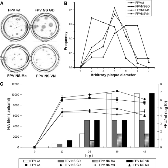
By reverse genetics, we generated the reassortants FPV NS GD, FPV NS Ma, and FPV NS VN, carrying the NS segment of A/Goose/Guangdong/16 (GD; H5N1), A/Mallard/NL/12/2000 (Ma; H7N3), and A/Vietnam/1203/2004 (VN; H5N1) viruses, respectively, in the genetic background of A/FPV/Rostock/34 (FPV; H7N1). In order to investigate whether the different NS segments altered the replication ability of the wt FPV virus, the plaque sizes of the different viruses were analyzed using MDCK cells (Fig. 1A), and the determined plaque diameters are represented as distribution curves in Fig. 1B. The reassortant virus FPV NS Ma produced plaques similar in size to those produced by wt FPV, while those of FPV NS VN were much smaller and those of FPV NS GD were much bigger than those of wt FPV. This indicates that the FPV NS GD virus has a propagation advantage in MDCK cells than do the other three viruses, confirming previous findings (26). In contrast, the FPV NS VN virus seems to replicate less efficiently in MDCK cells than do the other viruses.
FIG. 1.
Phenotype of foci, infectious titer, and HA titer of recombinant FPV viruses. (A) Focus assay showing plaque sizes. MDCK cell monolayers were infected with the different recombinant viruses at an MOI of 0.001 and stained using a monoclonal antibody against NP. (B) Distribution curve of the average diameters of foci. From each recombinant virus, 50 to 78 foci were measured. (C) Virus growth curves and HA titers at an MOI of 0.01. Virus growth curves (lines) were determined by focus assays in MDCK cells and measured in focus-forming units (FFU)/ml. HA titers (bars) were determined using 1.5% chicken red blood cells and measured in HA units/ml. Results represent the averages from three independent experiments.
Viral growth curves and the HA titers of the different viruses in MDCK cells were also determined (Fig. 1C). This analysis demonstrated that FPV NS GD replicates to higher titers than wt FPV, most conclusively at 36 h p.i., while FPV NS VN and FPV NS Ma replicated to lower titers (Fig. 1C, lines). This shows that the NS segments of both H7- and H5-type HPAIV can affect the virus replication efficiency of FPV in MDCK cells and corresponds to the results obtained from the plaque phenotypes. Although both the Ma and GD NS segments are grouped in allele B, the differences in the infectious titers between these two viruses indicate that the effects of the NS gene on virus replication efficiency are not dependent simply on the allele type. In addition, these results suggest that not all H5-sourced NS segments necessarily increase the H7-type HPAIV infectious titer, as both the GD and VN NS segments are from H5N1 viruses and only the FPV NS GD virus had an increased titer.
In order to determine whether the NS segments could affect the propagation of FPV in host cells of different origins, virus growth curves in two other mammalian cell lines and two avian cell lines were investigated (see Fig. S1 in the supplemental material). Results similar to those found in MDCK cells (Fig. 1C) were obtained for the mammalian A549 and 293T cell lines. However, in avian LMH and QT6 cells, the NS reassortants showed different growth curves. The wt FPV, FPV NS GD, and FPV NS Ma viruses had similar titers between 24 h and 36 h p.i., while FPV NS VN had a significantly lower titer (see Fig. S1 in the supplemental material). Even though the parental strain (A/VN/1203/2004) is known to replicate well in chickens, ducks, mice, ferrets, and humans (27), the reassortant FPV NS VN virus appears to be attenuated in the mammalian and avian cell lines tested. In contrast, the FPV NS Ma and wt FPV viruses replicate as efficiently as FPV NS GD in avian cells but less well in the mammalian cells employed in our study. It is therefore possible that NS segment reassortment can affect virus propagation in cell lines of different host origin.
Furthermore, the HA titers of the viruses differed from the infectious virus titers (Fig. 1C, bars), which may indicate that different NS segments can affect the formation of virus particles and change the ratio of defective particles to infectious particles. For example, at 48 h p.i., FPV NS VN has the highest HA titer of the reassortant viruses, equivalent to that of wt FPV; however, this virus has a much lower infectious titer than that of the wt virus in MDCK cells (Fig. 1C). FPV NS VN also has a low titer in the other mammalian and avian cell lines tested, while it produces HA titers higher than or similar to those of the other reassortant viruses (see Fig. S1 in the supplemental material), suggesting that this virus produces large numbers of noninfectious particles.
NS segment reassortment can affect viral genome replication and transcription.
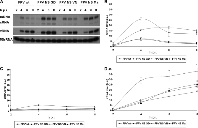
In order to analyze whether NS segment exchange affects the viral genome replication and transcription, MDCK cells were infected with the different viruses, RNA was harvested at various time points p.i., and NA segment-specific primer extensions were performed (Fig. 2). The results showed that NS segment reassortment altered the relative accumulation levels of viral RNA species for the different viruses. We show that the FPV NS GD virus had increased accumulation of mRNA, cRNA, and vRNA compared to that of the wt FPV, FPV NS Ma, and FPV NS VN viruses (Fig. 2B, C, D). Interestingly, the FPV NS Ma virus also produced higher levels of mRNA compared to the wt FPV and VN reassortants and showed a difference in the kinetics of mRNA accumulation. mRNA levels for FPV NS Ma peaked at 6 h p.i., while for the other three viruses they peaked at 4 h p.i. (Fig. 2B). Similar levels of accumulation of cRNA and vRNA were found for all of the viruses, with the exception of FPV NS GD (Fig. 2C and D). In order to determine whether the effects on replication and transcription were segment specific, we also analyzed viral RNAs derived from the NP segment. The results obtained (data not shown) show relative RNA accumulation levels similar to those obtained for the NA gene, indicating that the effect of NS segment exchange on the production of RNA species is not segment specific. Overall, these results suggest that the proteins expressed from the NS segment play a role in the regulation of viral replication and transcription.
FIG. 2.
Comparison of the accumulation of viral RNA species during infection by an NA gene-specific primer extension. (A) Primer extension analysis. MDCK cells were infected at an MOI of 1.0 and total RNA was harvested at the time points indicated. 32P-labeled NA gene-specific primers were used in a reverse transcription reaction and the products were analyzed by PAGE (6 M urea). The results are the averages from three independent experiments. a.u., arbitrary units. (B) Quantification of the accumulation of mRNA species for each recombinant virus. RNA levels were normalized using the 5S rRNA control, and results represent the averages from three independent experiments. Error bars represent standard deviations. (C) Quantification of the accumulation of cRNA species for each recombinant virus; (D) quantification of the accumulation of vRNA species for each recombinant virus.
Expression of the NS1 and NS2/NEP proteins affects the accumulation of viral RNAs in an RNP reconstitution assay.
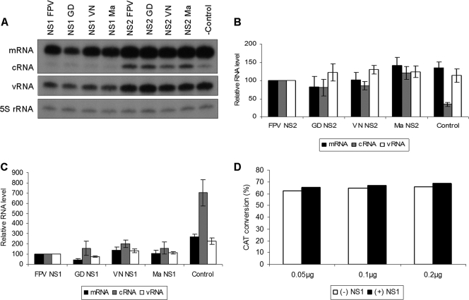
Having shown that NS segment exchange can affect the transcription and replication of the viral RNA, we set out to further investigate the role of individually expressed NS1 and NS2/NEP proteins on these processes. An RNP reconstitution assay was employed in which the four different NS1 or NS2/NEP proteins were individually coexpressed with the FPV polymerase and NP proteins and a viral RNA template (41). The results show that the transient expression of both NS1 and NS2/NEP proteins affected the accumulation of viral RNAs (Fig. 3). Expression of all NS2/NEP proteins resulted in an increase in cRNA levels compared to that of the negative control, consistent with a previous report (46) (Fig. 3B). However, the NS2/NEP proteins of GD, Ma, and VN did not produce significant changes in the accumulation of viral RNAs compared to the FPV NS2/NEP proteins, suggesting that this protein is not responsible for the differences in transcription and replication between the reassortant viruses. This is also supported by the results of a transfection/infection experiment (see Fig. S2 in the supplemental material), where we analyzed virus replication in MDCK cells transiently expressing the different NS2/NEP proteins after superinfection with wt FPV. Here, no specific effect by different NS2/NEP proteins on the virus titer was observed. Expression of all NS1 proteins resulted in a decrease in the accumulation levels of all viral RNAs compared to the levels in the negative control (Fig. 3C). Compared to FPV NS1, GD NS1 downregulated the accumulation of mRNA, while the Ma and VN NS1 proteins had little or no effect. The differential effect of the GD NS1 protein on the accumulation levels of viral mRNA may indicate a role for NS1 in viral transcription and replication.
FIG. 3.
Comparison of the accumulation of viral RNA species during RNP reconstitution by NA gene-specific primer extension. (A) Primer extension analysis. 293T cells were transfected with plasmids expressing the PB1, PB2, PA, and NP proteins of FPV and FPV NA vRNA. Either empty vector or plasmids encoding the various NS1 or NS2/NEP proteins were cotransfected into the cells. Total RNA was harvested after 48 hours and analyzed by NA gene-specific primer extension. (B) Quantification of RNA levels following expression of different NS2/NEP proteins. mRNA, cRNA, and vRNA levels were calculated from the results of three independent experiments and expressed as a percentage of the values in the presence of FPV NS2/NEP (which were set to 100%). (C) Quantification of RNA levels following expression of different NS1 proteins. mRNA, cRNA, and vRNA levels were calculated from the results of three independent experiments and expressed as a percentage of the values in the presence of FPV NS1 (which were set to 100%). (D) CAT assay testing the effect of the GD NS1 protein on CAT expression. 293T cells were transfected with plasmids expressing CAT at different concentrations (pcDNA3-CAT, 0.05 μg, 0.1 μg, 0.2 μg) and GD NS1 (pcDNA3-GD NS1, 4.0 μg). After 48 h, cell extracts were prepared and tested for CAT activity in a 1:10 dilution.
It has been reported previously that the NS1 protein can inhibit cellular mRNA processing through an interaction with CPSF30 (22, 36, 53) and PABII (6). To address the possibility that viral polymerase proteins and NP in the RNP reconstitution assay expressed from Pol II promoter-driven constructs were affected by NS1 protein expression, we transfected cells with a Pol II-driven reporter (pcDNA3.0-CAT) and an NS1 expression construct (pcDNA-GD-NS1). GD NS1 protein expression did not affect CAT protein expression (Fig. 3D), and therefore we conclude that viral protein expression from Pol II promoter-driven plasmids in the RNP reconstitution assay is unlikely to be affected by the NS1 protein.
To investigate the effect of different NS1 proteins on polymerase function, plasmids expressing polymerase proteins and nucleoprotein from either FPV, GD, or A/Puerto Rico/8/34 (PR8) viruses, together with a CAT reporter vector (41) and plasmids expressing different NS1 proteins (FPV,GD, and PR8), were transfected into 293T cells. We found that the FPV and GD NS1 proteins decreased the reporter gene expression in all three virus systems (see Fig. S3 in the supplemental material). These results are in agreement with the observation from the RNP reconstitution assay (Fig. 3B) that the various NS1 proteins can decrease the accumulation of all viral RNA products. Surprisingly, the PR8 NS1 protein enhanced CAT expression in the homologous PR8 system but not in the FPV or GD systems (see Fig. S3 in the supplemental material). Taken together, these data suggest that the NS1 proteins may affect the RNP complex and regulate transcription and replication without the help of other viral proteins.
NS1 protein localization does not correlate with infectious viral titer.
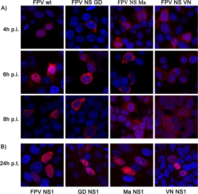
NS1 contains two nuclear localization signals and one nuclear export signal, which are responsible for the transport of the protein between the nucleus and cytoplasm. The NS1 protein can therefore be present in the nucleus, the cytoplasm, or both (14, 43). Our amino acid sequence comparison (see Fig. S4 in the supplemental material) showed that the sequence of both the NLS1 (34 to 39 amino acids [aa]) and NLS2 (216 to 220 aa) are conserved between the four NS1 proteins; however, the nuclear export sequence (NES) (138 to 147 aa) differs at three amino acid positions between the allele A and B NS1 proteins. Therefore, we investigated the localization of the NS1 proteins of the different reassortant viruses in both infected and transfected MDCK cells. At all time points of infection, it was found that FPV NS1 was predominantly nuclear, GD NS1 and Ma NS1 were predominantly cytoplasmic, and VN NS1 showed a cytoplasmic and nuclear localization (Fig. 4A). There was therefore no correlation between the infectious titer of the reassortant viruses and NS1 localization during infection. Interestingly, when NS1 localization was investigated in transfected MDCK cells after 24 h, NS1 proteins of all viruses were located exclusively in the nucleus (Fig. 4B).
FIG. 4.
Immunofluorescence of NS1 protein localization. (A) MDCK cells were infected with recombinant viruses at an MOI of 0.1. At 4 h, 6 h, and 8 h p.i., the cells were fixed for immunofluorescence analysis. (B) MDCK cells were transfected with plasmids expressing the different NS1 proteins and fixed at 24 h posttransfection for immunofluorescence analysis. Immunofluorescence was carried out using antibodies against the NS1 protein (red) and DAPI (blue) for the nucleus.
NS segment exchange alters RNP export.
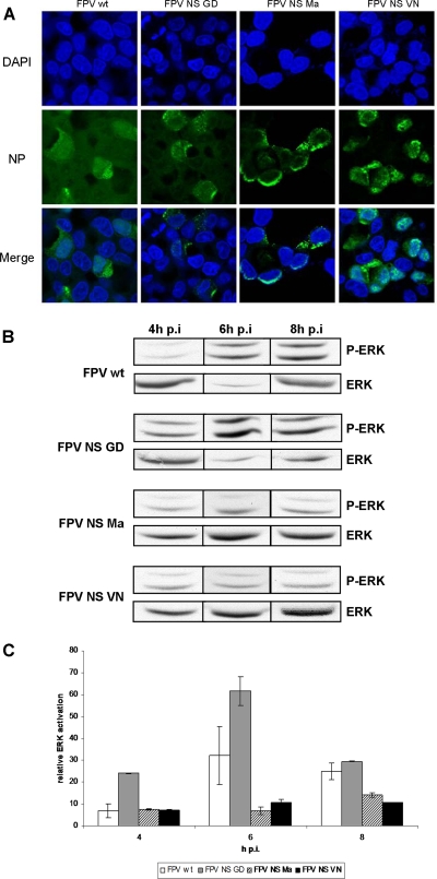
Efficient nuclear export of RNPs is important for the production of infectious virus (42). In order to investigate whether the NS segment exchange affected viral RNP export patterns, NP localization was analyzed by immunofluorescence microscopy (Fig. 5A). At 8 h p.i., most of the NP of wt FPV was located in the cytoplasm, as was that of FPV NS GD, while FPV NS Ma and FPV NS VN showed a more nuclear localization. These results indicate that NS segment exchange can alter RNP export and suggest a correlation between rapid RNP export and higher infectious virus titers.
FIG. 5.
Recombinant viruses show differences in the nuclear export of RNPs. (A) Immunofluorescence showing RNP export of the different recombinant viruses. MDCK cells were infected with recombinant viruses at an MOI of 0.1, and at 8 h p.i. cells were stained with an anti-NP antibody (green) and DAPI (blue) for the nuclei. (B) Cell lysates of MDCK cells infected with the different viruses at an MOI of 1 were analyzed for ERK activation by Western blotting using a phosphospecific anti-ERK and an anti-ERK2 (control) monoclonal antibody. (C) Graph showing ERK activation by different recombinant viruses. MDCK cells were infected with recombinant viruses at an MOI of 1, and cell lysates were prepared at 4 h, 6 h, and 8 h p.i. Samples were analyzed for ERK activation by Western blotting using a phosphospecific anti-ERK and an anti-ERK2 (control) monoclonal antibody. The relative ERK activation values from two independent experiments were normalized to the loading control (ERK2).
It has previously been shown that virus-induced activation of the Raf/MEK/ERK (mitogen-activated protein kinase [MAPK]) signal transduction cascade is an essential prerequisite for efficient nuclear RNP export (42). Membrane accumulation of the viral hemagglutinin (HA) glycoprotein in lipid-raft domains seems to be an important trigger that coordinates this signal-induction event with packaging of new RNPs into progeny virions (29). Induction or inhibition of the cascade will increase or reduce progeny virus titers, respectively (25, 30, 42). Therefore, we investigated the extent to which the viruses activated the MAPK cascade by detection of activated ERK (P-ERK) (Fig. 5B and 5C). The highest level of phosphorylated ERK was detected for the FPV NS GD virus, followed by that detected for wt FPV, with lower levels detected for FPV NS Ma and FPV NS VN. These results correlate with the finding that the RNPs of the wt FPV and GD NS viruses are transported more efficiently than those of the FPV NS Ma and VN viruses.
NS segment exchange affects IFN-β levels and apoptosis levels.
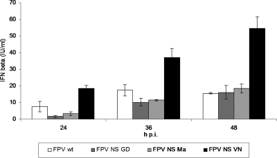
In order to test the ability of the different NS1 proteins to counteract the host innate immune response to support virus propagation, IFN-β levels in A549 cells infected with reassortant viruses at an MOI of 0.01 at various time points (24 h, 36 h, and 48 h p.i.) were measured by ELISA. We found that FPV NS VN induced the highest IFN-β response at all time points compared to that of the other three viruses, and FPV induced the second-highest response at 24 and 36 h p.i. (Fig. 6). When comparing these results to the infectious titers of the various viruses, it can be concluded that in general higher IFN-β levels correlate with lower infectious titers of the reassortant viruses.
FIG. 6.
Recombinant viruses induce different levels of IFN-β. A549 cells were infected with the recombinant viruses at an MOI of 0.01. Supernatants were harvested at 24, 36, and 48 h p.i., and measurements using an IFN-β ELISA kit were performed. Results represent the averages from three independent experiments.
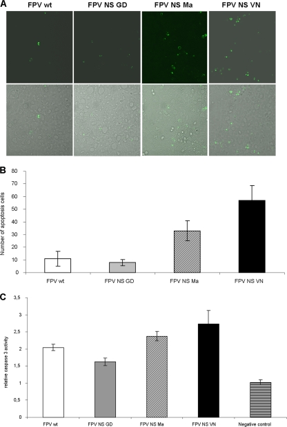
During the course of our experiments we observed that the FPV NS VN virus appeared to show a different cytopathic effect (CPE) in MDCK cells than the wt FPV, FPV NS GD, and FPV NS Ma viruses. Cells infected with FPV NS VN rounded up but did not detach from the dishes, while infection with the other three viruses resulted in the cells becoming detached. We hypothesized that the FPV NS VN virus might induce different levels of apoptosis compared to the other viruses. In order to determine the extent of apoptosis induced in cells infected with the different viruses, a TUNEL assay was performed in which MDCK cells were infected and examined by confocal microscopy (7). The number of apoptotic cells was counted in five random viewing fields, and the results were represented as a graph (Fig. 7). We observed that the FPV NS VN virus induced the highest levels of apoptosis, followed by FPV NS Ma, wt FPV, and FPV NS GD (Fig. 7B). The additional determination of caspase3 activity at 8 h p.i. (Fig. 7C) confirmed these results. Reassortant viruses containing different NS segments therefore differ in their abilities to induce cell death. When the extent of apoptosis is compared to the infectious titers of the various viruses, it can be concluded that the higher levels of apoptosis correlate with lower virus titers.
FIG. 7.
Level of apoptosis induced by the recombinant viruses. (A) MDCK cells were infected with recombinant viruses (MOI of 1). Ten hours p.i., the cells were fixed and permeabilized before being processed using an in situ cell death detection kit (Roche) according to the manufacturer's instructions. The specimens were then examined by confocal microscopy. Green dots represent apoptotic cells. The top panels were viewed using the laser channel alone, while the bottom panels were viewed using both light and the laser channel. (B) Quantification of the number of apoptotic cells. The number of green cells in five random views was counted in three independent experiments. (C) Relative caspase3 activity. MDCK cells were infected with recombinant viruses (MOI of 1). Eight hours p.i., the cells were analyzed for caspase3 activity. The results are based on averages from three independent experiments.
DISCUSSION
In this report, we showed that replacement of the NS segment of the A/FPV/Rostock/34 (H7N1) influenza virus with NS segments from a variety of other H5- and H7-type viruses resulted in changes in the viral characteristics in mammalian and avian cell culture. In order to elucidate how NS segment reassortment altered virus propagation, multiple aspects of the interaction between the reassortant viruses and the host were examined.
We show that the FPV NS GD virus, which has an allele B NS segment, replicated more efficiently than the wt FPV virus (allele A) in mammalian cell culture systems. This result and the previous finding that the FPV NS GD virus is able to replicate in mice in contrast to wt FPV (26) suggest that the GD NS segment may help the virus cross the host barrier to mammals. However, the general effect of NS exchange on the replication efficiency is independent of the genetic NS group, as the second virus with an allele B NS segment, FPV NS Ma, had a lower replication titer than wt FPV. Also, the effect of NS gene exchange on the propagation of recombinant viruses was not caused by differences in the NS1 protein expression levels (see Fig. S5 in the supplemental material), as the different properties of the recombinant viruses do not correlate with the different NS1 expression levels. Similar results were obtained for the NP expression levels (data not shown).
We also addressed the question of whether H5-sourced NS segments, particularly those from viruses that emerged after 1998, increased viral growth of an H7-type HPAIV in mammalian cells. In a previous study by Twu et al. (52), a series of reassortants of the human A/Udorn/72 (Ud; H3N2) virus carrying different NS segments were generated. It was shown that the reassortant with the NS segment of A/Hong Kong/483/97 (HK97; H5N1) virus replicated less efficiently, while the reassortant with the A/Vietnam/1203/04 (VN04; H5N1) virus NS segment replicated as efficiently as that of wt Ud. It was concluded that H5N1 virus NS genes selected after 1998 seemed to enhance viral replication in mammalian cells. However, in our study, we observed that the NS segment from the VN virus isolated in 2004 decreased the replication of the reassortant avian H7-type virus (FPV), while the NS segment from the GD virus, isolated in 1996, enhanced FPV replication. This shows that H5-sourced NS segments do not necessarily increase the replication of avian IV and the year of emergence does not appear to play a major role. Furthermore, it is possible that the same NS segment may have different effects on virus replication depending on the specific genetic background.
The replication differences of the reassortant viruses in mammalian and avian cells (see Fig. S1 in the supplemental material) suggest that the effect of NS segments on viral propagation also depends on specific host factors. In avian cells, wt FPV, FPV NS GD, and FPV NS Ma had similar titers but FPV NS VN had a significantly (2 logs) lower titer, whereas in mammalian cells FPV NS GD replicated to titers of almost 1 log higher than that of wt FPV and 2 logs higher than that of FPV NS Ma. The NS segments of GD, FPV, and Ma originated from avian viruses, and therefore it is tempting to speculate that they all could have a replication advantage in avian cells. In this context, it should be noted that in a previous study, the NS segment of the 1918 IV (H1N1) was set in the genetic background of A/WSN/33 (H1N1). The virus replicated well in tissue culture (MDCK cells) but was attenuated in mice compared with the control viruses (2). It was suggested that the attenuation in mice may be related to the human origin of the 1918 NS1 gene and that interaction of the NS1 protein with host-cell factors plays a significant role in viral pathogenesis. Taken together, for the effects exerted by the NS segment exchange, it seems to be relevant from which host the donor strain was isolated and in which host the recipient is tested.
IV with C-terminally truncated forms of NS1 have previously been found to produce less vRNA than wild-type viruses, suggesting that the NS1 protein is involved in viral replication (10). In our report, the C terminus of the VN NS1 is truncated by 10 aa, but the results in Fig. 2 show similar vRNA and cRNA production for wt FPV and FPV NS VN. The GD and Ma NS segments affected both transcription and replication and enhanced the accumulation of viral mRNAs compared to that of wt FPV and FPV NS VN in infected cells. In addition, FPV NS Ma had a delayed peak of viral mRNA accumulation, suggesting that NS segments can influence the time course of RNA synthesis. Similar results were observed for both the NA and NP gene segments. In vitro RNP reconstitution experiments (Fig. 3C) indicated that the NS1 protein was likely to have an effect on viral RNA production, as an increase in the accumulation of cRNA amounts by GD NS, VN NS, and Ma NS was observed compared to that of FPV NS. However, during infection, GD and Ma NS viruses showed increased accumulation of viral mRNAs, suggesting that the effect of NS segments on the viral transcription and replication in infected cells may not be a direct effect and might depend on other viral factors.
The IV polymerase can transcribe and replicate viral RNA in the absence of host factors (38), but it has also been shown that host factors may regulate or enhance viral RNA transcription and replication in infected cells (17, 35). Recently, the influenza A virus NS1 protein was reported to form a complex with CPSF-30 and the viral RNPs (20). Since CPSF-30 is present in both the infection and RNP reconstitution assays and our previous data showed that GD NS1 and FPV NS1 have similar binding affinities for the F2F3 domain of CPSF-30 (26), this interaction is unlikely to be responsible for the different effects observed during infection and RNP reconstitution.
It has been suggested that the transiently expressed PR8 NS1 protein can enhance CAT reporter gene expression by increasing overall translation (47). Our in vitro RNP reconstitution experiments using a CAT reporter gene (see Fig. S3 in the supplemental material) showed that the NS1 effect on viral transcription and replication depends on the strain background of the viral RNP complex, because the PR8 NS1 protein enhanced CAT expression of only the PR8 RNP complex but not the FPV and GD systems. The observation that NS1 proteins can form complexes containing CPSF30 with only cognate RNP complexes rather than RNP complexes of other viral strains (20) further supports this conclusion.
Although it is not clear how NS1 affects viral transcription and replication, we observed that all NS1 proteins were exclusively located in the nuclei of transfected cells, while a proportion of the GD and Ma NS1 proteins in infected cells also resided in the cytoplasm (Fig. 4). It has been suggested that the NES of NS1 is masked by an adjacent inhibitory amino acid sequence in transfected cells, while it is unmasked during infection due to specific interaction with another viral protein (23). It is therefore possible that the GD and Ma NS1 protein can enhance RNA-dependent RNA polymerase (RdRp) replication and transcription activity during infection but have reduced activity in RNP reconstitution experiments due to nuclear retention resulting in a direct or indirect decrease of polymerase activity.
Despite there being no obvious correlation of NS1 localization with infectious virus titers, the different localization patterns of the NS1 proteins during infection (Fig. 4) may be contributing to the effects we observed on virus replication and/or host immune responses. As the NS1 protein has diverse roles in both the cytoplasm and nuclei of infected cells, we cannot exclude the possibility that different localizations of the NS1 proteins studied may play a role in the downstream effects observed.
The observation that more rapid RNP export correlated with higher virus titers (Fig. 5) indicates that less efficient RNP export leads to lower infectious titers by limiting RNPs available for packaging (42). NS2/NEP can mediate RNP export by interaction with the M1 protein. The amino acid comparison of NS2/NEP (see Fig. S4C in the supplemental material) shows that there are only two amino acid differences between the GD and Ma NS2/NEP protein sequences. Therefore, if NS2/NEP alone confers the demonstrated difference in RNP export, it should be based on this two-amino-acid difference. Even though it is unlikely that NS2/NEP is responsible for the observed differences, further investigation is needed.
The NS1 protein is known to impair host immune responses. In this study, we show that NS segment exchange in an FPV background altered IFN-β production and that IFN-β levels were inversely correlated to the infectious virus titer (Fig. 6). It is also known that the NS1 protein is involved in the regulation of apoptosis; however, it is not clear whether it plays a pro- or antiapoptotic role (16). Nevertheless, in our experiments, we found that the levels of apoptosis induced were inversely correlated to the infectious virus titers (Fig. 7). IFN can make cells more sensitive to apoptosis (1), but it is still not clear how viral replication/transcription and the antiviral response are related.
In an overall comparison of the different NS proteins, we found that the GD NS1 protein sequence is similar to that of Ma NS1, differing by only 8 aa (see Fig. S4B in the supplemental material). Notable differences are found at (i) position 41, which is reported to be important for dsRNA binding (9, 54), is part of NLS1 (31), and comprises an ISG15 acceptor site (56); (ii) position 166, which is implicated in the interaction with PI3K (49); and (iii) position 224, which is located in the PABII binding region (6) and is important for the nucleolar targeting function of the NS1 protein (31). The VN NS1 protein is truncated at the C terminus by 10 aa and also contains a 5-aa internal deletion (see Fig. S4B in the supplemental material). As a consequence of the C-terminal truncation, the VN NS1 protein lacks a PDZ domain (227 to 230 aa) and a PABII-binding region (223 to 230 aa). NS2/NEP amino acid sequences are more conserved than that of NS1 (see Fig. S4C in the supplemental material). The majority of differences are found between the two allele A and the two allele B segments. Interestingly, a single amino acid difference between FPV and VN NS2/NEP that is located at position 14 is in the nuclear export sequence (NES), which is suggested to be a Crm-1 binding site (37). All remaining differences are in uncharacterized regions. To which extent these differences contribute to the different viral characteristics is currently under investigation.
The NS1 protein plays a very important and multifactorial role in the battle between IV and its host. Our results show that the exchange of NS segments can have a significant effect on the replication ability of the virus and supports the notion that the protein products of the NS segment are involved in many stages of the virus life cycle. Surprisingly, as shown here for the first time, this includes not only the inhibition of the host response and induction of apoptosis but also accumulation of viral RNAs and vRNP export. Presently, it is not possible to conclude which of these effects is the most important in determining the replication ability of viruses. The effects of different NS segments on the replication of reassortant viruses are likely to be due to multiple effects of the NS1 protein on multiple viral and host processes.
Biosafety.
All experiments with infectious virus were performed according to German regulations for the propagation of influenza A viruses. All experiments involving highly pathogenic influenza A viruses were performed in a biosafety level 3 (BSL3) containment laboratory approved for such use by the local authorities (RP, Giessen, Germany).
Supplementary Material
Acknowledgments
We thank J. Pavlovic (Institute of Virology, Zurich, Switzerland) for providing the plasmids pHMG-PB1, -PB2, -PA, and -NP and K. Saksela (Helsinki, Finland) for providing the pPolI-NS plasmid encoding A/Mallard/NL/12/2000 NS vRNA. In addition, we thank M. Stein for his excellent technical assistance.
This work was supported in part by grants from the DFG (GRK1384, fellowship to Z.W.), from the European Specific Targeted Research Project, EuroFlu—Molecular Factors and Mechanisms of Transmission and Pathogenicity of Highly Pathogenic Avian Influenza Virus, funded by the 6th Framework Program (FP6) of the EU (grant SP5B-CT-2007-044098, to S.P.), from the FluResearchNet—Molecular Signatures Determining Pathogenicity and Species Transmission of Influenza A Viruses (grant 01 KI 07136, to S.P., and grant 01 KI 07132, to T.W.), and from the MRC (grant G0700848, to E.F.).
Footnotes
Published ahead of print on 25 August 2010.
Supplemental material for this article may be found at http://jvi.asm.org/.
REFERENCES
- 1.Balachandran, S., P. C. Roberts, L. E. Brown, H. Truong, A. K. Pattnaik, D. R. Archer, and G. N. Barber. 2000. Essential role for the dsRNA-dependent protein kinase PKR in innate immunity to viral infection. Immunity 13:129-141. [DOI] [PubMed] [Google Scholar]
- 2.Basler, C. F., A. H. Reid, J. K. Dybing, T. A. Janczewski, T. G. Fanning, H. Zheng, M. Salvatore, M. L. Perdue, D. E. Swayne, A. Garcia-Sastre, P. Palese, and J. K. Taubenberger. 2001. Sequence of the 1918 pandemic influenza virus nonstructural gene (NS) segment and characterization of recombinant viruses bearing the 1918 NS genes. Proc. Natl. Acad. Sci. U. S. A. 98:2746-2751. [DOI] [PMC free article] [PubMed] [Google Scholar]
- 3.Bergmann, M., A. Garcia-Sastre, E. Carnero, H. Pehamberger, K. Wolff, P. Palese, and T. Muster. 2000. Influenza virus NS1 protein counteracts PKR-mediated inhibition of replication. J. Virol. 74:6203-6206. [DOI] [PMC free article] [PubMed] [Google Scholar]
- 4.Chen, H., G. Deng, Z. Li, G. Tian, Y. Li, P. Jiao, L. Zhang, Z. Liu, R. G. Webster, and K. Yu. 2004. The evolution of H5N1 influenza viruses in ducks in southern China. Proc. Natl. Acad. Sci. U. S. A. 101:10452-10457. [DOI] [PMC free article] [PubMed] [Google Scholar]
- 5.Chen, H., G. J. Smith, K. S. Li, J. Wang, X. H. Fan, J. M. Rayner, D. Vijaykrishna, J. X. Zhang, L. J. Zhang, C. T. Guo, C. L. Cheung, K. M. Xu, L. Duan, K. Huang, K. Qin, Y. H. Leung, W. L. Wu, H. R. Lu, Y. Chen, N. S. Xia, T. S. Naipospos, K. Y. Yuen, S. S. Hassan, S. Bahri, T. D. Nguyen, R. G. Webster, J. S. Peiris, and Y. Guan. 2006. Establishment of multiple sublineages of H5N1 influenza virus in Asia: implications for pandemic control. Proc. Natl. Acad. Sci. U. S. A. 103:2845-2850. [DOI] [PMC free article] [PubMed] [Google Scholar]
- 6.Chen, Z., Y. Li, and R. M. Krug. 1999. Influenza A virus NS1 protein targets poly(A)-binding protein II of the cellular 3′-end processing machinery. EMBO J. 18:2273-2283. [DOI] [PMC free article] [PubMed] [Google Scholar]
- 7.Cilloniz, C., K. Shinya, X. Peng, M. J. Korth, S. C. Proll, L. D. Aicher, V. S. Carter, J. H. Chang, D. Kobasa, F. Feldmann, J. E. Strong, H. Feldmann, Y. Kawaoka, and M. G. Katze. 2009. Lethal influenza virus infection in macaques is associated with early dysregulation of inflammatory related genes. PLoS Pathog 5:e1000604. [DOI] [PMC free article] [PubMed] [Google Scholar]
- 8.Dauber, B., L. Martinez-Sobrido, J. Schneider, R. Hai, Z. Waibler, U. Kalinke, A. Garcia-Sastre, and T. Wolff. 2009. Influenza B virus ribonucleoprotein is a potent activator of the antiviral kinase PKR. PLoS Pathog. 5:e1000473. [DOI] [PMC free article] [PubMed] [Google Scholar]
- 9.Donelan, N. R., C. F. Basler, and A. Garcia-Sastre. 2003. A recombinant influenza A virus expressing an RNA-binding-defective NS1 protein induces high levels of beta interferon and is attenuated in mice. J. Virol. 77:13257-13266. [DOI] [PMC free article] [PubMed] [Google Scholar]
- 10.Falcon, A. M., R. M. Marion, T. Zurcher, P. Gomez, A. Portela, A. Nieto, and J. Ortin. 2004. Defective RNA replication and late gene expression in temperature-sensitive influenza viruses expressing deleted forms of the NS1 protein. J. Virol. 78:3880-3888. [DOI] [PMC free article] [PubMed] [Google Scholar]
- 11.Gack, M. U., R. A. Albrecht, T. Urano, K. S. Inn, I. C. Huang, E. Carnero, M. Farzan, S. Inoue, J. U. Jung, and A. Garcia-Sastre. 2009. Influenza A virus NS1 targets the ubiquitin ligase TRIM25 to evade recognition by the host viral RNA sensor RIG-I. Cell Host Microbe 5:439-449. [DOI] [PMC free article] [PubMed] [Google Scholar]
- 12.Gambotto, A., S. M. Barratt-Boyes, M. D. de Jong, G. Neumann, and Y. Kawaoka. 2008. Human infection with highly pathogenic H5N1 influenza virus. Lancet 371:1464-1475. [DOI] [PubMed] [Google Scholar]
- 13.Garcia-Sastre, A. 2004. Identification and characterization of viral antagonists of type I interferon in negative-strand RNA viruses. Curr. Top. Microbiol. Immunol. 283:249-280. [DOI] [PubMed] [Google Scholar]
- 14.Greenspan, D., P. Palese, and M. Krystal. 1988. Two nuclear location signals in the influenza virus NS1 nonstructural protein. J. Virol. 62:3020-3026. [DOI] [PMC free article] [PubMed] [Google Scholar]
- 15.Guo, Y., M. Wang, Y. Kawaoka, O. Gorman, T. Ito, T. Saito, and R. G. Webster. 1992. Characterization of a new avian-like influenza A virus from horses in China. Virology 188:245-255. [DOI] [PubMed] [Google Scholar]
- 16.Hale, B. G., R. E. Randall, J. Ortin, and D. Jackson. 2008. The multifunctional NS1 protein of influenza A viruses. J. Gen. Virol. 89:2359-2376. [DOI] [PubMed] [Google Scholar]
- 17.Jorba, N., S. Juarez, E. Torreira, P. Gastaminza, N. Zamarreno, J. P. Albar, and J. Ortin. 2008. Analysis of the interaction of influenza virus polymerase complex with human cell factors. Proteomics 8:2077-2088. [DOI] [PubMed] [Google Scholar]
- 18.Koopmans, M., B. Wilbrink, M. Conyn, G. Natrop, H. van der Nat, H. Vennema, A. Meijer, J. van Steenbergen, R. Fouchier, A. Osterhaus, and A. Bosman. 2004. Transmission of H7N7 avian influenza A virus to human beings during a large outbreak in commercial poultry farms in the Netherlands. Lancet 363:587-593. [DOI] [PubMed] [Google Scholar]
- 19.Krug, R. M., W. Yuan, D. L. Noah, and A. G. Latham. 2003. Intracellular warfare between human influenza viruses and human cells: the roles of the viral NS1 protein. Virology 309:181-189. [DOI] [PubMed] [Google Scholar]
- 20.Kuo, R. L., and R. M. Krug. 2009. Influenza A virus polymerase is an integral component of the CPSF30-NS1A protein complex in infected cells. J. Virol. 83:1611-1616. [DOI] [PMC free article] [PubMed] [Google Scholar]
- 21.Li, K. S., Y. Guan, J. Wang, G. J. Smith, K. M. Xu, L. Duan, A. P. Rahardjo, P. Puthavathana, C. Buranathai, T. D. Nguyen, A. T. Estoepangestie, A. Chaisingh, P. Auewarakul, H. T. Long, N. T. Hanh, R. J. Webby, L. L. Poon, H. Chen, K. F. Shortridge, K. Y. Yuen, R. G. Webster, and J. S. Peiris. 2004. Genesis of a highly pathogenic and potentially pandemic H5N1 influenza virus in eastern Asia. Nature 430:209-213. [DOI] [PubMed] [Google Scholar]
- 22.Li, Y., Z. Y. Chen, W. Wang, C. C. Baker, and R. M. Krug. 2001. The 3′-end-processing factor CPSF is required for the splicing of single-intron pre-mRNAs in vivo. RNA 7:920-931. [DOI] [PMC free article] [PubMed] [Google Scholar]
- 23.Li, Y., Y. Yamakita, and R. M. Krug. 1998. Regulation of a nuclear export signal by an adjacent inhibitory sequence: the effector domain of the influenza virus NS1 protein. Proc. Natl. Acad. Sci. U. S. A. 95:4864-4869. [DOI] [PMC free article] [PubMed] [Google Scholar]
- 24.Ludwig, S., U. Schultz, J. Mandler, W. M. Fitch, and C. Scholtissek. 1991. Phylogenetic relationship of the nonstructural (NS) genes of influenza A viruses. Virology 183:566-577. [DOI] [PubMed] [Google Scholar]
- 25.Ludwig, S., T. Wolff, C. Ehrhardt, W. J. Wurzer, J. Reinhardt, O. Planz, and S. Pleschka. 2004. MEK inhibition impairs influenza B virus propagation without emergence of resistant variants. FEBS Lett. 561:37-43. [DOI] [PubMed] [Google Scholar]
- 26.Ma, W., D. Brenner, Z. Wang, B. Dauber, C. Ehrhardt, K. Hogner, S. Herold, S. Ludwig, T. Wolff, K. Yu, J. A. Richt, O. Planz, and S. Pleschka. 2010. The NS segment of an H5N1 highly pathogenic avian influenza virus (HPAIV) is sufficient to alter replication efficiency, cell tropism, and host range of an H7N1 HPAIV. J. Virol. 84:2122-2133. [DOI] [PMC free article] [PubMed] [Google Scholar]
- 27.Maines, T. R., X. H. Lu, S. M. Erb, L. Edwards, J. Guarner, P. W. Greer, D. C. Nguyen, K. J. Szretter, L. M. Chen, P. Thawatsupha, M. Chittaganpitch, S. Waicharoen, D. T. Nguyen, T. Nguyen, H. H. Nguyen, J. H. Kim, L. T. Hoang, C. Kang, L. S. Phuong, W. Lim, S. Zaki, R. O. Donis, N. J. Cox, J. M. Katz, and T. M. Tumpey. 2005. Avian influenza (H5N1) viruses isolated from humans in Asia in 2004 exhibit increased virulence in mammals. J. Virol. 79:11788-11800. [DOI] [PMC free article] [PubMed] [Google Scholar]
- 28.Marion, R. M., T. Zurcher, S. de la Luna, and J. Ortin. 1997. Influenza virus NS1 protein interacts with viral transcription-replication complexes in vivo. J. Gen. Virol. 78:2447-2451. [DOI] [PubMed] [Google Scholar]
- 29.Marjuki, H., M. I. Alam, C. Ehrhardt, R. Wagner, O. Planz, H. D. Klenk, S. Ludwig, and S. Pleschka. 2006. Membrane accumulation of influenza A virus hemagglutinin triggers nuclear export of the viral genome via protein kinase Calpha-mediated activation of ERK signaling. J. Biol. Chem. 281:16707-16715. [DOI] [PubMed] [Google Scholar]
- 30.Marjuki, H., H. L. Yen, J. Franks, R. G. Webster, S. Pleschka, and E. Hoffmann. 2007. Higher polymerase activity of a human influenza virus enhances activation of the hemagglutinin-induced Raf/MEK/ERK signal cascade. Virol. J. 4:134. [DOI] [PMC free article] [PubMed] [Google Scholar]
- 31.Melen, K., L. Kinnunen, R. Fagerlund, N. Ikonen, K. Y. Twu, R. M. Krug, and I. Julkunen. 2007. Nuclear and nucleolar targeting of influenza A virus NS1 protein: striking differences between different virus subtypes. J. Virol. 81:5995-6006. [DOI] [PMC free article] [PubMed] [Google Scholar]
- 32.Mibayashi, M., L. Martinez-Sobrido, Y. M. Loo, W. B. Cardenas, M. Gale, Jr., and A. Garcia-Sastre. 2007. Inhibition of retinoic acid-inducible gene I-mediated induction of beta interferon by the NS1 protein of influenza A virus. J. Virol. 81:514-524. [DOI] [PMC free article] [PubMed] [Google Scholar]
- 33.Min, J. Y., and R. M. Krug. 2006. The primary function of RNA binding by the influenza A virus NS1 protein in infected cells: inhibiting the 2′-5′ oligo (A) synthetase/RNase L pathway. Proc. Natl. Acad. Sci. U. S. A. 103:7100-7105. [DOI] [PMC free article] [PubMed] [Google Scholar]
- 34.Min, J. Y., S. Li, G. C. Sen, and R. M. Krug. 2007. A site on the influenza A virus NS1 protein mediates both inhibition of PKR activation and temporal regulation of viral RNA synthesis. Virology 363:236-243. [DOI] [PubMed] [Google Scholar]
- 35.Nagata, K., A. Kawaguchi, and T. Naito. 2008. Host factors for replication and transcription of the influenza virus genome. Rev. Med. Virol. 18:247-260. [DOI] [PubMed] [Google Scholar]
- 36.Nemeroff, M. E., S. M. Barabino, Y. Li, W. Keller, and R. M. Krug. 1998. Influenza virus NS1 protein interacts with the cellular 30 kDa subunit of CPSF and inhibits 3′-end formation of cellular pre-mRNAs. Mol. Cell 1:991-1000. [DOI] [PubMed] [Google Scholar]
- 37.Neumann, G., M. T. Hughes, and Y. Kawaoka. 2000. Influenza A virus NS2 protein mediates vRNP nuclear export through NES-independent interaction with hCRM1. EMBO J. 19:6751-6758. [DOI] [PMC free article] [PubMed] [Google Scholar]
- 38.Newcomb, L. L., R. L. Kuo, Q. Ye, Y. Jiang, Y. J. Tao, and R. M. Krug. 2009. Interaction of the influenza A virus nucleocapsid protein with the viral RNA polymerase potentiates unprimed viral RNA replication. J. Virol. 83:29-36. [DOI] [PMC free article] [PubMed] [Google Scholar]
- 39.Obenauer, J. C., J. Denson, P. K. Mehta, X. Su, S. Mukatira, D. B. Finkelstein, X. Xu, J. Wang, J. Ma, Y. Fan, K. M. Rakestraw, R. G. Webster, E. Hoffmann, S. Krauss, J. Zheng, Z. Zhang, and C. W. Naeve. 2006. Large-scale sequence analysis of avian influenza isolates. Science 311:1576-1580. [DOI] [PubMed] [Google Scholar]
- 40.O'Neill, R., R. Jaskunas, G. Blobeli, P. Palese, and J. Moroianu. 1995. Nuclear import of influenza virus RNA can be mediated by viral nucleoprotein and transport factors required for protein import. J. Biol. Chem. 270:22701-22704. [DOI] [PubMed] [Google Scholar]
- 41.Pleschka, S., R. Jaskunas, O. G. Engelhardt, T. Zurcher, P. Palese, and A. Garcia-Sastre. 1996. A plasmid-based reverse genetics system for influenza A virus. J. Virol. 70:4188-4192. [DOI] [PMC free article] [PubMed] [Google Scholar]
- 42.Pleschka, S., T. Wolff, C. Ehrhardt, G. Hobom, O. Planz, U. R. Rapp, and S. Ludwig. 2001. Influenza virus propagation is impaired by inhibition of the Raf/MEK/ERK signaling cascade. Nat. Cell Biol. 3:301-305. [DOI] [PubMed] [Google Scholar]
- 43.Qian, X. Y., F. Alonso-Caplen, and R. M. Krug. 1994. Two functional domains of the influenza virus NS1 protein are required for regulation of nuclear export of mRNA. J. Virol. 68:2433-2441. [DOI] [PMC free article] [PubMed] [Google Scholar]
- 44.Reference deleted.
- 45.Rehwinkel, J., C. P. Tan, D. Goubau, O. Schulz, A. Pichlmair, K. Bier, N. Robb, F. Vreede, W. Barclay, E. Fodor, and C. Reis e Sousa. 2010. RIG-I detects viral genomic RNA during negative-strand RNA virus infection. Cell 140:397-408. [DOI] [PubMed] [Google Scholar]
- 46.Robb, N. C., M. Smith, F. T. Vreede, and E. Fodor. 2009. NS2/NEP protein regulates transcription and replication of the influenza virus RNA genome. J. Gen. Virol. 90:1398-1407. [DOI] [PubMed] [Google Scholar]
- 47.Salvatore, M., C. F. Basler, J. P. Parisien, C. M. Horvath, S. Bourmakina, H. Zheng, T. Muster, P. Palese, and A. Garcia-Sastre. 2002. Effects of influenza A virus NS1 protein on protein expression: the NS1 protein enhances translation and is not required for shutoff of host protein synthesis. J. Virol. 76:1206-1212. [DOI] [PMC free article] [PubMed] [Google Scholar]
- 48.Satterly, N., P. L. Tsai, J. van Deursen, D. R. Nussenzveig, Y. Wang, P. A. Faria, A. Levay, D. E. Levy, and B. M. Fontoura. 2007. Influenza virus targets the mRNA export machinery and the nuclear pore complex. Proc. Natl. Acad. Sci. U. S. A. 104:1853-1858. [DOI] [PMC free article] [PubMed] [Google Scholar]
- 49.Shin, Y. K., Y. Li, Q. Liu, D. H. Anderson, L. A. Babiuk, and Y. Zhou. 2007. SH3 binding motif 1 in influenza A virus NS1 protein is essential for PI3K/Akt signaling pathway activation. J. Virol. 81:12730-12739. [DOI] [PMC free article] [PubMed] [Google Scholar]
- 50.Suarez, D. L., and M. L. Perdue. 1998. Multiple alignment comparison of the non-structural genes of influenza A viruses. Virus Res. 54:59-69. [DOI] [PubMed] [Google Scholar]
- 51.Treanor, J. J., M. H. Snyder, W. T. London, and B. R. Murphy. 1989. The B allele of the NS gene of avian influenza viruses, but not the A allele, attenuates a human influenza A virus for squirrel monkeys. Virology 171:1-9. [DOI] [PubMed] [Google Scholar]
- 52.Twu, K. Y., R. L. Kuo, J. Marklund, and R. M. Krug. 2007. The H5N1 influenza virus NS genes selected after 1998 enhance virus replication in mammalian cells. J. Virol. 81:8112-8121. [DOI] [PMC free article] [PubMed] [Google Scholar]
- 53.Twu, K. Y., D. L. Noah, P. Rao, R. L. Kuo, and R. M. Krug. 2006. The CPSF30 binding site on the NS1A protein of influenza A virus is a potential antiviral target. J. Virol. 80:3957-3965. [DOI] [PMC free article] [PubMed] [Google Scholar]
- 54.Wang, W., K. Riedel, P. Lynch, C. Y. Chien, G. T. Montelione, and R. M. Krug. 1999. RNA binding by the novel helical domain of the influenza virus NS1 protein requires its dimer structure and a small number of specific basic amino acids. RNA 5:195-205. [DOI] [PMC free article] [PubMed] [Google Scholar]
- 55.WHO. 2008. Cumulative number of confirmed human cases of avian influenza A (H5N1) reported to WHO. World Health Organization, Geneva, Switzerland.
- 56.Zhao, C., T. Y. Hsiang, R. L. Kuo, and R. M. Krug. 2010. ISG15 conjugation system targets the viral NS1 protein in influenza A virus-infected cells. Proc. Natl. Acad. Sci. U. S. A. 107:2253-2258. [DOI] [PMC free article] [PubMed] [Google Scholar]
Associated Data
This section collects any data citations, data availability statements, or supplementary materials included in this article.


