Abstract
Background
Upper extremity weight-bearing exercises are routinely used in physical therapy for patients with shoulder pathology. However, little evidence exists regarding the demand on the shoulder musculature.
Objective
To examine changes in shoulder muscle activity and center of pressure during upper extremity weight-bearing exercises of increasing difficulty.
Methods
Electromyographic (EMG) and kinetic data were recorded from both shoulders of 15 healthy subjects (10 male and 5 female). Participants were tested in a modified tripod position under three conditions of increasing difficulty: (1) hand directly on the force plate, (2) on a green Stability Trainer™ and (3) on a blue Stability Trainer™. Ground reaction forces were recorded for each trial. Surface EMG was recorded from the serratus anterior, pectoralis major, upper trapezius, lower trapezius, infraspinatus, anterior deltoid, posterior deltoid, and the lateral head of the triceps muscles.
Results
Mean deviation from center of pressure significantly increased when using the Stability Trainer™ pads. The activities of the triceps, serratus anterior, and anterior deltoid muscles significantly increased as each trial progressed, irrespective of stability condition. Additionally, activity in the anterior deltoid, lower trapezius, and serratus anterior muscles significantly decreased with increasing difficulty, whereas activity in the triceps muscles significantly increased.
Discussion and Conclusion
Balancing on a foam pad made it more difficult to maintain the upper extremity in a stable position. However, this activity did not alter the proprioceptive stimulus enough to elicit an increase in shoulder muscle activation. While the results on this study support the use of different level Stability Trainers™ to facilitate neuromuscular re-education, a less compliant unstable surface may produce larger training effects.
Keywords: closed chain, shoulder, muscle activity
INTRODUCTION
During activities of daily living and sports, the upper extremity is used in both open kinetic chain and closed kinetic chain positions. Examples of closed kinetic chain activities of the shoulder include pushing oneself up from a chair or pass blocking a rushing defender during a football game. Therefore, both open and closed chain exercises should be integrated into a comprehensive rehabilitation program. Examples of upper extremity weight bearing exercises include push-ups with or without modifications and quadruped, prayer, and tripod positions. The rationale for these exercises is to improve proprioception, joint stability, and strength.1–4 In addition, a progression from a stable surface to an unstable surface is a standard method of increasing the difficulty of the exercise. Despite the large use of upper extremity weight bearing exercises in the clinical setting little is known about the demand on the shoulder musculature.5
The purpose of this study was to examine changes in the deviation of the hand center of pressure and activity of shoulder musculature during three different upper extremity weight-bearing positions of increasing difficulty. The hypothesis to be tested is that with an increasingly compliant surface (less stability), both the mean deviation of the center of pressure and shoulder muscle activity will increase. These findings would support the use of such exercises for the purpose of increasing demand on the shoulder by using increasingly compliant surfaces.
METHODS
Subjects
Electromyographic and kinetic data were recorded from both shoulders of 15 healthy subjects (10 male and 5 female) (age: 30 ± 6 years; height: 171 ± 8 cm; weight: 76 ± 19 kg). Prior to participation, subjects provided informed consent and the study was approved by the Lenox Hill Hospital Institutional Review Board. Subjects were included if they were without a history of upper extremity pathology, had bilateral shoulder strength of ⅘ or greater in all shoulder girdle manual muscle testing positions, and were able to maintain the modified tripod test position for > 20 seconds.
Instrumentation
The subject's skin was prepared in a standard fashion prior to electrode application to minimize electrical impedance.6 After cleaning and abrading the skin, bipolar surface electrodes (Ag/AgCl) were placed over the serratus anterior, pectoralis major, upper trapezius, lower trapezius, infraspinatus, anterior deltoid, posterior deltoid, and the lateral head of the triceps muscles using a standardized methodology.2,6–10 Serratus anterior electrodes were placed below the axilla, anterior to the latissimus, and placed vertically over the ribs4–6, 9 The pectoralis major electrodes were positioned one-third of the distance from the greater tuberosity to the xiphoid process with the arm abducted to 90°.2,8,10 Upper trapezius electrodes were located one-third of the distance between the spinous process of the C7 vertebra and the distal clavicle.8 For the lower trapezius, subjects were lying prone with the arm extended overhead. Electrodes were placed at the level of the inferior angle of the scapula, 2 cm from the vertebral column.7,8 Infraspinatus electrodes were placed one-half the distance from the inferior angle to the scapular spine root, 2 cm lateral from the scapula's medial border.2,8 Anterior deltoid electrode placement was two to three finger-breadths below the acromion process, over the muscle belly, in line with the fibers.6,9 Posterior deltoid electrode placement was three finger-widths behind the angle of the acromion, over the muscle belly, in line with the fibers.6,9 The location of the triceps electrodes was 4 cm distal to the axillary fold.7,8
Subjects performed maximal volitional contractions (MVC) against manual resistance to determine the maximum activation for each muscle in a standard manual muscle test position.11 Muscle activity was recorded at 1000 Hz with an eight-channel telemetry system (Noraxon Telemyo). To compute the linear envelope of the electromyography (EMG),12 data from each muscle was full-wave rectified and low-pass filtered using a fourth-order Butterworth filter with a 10 Hz cutoff frequency (the same processing was applied to the EMG from each trial described later in the methods). The maximal value for EMG from each muscle (during the appropriate test) was used to normalize the EMG data for analysis.
Test Protocol
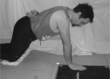
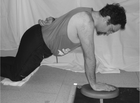
The subjects performed three trials for each of the three conditions, for a total of nine trials for both arms. The testing position was a modified tripod position.2 Subjects were on both knees with one hand on the force plate (Multicomponent Force Plate for Biomechanics, Model #9286, Kistler, Amherst, NY) and the opposite hand on the lower back. To standardize the position, the subjects were instructed to maintain 70° of shoulder flexion, neutral shoulder horizontal abduction/adduction, and 50° of hip flexion throughout data collection. The tester documented this position with goniometric measurements at the start of each trial. Force plate and EMG data were recorded as subjects held the test position under three different conditions: the subject's hand resting directly on the force plate (floor) (Figure 1), on a green Thera-Band® (The Hygenic Corp., Akron, OH) Stability Trainer™ (75% deformable under 1000lb. load) over the force plate, (Figure 2) and on a blue Stability Trainer™ (61% deformable under 1000 lb. load) over the force plate. The order of these positions was randomized for each subject to reduce fatigue or learning effects. Each trial lasted twenty seconds and a one-minute rest was given between trials. Both the dominant and non-dominant arms of each subject were tested. The dominant arm was defined as the arm with which the subjects would throw a ball.
Figure 1:
Subject testing position directly on force plate.
Figure 2:
Subject testing position on green Thera-Band® stability trainer.
Data Analysis
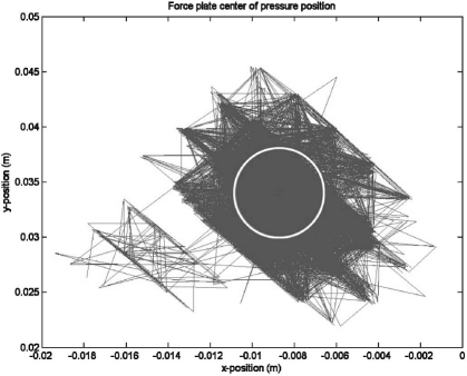
The average location of the center of pressure for each trial was calculated from the ground reaction forces. The mean deviation from the center of pressure was defined as the average distance of the instantaneous center of pressure from the mean location for the entire trial (Figure 3). This distance gives a region where the center of pressure can be expected to be located. To assess the main effects and any interactions, a 2 (hand dominance) × 3 (test condition) repeated-measures analysis of variance (ANOVA) was performed on this measurement.
Figure 3:
The mean deviation from the center of pressure was defined as the average distance the instantaneous center of pressure traveled from its mean location for the entire trial.
The linear envelope (rectified, smoothed) EMG activity was normalized to the maximal activation level determined for each muscle, as described above. Each 20-second trial was divided into three equal parts to examine potential changes in muscle activity over time. The average value over each third of each trial was used for analysis. Repeated-measures ANOVA {2 (hand dominance) × 3 (test condition) × 3 (time)} was then performed to assess the main effects and any interactions of hand dominance, test condition, and test duration on the EMG data from each muscle. Pairwise post-hoc t-tests with Bonferroni corrections were applied where significant main effects were found. Any p values less than 0.05 were considered significant.
RESULTS
Mean Deviation of the Center of Pressure
Statistical analysis revealed a significant main effect of the stability condition on the mean deviation of the center of pressure (p= 0.015). The mean deviation of the center of pressure was lower for the floor condition compared to either of the Stability Trainers (p = 0.04). No difference in the mean deviation existed between the blue and green Stability Trainers (p = 0.977). Additionally, no effect of hand-dominance was found for this measurement (p=0.99).
EMG Data
Statistical analysis revealed a significant main effect of time (p=0.005) on overall muscle activity. Further analysis revealed that the activity of the triceps, serratus anterior, and anterior deltoid muscles increased as each trial progressed (p=0.001, p = 0.025, p=0.002, respectively) (Table 1), irrespective of the stability condition utilized. A significant condition by muscle interaction (p=0.015) on overall muscle activity also occurred. Activity in the anterior deltoid, lower trapezius, and serratus anterior muscles significantly decreased with decreasing stability (Main Effect of Condition: p= 0.023, p=0.029, p=0.001, respectively), whereas, activity in the triceps significantly increased (p = 0.002) (Table 2).
Table 1:
Change in shoulder muscle activity (%) over time (beginning of trial to end of trial) irrespective of stability condition.
| Muscle | % Change (SD) | Time Main Effect (p-value) |
|---|---|---|
| Anterior Deltoid | 13.5 (20.6) | 0.002* |
| Posterior Deltoid | −0.7 (15.0) | 0.652 |
| Infraspinatus | 2.5 (19.9) | 0.941 |
| Lower Trapezius | 11.9 (21.4) | 0.457 |
| Upper Trapezius | 4.5 (20.0) | 0.383 |
| Serratus Anterior | 9.1 (17.3) | 0.025* |
| Pectoralis | 10.8 (30.4) | 0.115 |
| Triceps | 11.3 (13.5) | <0.001* |
Significant
Table 2:
Effect of stability condition on shoulder muscle activation.
| Muscle | Floor %MVC (SD) | Green %MVC (SD) | Blue %MVC (SD) | Condition Main Effect (p-value) |
|---|---|---|---|---|
| Anterior Deltoid | 11.0 (7.2) | 9.9 (7.0) | 9.7 (7.0) | 0.023* |
| Posterior Deltoid | 12.5 (8.5) | 12.1 (9.1) | 12.0 (8.3) | 0.506 |
| Infraspinatus | 26.3 (10.0) | 26.0 (9.9) | 25.4 (10.5) | 0.656 |
| Lower Trapezius | 16.2 (8.9) | 15.4 (9.4) | 14.5 (8.2) | 0.029* |
| Upper Trapezius | 4.6 (4.6) | 4.3 (4.1) | 4.3 (4.1) | 0.103 |
| Serratus Anterior | 15.0 (9.3) | 13.5 (9.2) | 13.0 (9.6) | <0.001* |
| Pectoralis | 8.7 (7.2) | 9.6 (8.6) | 9.6 (8.1) | 0.135 |
| Triceps | 23.1 (11.6) | 25.1 (12.8) | 25.1 (12.5) | 0.002# |
EMG activity decreased as task stability decreased.
EMG activity increased as task stability decreased.
DISCUSSION
When complementing common open chain therapeutic exercises with closed chain therapeutic exercise during shoulder rehabilitation, the demand placed on the surrounding shoulder musculature during these exercises should be understood. The hypothesis to be tested was that with an increasingly compliant surface, stability would decrease (as evidenced by the increased deviation of the center of pressure) and muscle activity would increase (as evidenced by increased EMG activity). The increase in the mean deviation of the center of pressure indicates that balancing on a foam pad made it more difficult for the subject to maintain the upper extremity in a stable position. The EMG data, however, was less conclusive. Anterior deltoid, upper trapezius, lower trapezius, and serratus anterior muscles demonstrated small decreases in muscle activity with decreasing stability, while the triceps showed a small increase. These findings seem to indicate that the increase in center of pressure deviation produced by balancing on the Stability Trainers was not large enough to require an increase in shoulder muscle activation. Compared to balancing on the floor, balancing on the Stability Trainers most likely changed the position of the hand and the force distribution under it. These changes may have produced the need for more stability at the elbow, hence the increase in triceps activity.
Muscle activity increased throughout each trial, which may indicate muscle fatigue.5 While muscle fatigue was not explicitly measured, but seems to be a logical conclusion based on the increase in EMG activity over the length of the trial. Physiologically, as a muscle fatigues, more motor units are recruited in order to maintain the specific force output. This results in an increase in action potentials along the muscle (i.e. increased EMG activity). In order to support the weight of the body and maintain stability, more motor units were recruited as the prime support muscles fatigued. Considering that the activity is essentially an isometric exercise, the increase in EMG activity could not have been due to muscle length changes or to a change in contraction velocity.
Few studies have examined upper extremity weight bearing exercises. Lear et al5 supported incorporating push-up progressions into upper extremity rehabilitation for advanced training of the scapular stabilizers (serratus anterior, upper and lower trapezius muscles) using the push-up “plus” (“plus” indicating active scapular protraction at the end of the up phase). Lear et al5 chose to vary the exercise by elevating the subject's feet and having the subjects place their hands on a mini trampoline. The authors found that elevating the subject's feet had a significant effect on serratus anterior and upper trapezius muscle activity but no significant effect on lower trapezius activity. Placing the hands on an unstable surface also increased activity of the serratus anterior and upper trapezius but did not increase lower trapezius activity. In contrast, the present study demonstrated an increase in triceps activity while anterior deltoid, upper and lower trapezius, and serratus anterior muscle activity decreased with decreasing stability.
The use of the “plus” phase of the push-up in the Lear et al5 study is likely to explain the increase in muscle activity of the serratus anterior. In the current study, subjects were not instructed to hold a protracted position of the scapula during the trials. The low activation levels of the serratus anterior may be explained by the difficulty of protracting the scapula in a unilateral, close kinetic chain position. Additionally, changes in surface compliance may not have provided a strong enough stimulus to require an increase in serratus anterior muscle activity.
While the push-up plus position in rehabilitation is one of the greatest activators of the serratus anterior muscle, the purpose for this study was not to determine what muscles would activate the most, but to see what muscles are activated and to what degree during a standard rehabilitation progression of a stable surface to an unstable surface. Clinically, a patient is not typically placed in a closed chain “plus” position when the program is initiated. This position would be more advanced and would be added at a later time with this current progression. In terms of maintaining “neutral” position, human positioning is always a difficult thing to standardize, especially in the shoulder. While the subject maintained the tripod position, verbal feedback was provided from the investigators when the subject began to shift into a retracted or protracted position (retracted was more common). At this point, the subject was cued to maintain their shoulders parallel to the floor.
Uhl et al2 also sought to determine the demand on shoulder muscles with weight-bearing exercises, and the relationship between increased weight-bearing posture and shoulder muscle activation of the anterior and posterior deltoid, infraspinatus, pectoralis major, and supraspinatus muscles in a progression of seven static upper extremity weight-bearing exercises. The authors found that force, measured through household bathroom scales, significantly increased with an increase in weight-bearing position (r = 0.97, p < 0.01). They also found that muscle activity changed with position and increased with the progression of exercises. Similar to the present findings, the infraspinatus had the highest EMG activity in all conditions. Additionally, the standard push-up had the highest levels of muscle activation, with values significantly higher than the majority of other exercises. Uhl et al2 concluded that alterations of weight-bearing exercises, by varying the amount of arm support and force, resulted in very different demands on the shoulder musculature.
A properly designed shoulder rehabilitation program needs to encompass both open and closed chain therapeutic exercises. Previous studies have provided insight into the muscle activity generated by various open chain exercises.13–16 Complementing these exercises with closed chain training, will offer a well-balanced rehabilitation program. However, only a few studies have offered insight into the muscle activity generated by upper extremity closed chain exercises.2,3,5 Understanding a progression of when and how to incorporate these closed chain exercises into a rehabilitation program is very important. This study has demonstrated that by increasing the compliance of the surface during a tripod closed chain exercise, the ability to maintain stability is challenged. Interestingly, no significant difference was noted between maintaining stability with the green or the blue Thera-Band® Stability Trainer. As noted earlier, balancing on the Stability Trainers may have altered hand position and distribution of forces acting on the hand. While this altered hand position changes the deviation of the center of pressure, an increase in shoulder muscle activity was not necessary to maintain stability at the level of the shoulder. Also, because the Stability Trainers were compliant, subjects tended to “sink” into the surface. Therefore, stability may have been added to the system due to the fact that the foam conformed around the hand as subjects held the tripod position. While the foam Stability Trainers have been shown to increase the deviation of the center of pressure, using a less compliant but unstable surface, such as a balance board, may be more successful in eliciting an increase in shoulder muscle activity.
This study utilized healthy subjects with no history of upper extremity involvement; future work should investigate the utilization of upper extremity closed chain exercises for subjects with shoulder pathology. In choosing appropriate interventions for individuals with impairment, it will be necessary to consider the different forces being transmitted through the shoulder with these exercises in addition to desired muscle activity, selective muscle activation, and patient tolerance to positions.
CONCLUSION
The use of unstable surfaces was shown to progressively challenge proprioception and joint stability in the upper extremity. However, providing progressively more unstable surfaces did not lead to a progressive and selective increase in muscle activity. Although activity increased in most muscles throughout the exercise duration irrespective of the stability condition, increasing the difficulty of the task did not have a similar effect. The compliance of the foam Stability Trainer pads may not have provided enough propioceptive stimulus to elicit an increase in shoulder muscle activation. Using a less compliant unstable surface may produce the desired increases in shoulder muscle activation; however, further investigation needs to determine a safe and selective progression of treatment.
REFERENCES
- 1.Ellenbecker TS, Cappel K. Clinical application of closed kinetic chain exercises in the upper extremities. Orthop Phys Ther Clin N Am. 2000;9: 231–245 [Google Scholar]
- 2.Uhl TL, Carver TJ, Mattacola CG, et al. Shoulder musculature activation during upper extremity weight-bearing exercise. J Orthop Sports Phys Ther. 2003;33:109–117 [DOI] [PubMed] [Google Scholar]
- 3.Ludewig PM, Hoff MS, Osowski EE, et al. Relative balance of serratus anterior and upper trapezius muscle activity during push-up exercises. Am J Sports Med. 2004;32:484–493 [DOI] [PubMed] [Google Scholar]
- 4.Kibler WB, Livingston B. Closed-chain rehabilitation for upper and lower extremities. J Am Acad Orthop Surg. 2001;9:412–21 [DOI] [PubMed] [Google Scholar]
- 5.Lear LJ, Gross MT. An electromyographical analysis of the scapular stabilizing synergists during a push-up progression. J Orthop Sports Phys Ther. 1998;28:146–57 [DOI] [PubMed] [Google Scholar]
- 6.Basmajian JV, Deluca CJ. Muscles Alive: Their Functions Revealed by Electromyography. 5th edition.Baltimore: Williams & Watkins; 1985 [Google Scholar]
- 7.Arwart HJ, de Groot J, Van Woensel WW, et al. Electromyography of shoulder muscles in relation to force direction. J Shoulder Elbow Surg. 1997;6:360–370 [DOI] [PubMed] [Google Scholar]
- 8.Zipp P. Recommendations for the standardization of lead positions in surface electromyography. Eur J Appl Physiol Occup Physiol. 1982;50:41–54 [Google Scholar]
- 9.Decker MJ, Hintermeister RA, Faber KJ, et al. Serratus anterior muscle activity during selected rehabilitation exercises. Am J Sports Med. 1999;27:784–791 [DOI] [PubMed] [Google Scholar]
- 10.Wise MB, Uhl TL, Mattacola CG, et al. The effect of limb support on muscle activation during shoulder exercises. J Shoulder Elbow Surg. 2004;13:614–620 [DOI] [PubMed] [Google Scholar]
- 11.Kendall FP, McCreary EK, Provance PG. Muscle Testing and Function. Baltimore: Williams and Wilkins; 1993 [Google Scholar]
- 12.Kamen G, Caldwell GE. Physiology and interpretation of the electromyogram. J Clin Neurophyiol. 1996;13:366–84 [DOI] [PubMed] [Google Scholar]
- 13.Townsend H, Jobe FW, Pink M, et al. Electromyographic analysis of the glenohumeral muscles during a baseball rehabilitation program. Am J Sports Med. 1991;3:264–272 [DOI] [PubMed] [Google Scholar]
- 14.Blackburn TA, McLeod WD, White B, et al. EMG analysis of posterior rotator cuff exercises. Athletic Training. 1990;40–45 [Google Scholar]
- 15.Moseley JB, Jobe FW, Pinks M, et al. EMG analysis of the scapular muscles during a shoulder rehabilitation program. Am J Sports Med. 1992;20:128–134 [DOI] [PubMed] [Google Scholar]
- 16.Reinold MM, Wilk KE, Fleisig GS, et al. Electromyographic analysis of the rotator cuff and deltoid musculature during common shoulder external rotation exercises. J Orthop Sports Phys Ther. 2004;34:385–94 [DOI] [PubMed] [Google Scholar]


