Abstract
Background
Although anterior cruciate ligament (ACL) sprains usually occur during the initial phase of the landing cycle (less than 40° knee flexion), the literature has focused on peak values of knee angles, vertical ground reaction force (VGRF), and muscle activity even though it is unclear what occurs during the initial phase of landing.
Objectives
The objectives of this study were to determine the effects of sex (male and female) and fatigue (prefatigue/post-fatigue) on knee flexion angles at the occurrence of peak values of biomechanical variables [knee valgus angle, VGRF, and normalized electromyographic amplitude (NEMG) of the quadriceps and hamstring muscles] during a bilateral drop landing task.
Methods
Knee valgus angle, VGRF, and NEMG of the quadricep and hamstring muscles were collected during bilateral drop landings for twenty-nine recreational athletes before and after a fatigue protocol.
Results
Peak values of knee valgus, VGRF, and NEMG of medial and lateral hamstring muscles occurred during the late phase of the landing cycle (>40° of knee flexion). Females in the post-fatigue condition exhibited peak VGRF at significantly less knee flexion than in the pre-fatigue condition. Males in the post-fatigue condition exhibited peak lateral hamstring muscles NEMG at significantly higher knee flexion than in the pre-fatigue condition.
Discussion and Conclusion
Peak values of biomechanical variables that have been previously linked to ACL injury did not occur during the initial phase of landing when ACL injuries occur. No biomechanical variables peaked during the initial phase of landing; therefore, peak values may not be an optimal indicator of the biomechanical factors leading to ACL injury during landing tasks.
INTRODUCTION
Sprains of the anterior cruciate ligament (ACL) are often season-ending injuries that cause significant physical and emotional burden on the injured athlete. These ACL injuries also create substantial financial impact with costs related to orthopaedic care and rehabilitation in the US reaching approximately $850 million each year.1
Research efforts aimed at the prevention of ACL sprains have focused on improving the understanding of the biomechanics at the moment of injury. Previous researchers have analyzed ACL sprains captured on video tape in a variety of sports such as team handball, basketball, soccer, and volleyball. These studies reported that the majority of ACL injuries occur during the initial phase of landing when the knee is flexed less than 40°.2–4 Additional evidence suggesting that the initial phase of landing (less than 40° of knee flexion) represents the most vulnerable range for ACL tears comes from cadaveric,5,6 “in vivo,”7,8 and computer simulation9,10 studies of ACL strain or force. These studies form a remarkable consensus within the literature, suggesting that the initial phase of landing from a jump may be the most appropriate focus of biomechanical studies attempting to clarify the mechanism of ACL injury. Despite this consensus, current biomechanical studies11–15 have focused analysis on variables without verifying that these variables occur in the initial phase of landing.
Landing from a jump has been cited as one of the most common athletic maneuvers to cause ACL injuries,1,3,4,16–20 and several researchers have investigated the biomechanics of landing.11–15 Although studies have used different methodological approaches,11,12 many studies have used analysis of peak values of lower extremity joint angles and vertical ground reaction force (VGRF) without regard to the degree of knee flexion.11,14,15 This approach assumes that peak values are valid indicators of important biomechanical events regardless of the degree of knee flexion at which they occur. Consequently, existing studies have not described when peak values occur within the landing cycle relative to the natural progression of knee flexion from initial contact to peak knee flexion. It is unknown if the peak values reported in the previous literature11,14,15 occur during the initial phase of landing when injury risk and ACL strain are greatest, or during the latter stages of the landing cycle, when studies suggest ACL injury risk 2–8 is decreased. If peak values occur during the initial stages of landing, previous studies would be supported by suggesting that peak values occur at angles of knee flexion that are associated with increased ACL injury risk. However, if peak values do not occur during the initial phase of landing, it would suggest that the methods in previous studies focusing only on peak values measured across the entire landing cycle may have inadequately addressed an important factor in ACL injury risk and strain, namely, degree of knee flexion. Therefore, it is important to determine when peak values of key biomechanical variables that have been linked to injury (knee valgus angle, VGRF) occur relative to knee flexion angles during the landing cycle.
Additionally, although several studies have investigated the effect of sex (male vs female)11–15 and fatigue12,21–25 on the biomechanics of landing, none have described if differences exist in the angle of knee flexion at peak values relative to sex and fatigue. Such differences may influence the interpretation of data to date and future data. For example, if women have a peak value which is significantly higher than men, but the peak value for women occurs much later in the cycle than the peak value for men, direct comparison of these values for injury risk may be not be valid given the influence of knee joint angle on ACL forces and relative function of various muscles about the knee. Therefore, this study will also examine differences in knee flexion at the occurrence of peak biomechanical values relative to sex and fatigue.
Biomechanical variables that have been linked to knee injury by investigators include knee valgus, VGRF, and lower extremity muscle activity (LEMG). In a prospective biomechanical-epidemiological study,26 athletes with higher knee valgus angles during landing were at higher risk of suffering an ACL sprain. High VGRF has been linked to injury risk as the forces are being transferred up the kinetic chain, thereby, increasing the moments and forces in the joints.27 Strong contractions of the quadricep muscles have been shown to increase anterior translation of the tibia and place increased demands on the ACL9,28 while hamstrings muscle contractions reduce the force within the ACL.9 Most ACL injuries occur during closed kinetic chain activities with the foot planted and the quadricep muscles eccentrically contracting such as during landing from a jump or cutting.
Given this information presented, the objectives of the present study were:
To describe the knee flexion angles at which peak bio-mechanical variables (knee valgus, VGRF, and NEMG of the rectus femoris, vastus medialis, medial hamstring, and lateral hamstring muscles) occur during bilateral drop landings from a 40 cm platform.
To statistically evaluate the difference between sex and fatigue status on the knee flexion angles at which peak biomechanical variables occur.
METHODS
Subjects
Twenty-nine recreational athletes (14 females and 15 females) between the ages of 20-40 years were recruited. The inclusion criteria were willingness to participate in the study and participation in recreational sports at least twice/week for a minimum of 45 minutes per practice session. Exclusion criteria were: obesity (body mass index greater than 30 kg/m2); a history of injuries or diseases that would render unsafe the execution of the protocol; and a history of injuries or diseases that could affect the biomechanics of landing, such as lower extremity fractures. Subjects were excluded if they had received specialized training in jumping and landing techniques as could occur through participation in gymnastics or dance.
Instrumentation
Electromyographic data were collected with the Noraxon Myosystem 1400 (Noraxon USA, Inc., Scottsdale, AZ). The electrodes were disposable, surface, passive electrodes (blue sensor, Ambu, Inc., Linthicum, MD). The skin was prepared and the surface electrodes were placed on the rectus femoris, vastus medialis, lateral hamstring, and medial hamstring muscles as in previous research.22 These sites of electrode placement are consistent with established guidelines29 and are located between the motor point and the distal tendon in order to improve intra and inter-subject comparison reliability.30 Two electrodes were placed on each muscle at a 20 mm inter-electrode distance and parallel to fiber orientation.29 Athletic tape was used to fixate the electrodes and decrease movement artifact.29
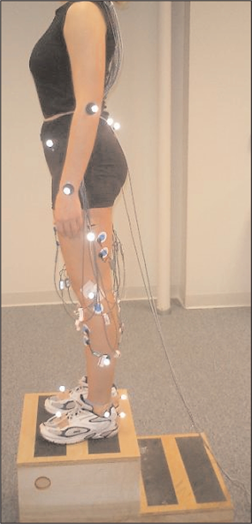
Kinematic data were collected with the use of eight Eagle cameras (Motion Analysis Corp. Santa Rosa, CA) and reflective markers were placed bilaterally as per established protocol31 on the second dorsal metatarsophalangeal joint, calcaneus, lateral malleolus, lateral femoral epicondyle, lateral mid-fibula (half way between the calcaneal and lateral femoral epicondyle markers), lateral mid-thigh (half way between the lateral femoral epicondyle and anterior superior iliac spine markers), anterior superior iliac spine, acromion, lateral humeral epicondyle, distal radioulnar joint, sacrum and left posterior superior iliac spine (offset) (Figure 1). The software for data collection was the EvaRT 4.0 (Motion Analysis Corp. Santa Rosa, CA).
Figure 1.

Participant with markers and electrodes preparing to perform a drop landing.
The force plate was an OR6-5 AMTI biomechanical platform (AMTI, Watertown, MA). The force platform was time synchronized to the electromyography (EMG) and the motion analysis system. The kinetic and EMG data were sampled at 1200 Hz and the kinematic data were sampled at 240 Hz as appropriate for fast athletic maneuvers.14
Experimental Protocol
Subjects were informed of the study protocol and total time needed for testing. All risks and possible harm as described in the consent form were verbally explained. All subjects completed a sports activity and medical history questionnaire, signed a consent form approved by the Institutional Review Board at New York University School of Medicine, and were measured for height, weight, foot width and length, and knee width.
Subjects completed the entire protocol (three landings pre-fatigue, fatigue protocol, and three landings post-fatigue) in a single session. The subjects were allowed two practice jumps and then performed three bilateral drop landings from a 40 cm platform. They were instructed to drop directly down off the box and land with both legs on the force plate. Subjects did not receive any instructions on the landing technique to avoid a coaching effect. The effect of the arms was minimized by asking the subjects to keep their arms crossed against their chest.32,33 Trials were repeated when they were judged as non-acceptable (such as when subjects lost their balance or did not land with both feet on the force plate) by the primary investigator who was observing the real-time data on the monitor, the research assistant who was closely monitoring the jumps, or the subject. Upon completion of three successful landings, the wires were disconnected from the electrodes (but the electrodes were not removed). The subjects then followed the fatigue protocol: they jumped over five consecutive 5-7 cm obstacles. This was repeated 20 times for a total of 100 jumps. Then, the subjects jumped maximally vertically 50 times. After the fatigue protocol was completed, the wires were re-connected to the EMG electrodes and the same procedure of landing assessment was repeated for the post-fatigue part of data collection. All subjects completed all post-fatigue trials within six minutes after the completion of the fatigue protocol.
Data Processing
The analysis of the data was performed with Orthotrak 5.0 (Motion Analysis Corp. Santa Rosa, CA). Kinematic data were smoothed using a Butterworth fourth order low pass filter with a cut-off frequency of 6 Hz. The EMG data were filtered through a 6th order Butterworth filter (10-500Hz).31 The EMG amplitude was normalized to the maximum linear-enveloped EMG of each muscle32,34–36 exhibited during the landing phase of bilateral landings from a 20 cm platform (mean of three trials). The VGRF was normalized to body weight as in previous studies.16,37
Statistical Analysis
This project utilized a repeated measures pre-fatigue and post-fatigue experimental design that used measures of NEMG, kinetic, and kinematic data. The knee flexion angles at which each biomechanical variable peaked were averaged for the three trials. The kinetic, kinematic, and NEMG data of the dependent variables relative to the different levels of the independent variables were entered into a statistical software package (SPSS 12.0, SPSS Inc., Chicago, IL, 60606).
The independent variables were sex (male/female) and level of fatigue (pre-fatigue/post-fatigue). The dependent variables were knee flexion angle at the occurrence of peak values for the following biomechanical variables: knee valgus angles; VGRF; and NEMG amplitude of the rectus femoris, vastus medialis, medial hamstring, and lateral hamstring muscles. All NEMG and kinematic measurements were in reference to the right lower extremity (which was the dominant leg determined by leg used for kicking a ball) for all participants. Descriptive statistics (mean and SD) were produced for the values of knee flexion angle at the occurrence of peak values of the dependent variables (four NEMG amplitudes, VGRF, and knee valgus angle). The data were inspected and tested to ensure that the assumptions for data normality and sphericity of the univariate and multivariate repeated measures analysis of variance (MANOVA) were not violated.
A MANOVA procedure was used to evaluate the effects of sex (male/female), fatigue (pre-fatigue/post-fatigue) and their interaction on knee flexion angle at the occurrence of peak values. Follow up analysis of variance (ANOVA) tests were performed when the MANOVA reached significance (p<0.05)38,39 to determine which of the variables achieved significance. Significance was accepted at p<0.05.
RESULTS
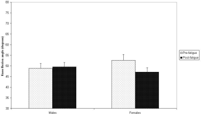
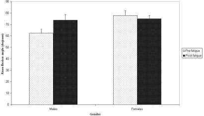
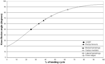
No landing trial had to be repeated due to subjects losing their balance or failing to follow the instructions. No differences between males and females existed in respect to weekly number of sports participation hours as reported by the volunteers [mean hours/wk (SD): males: 6.6 (3), females 7.1 (6), p = 0.77]. Peak values for all investigated biomechanical variables occurred when the knee was flexed more than 40° (Figures 2–5). The results of the MANOVA found that neither sex (df = 7:21; F = 2.44, p = 0.053) nor fatigue (df = 7:21; F = 1.91, p = 0.119) had a significant effect on knee flexion angle at occurrence of peak values of the biomechanical variables, however, the interaction of sex x fatigue was statistically significant (df = 7:20; F = 4.8, p < 0.05). Univariate repeated-measures ANOVA tests were performed for sex x fatigue and determined that two of the variables were significantly different: 1) knee flexion at peak VGRF (p < 0.05) - the knee angle increased in males by 1° but decreased by 5.6° in females in the post fatigue condition [mean (SD); non-fatigued males: 48.8° (±13), fatigued males: 49.6° (±15), non-fatigued females: 52.7° (±11), 47.1° (±11)] (see Figure 6); 2) knee flexion angle at peak lateral hamstring muscles NEMG (p = 0.003) - the knee angle increased by 11° in males but decreased in females by 3° in the post fatigue condition [mean (SD); non-fatigued males: 62.2° (±19), fatigued male: 73.7° (±21), non-fatigued females: 77.8° (±16), fatigued females: 75° (±15)] (Figure 7).
Figure 2.
Occurrence of peak values in the landing cycle: non-fatigued males
Figure 5.
Occurrence of peak values in the landing cycle: fatigued females
Figure 6.
Knee flexion angle at occurrence of peak vertical ground reaction force
Figure 7.
Knee flexion angle at occurrence of peak lateral hamstring muscles normalized electromyographic amplitude
Figure 3.
Occurrence of peak values in the landing cycle: fatigued males
Figure 4.
Occurrence of peak values in the landing cycle: non-fatigued females
DISCUSSION
The present study examined knee flexion angles at the occurrence of peak biomechanical values. The peak values of all variables, (including variables that have been previously cited as contributors to ACL injury: knee valgus, VGRF, and quadriceps activity)2,15,26–28 did not occur during the initial phase of the landing cycle (knee flexion angle less than 40°) when ACL injury risk is increased. Given the literature cited in the introduction, these findings suggest that the methods used in previous studies focusing only on peak values measured across the entire landing cycle may have inadequately addressed an important factor in ACL injury risk, namely, the degree of knee flexion at which peak biomechanical values occur. These studies have provided insight with regards to biomechanical differences between males and females; they found that females land with greater peak knee valgus,14,15,40 greater peak quadriceps NEMG amplitude,22 and greater peak VGRF41 compared to males. Previous findings on peak values42 also suggest that females exhibit greater knee valgus and VGRF than males, however, the differences due to sex on the effect on quadricep muscle NEMG did not reach statistical significance. Although peak values of biomechanical variables occur and can be analyzed after 40° of knee flexion, examining biomechanical variables at peak values may not be the optimal methodological approach given the potential for knee flexion to influence ACL injury risk.
The two variables that were significantly different due to the interaction of sex x fatigue were VGRF and NEMG of the lateral hamstring muscles. Although contraction of the hamstring muscles can effectively decrease anterior tibial translation and prevent excessive stress on the ACL,9 it is unclear if the observed increase of 11° in knee flexion angle at the peak NEMG of the lateral hamstrings in males represents a finding that is related to the ACL injury mechanism. More likely, this study's findings relative to lateral hamstring muscle NEMG may not be clinically relevant as peak values of lateral hamstring muscles occur very late in the landing cycle (more than 60° of knee flexion) where ACL injury risk is less. An alternative explanation of the effect of fatigue on lateral but not medial hamstring muscles may be related to an effort to resist a frontal plane or rotary force.
However, the findings relative to VGRF may have clinical relevance as VGRF has been identified as a variable important to ACL injuries15,27 and the peak values occurred at knee flexion angles which are much closer to the angles known to demonstrate increased risk. In the current study, after a fatiguing protocol, females decreased the amount of knee flexion at which peak VGRF occurred by 5.6° to a value of 47.1°, while men increased the amount of knee flexion by 0.8° to a value of 49.6°. It appears that peak VGRF in men tends to occur in similar or slightly higher knee flexion angles in the post-fatigue condition while in women peak VGRF tends to occur in lower knee flexion angles, thereby, placing their knees closer to knee flexion values known to be related to ACL injury risk. This effect may be magnified with a fatigue protocol that is either more vigorous or ensures that all subjects are fatigued to the same level and potentially cause peak VGRF in fatigued females to occur when the knee is flexed less than 40° and the ACL more vulnerable to trauma.
In addition to finding that all peak variables occurred after the initial phase of landing and that the interaction of sex (male vs female) x fatigue (pre-fatigue/post-fatigue) was significant for VGRF and NEMG of the lateral hamstrings muscle, the current study also found that knee flexion angle at which peak values occurred was not significantly different relative to the difference between sex or fatigue (Figures 2–5). Therefore, the findings of the current study suggest that peak values of key biomechanical variables occur at similar knee flexion angles in non-fatigued male and female athletes and in pre and post-fatigued athletes of the same sex. However, caution should be taken in regard to this interpretation as two important issues that can potentially diminish the validity of sex and fatigue comparisons without regard for knee flexion. First, the use of peak values at degrees of knee flexion beyond 40° may not adequately describe biomechanical strain on the ACL. Second, as found in this study, the interaction of sex x fatigue produce significant differences in some variables and, therefore, knee flexion angles may have an influence on biomechanical strain of the ACL when examining differences between males and females using a fatiguing protocol.
This specific fatigue protocol was chosen because the combination of tasks simulates activities commonly performed in sports and because an eccentric-concentric fatigue protocol is more effective in producing fatigue than a concentric fatigue protocol.43 The fatigue protocol was designed in a way that the fatigue-induced pattern was applicable to functional activities outside the laboratory setting. The protocol used in the present study was similar to fatigue protocols used in previous research.12,21 Other research21,44 has demonstrated that a fatigue protocol similar to the one used in the current study is sufficient to induce fatigue in a similar way to subjects of different training levels.45 Moreover, the demands of games such as soccer are very similar for males and females in terms of distance covered, sprint duration, and exercise intensity46 suggesting that laboratory fatigue protocols have greater applicability if they fatigue male and female athletes in a similar way as it occurs on the athletic field.
Implications for Future Research
As measurements of peak values occur late in the landing cycle when ACL injury risk is less, future biomechanical studies may be improved by examining biomechanical variables during the initial phase of landing. Future studies should determine if measurements at predefined knee flexion angles in the initial phase of landing are better predictors of ACL injury than peak values which occur after 40° of knee flexion. Future research should also investigate the differences between males and females using a more vigorous fatigue protocol in order to determine if increased fatigue may further alter the degree of knee flexion at which peak VGRF values occur. Considering the rapid proliferation of biomechanical studies of landing from a jump in recent years, the limited number of subjects in most studies, and the highly variable methodology across studies, methodology standardization may be needed to allow a meta-analysis investigation. The present study represents a first step towards standardization of methodology by suggesting that appropriate measures of biomechanical variables should occur during the initial phase of landing and by demonstrating that peak values do not occur until later in the landing cycle. Future studies should identify the variables that best predict ACL injury and the exact time in the initial phase of the landing cycle that the variable should be measured.
Limitations
Although all subjects were fatigued with the same fatigue protocol as opposed to normalizing the protocol to their athletic abilities, a specific measure of fatigue could have been used to ensure that all subjects had exceeded some minimum cut-off. Doing so might have allowed for a more meaningful interpretation of the effect of fatigue.
A general limitation of the present study is that the landing task may not adequately represent landing techniques on the athletic field because subjects were instructed to keep their arms crossed across their chest and jump down from a platform. Although these modifications were deemed necessary in order to have all subjects perform the same task with minimal variability, generalizability of the findings is decreased. In addition to drop landings, which have been used extensively in the literature of sports injury biomechanics,14,40,47 researchers have also used stop-jump and cutting maneuvers.11–13,48,49 Investigating both drop landings and continuous tasks such as cutting or stop-jump may have provided a more comprehensive picture of the effect of sex and fatigue on the biomechanical variables. Additionally, as with all biomechanics studies, direct implications to ACL injury cannot be made as no injuries occurred during the testing.
All subjects were recreational athletes who participated at least twice per week in a variety of sports that involved jumping. No differences existed between males and females in regards to hours of sports participation per week. However, this lack of a difference does not ensure equal proficiency in drop landings. Some subjects may have been more proficient than others in landing from a jump. A more homogenous group of subjects such as recreational basketball or volleyball players would make the findings of this study less generalizable but may increase its internal validity.
CONCLUSION
In summary, the present study demonstrated that peak values of the biomechanical variables that have been previously cited as contributors to ACL injury, such as knee valgus, VGRF, and quadricep muscles activity did not occur during the initial phase of the landing cycle when ACL injury risk is greatest. This finding suggests that analyses based only on peak values may not be adequately addressing the influence of knee flexion on ACL strain which is higher when the knee is in less than 40° of flexion.
REFERENCES
- 1.Griffin L, Agel J, Albohm M, et al. Noncontact anterior cruciate ligament injuries: Risk factors and prevention strategies. J Am Acad Orthop Surg. 2000;8:141–150 [DOI] [PubMed] [Google Scholar]
- 2.Olsen O, Myklebust G, Engebretsen L, et al. Injury mechanisms for anterior cruciate ligament injuries in team handball. Am J Sports Med. 2004;32:1002–1012 [DOI] [PubMed] [Google Scholar]
- 3.Griffin L. Prevention of Noncontact ACL Injuries. Rosemont, IL: American Academy of Orthopaedic Surgeons, 2001 [Google Scholar]
- 4.Boden BP, Dean GS, Feagin, JA, Jr., et al. Mechanisms of anterior cruciate ligament injury. Orthopedics. 2000;23:573–578 [DOI] [PubMed] [Google Scholar]
- 5.Kanamori A, Woo S, Ma B, et al. The forces in the anterior cruciate ligament and knee kinematics during a simulated pivot shift test: A human cadaveric study using robotic technology. Arthroscopy. 2000;16:633–639 [DOI] [PubMed] [Google Scholar]
- 6.Markolf K, O'Neil G, Jackson S. Effects of applied quadriceps and hamstrings muscle loads on forces in the anterior and posterior cruciate ligaments. Am J Sports Med. 2004;32:1144–1149 [DOI] [PubMed] [Google Scholar]
- 7.Pedowitz RA, O'Connor J, Akeson W. Daniel's Knee Injuries: Ligament and Cartilage Structure, Function, Injury and Repair. 2nd ed.Philadelphia, PA: Lippincott, Williams & Wilkins; 2003 [Google Scholar]
- 8.Heijne A, Fleming BC, Renstrom PA, et al. Strain on the anterior cruciate ligament during closed kinetic chain exercises. Med Sci Sports Exerc. 2004;36:935–941 [DOI] [PubMed] [Google Scholar]
- 9.Pandy MG, Shelburne KB. Dependence of cruciate-ligament loading on muscle forces and external load. J Biomech. 1997;30:1015–1024 [DOI] [PubMed] [Google Scholar]
- 10.Pflum M, Shelburne K, Torry M, et al. Model prediction of anterior cruciate ligament force during drop-landings. Med Sci Sports Exerc. 2004;36:1949–1958 [DOI] [PubMed] [Google Scholar]
- 11.Sell T, Ferris C, Abt J, et al. The effect of direction and reaction on the neuromuscular and biomechanical characteristics of the knee during tasks that simulate the noncontact anterior cruciate ligament injury mechanism. Am J Sports Med. 2006;34:43–54 [DOI] [PubMed] [Google Scholar]
- 12.Chappell J, Herman D, Knight B, et al. Effect of fatigue on knee kinetics and kinematics in stop-jump tasks. Am J Sports Med. 2005;33:1022–9 [DOI] [PubMed] [Google Scholar]
- 13.Chappell J, Yu B, Kirkendall D, et al. A comparison of knee kinetics between male and female recreational athletes in stop-jump tasks. Am J Sports Med. 2002;30:261–7 [DOI] [PubMed] [Google Scholar]
- 14.Ford K, Myer G, Hewett T. Valgus knee motion during landing in high school female and male basketball players. Med Sci Sports Exerc. 2003;35:1745–1750 [DOI] [PubMed] [Google Scholar]
- 15.Kernozek T, Torry M, Van Hoof H, et al. Gender differences in frontal and sagittal plane biomechanics during drop landings. Med Sci Sports Exerc. 2005;37:1003–1012 [PubMed] [Google Scholar]
- 16.Hewett T, Lindenfeld T, Riccobene J, et al. The effect of neuromuscular training on the incidence of knee injury in female athletes. Am J Sports Med. 1999;27:699–705 [DOI] [PubMed] [Google Scholar]
- 17.Kirkendall D, Garrett W. The anterior cruciate ligament enigma: Injury mechanisms and prevention. Clin Orthop. 2000;372:64–8 [DOI] [PubMed] [Google Scholar]
- 18.Arendt E, Dick R. Knee injury patterns among men and women in collegiate basketball and soccer NCAA data and review of literature. Am J Sports Med. 1995;23:694–701 [DOI] [PubMed] [Google Scholar]
- 19.Kirialanis P, Malliou P, Beneka A, et al. Occurence of acute lower limb injuries in artistic gymnasts in relation to event and exercise phase. Br J Sports Med. 2003;37:137–139 [DOI] [PMC free article] [PubMed] [Google Scholar]
- 20.Gray J, Taunton J, McKenzie D, et al. A survey of injuries to the anterior cruciate ligament of the knee in female basketball players. Int J Sports Med. 1985;6:314–316 [DOI] [PubMed] [Google Scholar]
- 21.Madigan M, Pidcoe P. Changes in landing biomechanics during a fatiguing landing activity. J Electromyography Kines. 2003;13:491–498 [DOI] [PubMed] [Google Scholar]
- 22.Fagenbaum R, Darling W. Jump landing strategies in male and female college athletes and the implications of such strategies for anterior cruciate ligament injury. Am J Sports Med. 2003;31:233–240 [DOI] [PubMed] [Google Scholar]
- 23.Rozzi S, Lephart S, Fu F. Effects of muscular fatigue on knee joint laxity and neuromuscular characteristics of male and female athletes. J Athl Train. 1999;34:106–114 [PMC free article] [PubMed] [Google Scholar]
- 24.Bonnard M, Sirin A, Oddsson L, et al. Different strategies to compensate for the effects of fatigue revealed by neuromuscular adaptation processes in humans. Neurosci Lett. 1994;166:101–5 [DOI] [PubMed] [Google Scholar]
- 25.McLean S, Felin R, Suedekum N, et al. Impact of fatigue on gender-based high-risk landing strategies. Med Sci Sports Exerc. 2007;39:502–514 [DOI] [PubMed] [Google Scholar]
- 26.Hewett T, Myer G, Ford K, et al. Biomechanical measures of neuromuscular control and valgus loading of the knee predict anterior cruciate ligament injury risk in female athletes. Am J Sports Med. 2005;33:492–501 [DOI] [PubMed] [Google Scholar]
- 27.Dufec J, Bates B. Biomechanical factors associated with injury during landing in jump sports. Sports Med. 1991; 12:326–337 [DOI] [PubMed] [Google Scholar]
- 28.DeMorat G, Weinhold P, Blackburn T, et al. Aggressive quadriceps loading can induce noncontact anterior cruciate ligament injury. Am J Sports Med. 2004;32: 477–483 [DOI] [PubMed] [Google Scholar]
- 29.Hermens H, Freriks B, Disselhorst-Klug C, et al. Development of recommendations for SEMG sensors and sensor placement procedures. J Electromyography Kinesiol. 2000;10:361–374 [DOI] [PubMed] [Google Scholar]
- 30.Basmajian J, DeLuca C. Muscles Alive. Baltimore, MD: Williams and Wilkins, 1985 [Google Scholar]
- 31.Richards J, Orthotrak 5.0. Santa Rosa, CA: Motion Analysis, 2002 [Google Scholar]
- 32.Rodacki A, Fowler N, Bennett S. Vertical jump coordination: Fatigue effects. Med Sci Sports Exerc. 2002; 34:105–116 [DOI] [PubMed] [Google Scholar]
- 33.Decker M, Torry M, Wyland D, et al. Gender differences in lower extremity kinematics, kinetics, and energy absorption during landing. Clin Biomech. 2003;18:662–669 [DOI] [PubMed] [Google Scholar]
- 34.Horita T, Komi P, Nicol C, et al. Effect of exhausting stretch-shortening cycle exercise on the time courseof mechanical behaviour in the drop jump: Possible role of muscle damage. Eur J Appl Physiol. 1999;79:160–167 [DOI] [PubMed] [Google Scholar]
- 35.Arampatzis A, Morey-Klasping G, Bruggemann G. The effect of falling height on muscle activity and foot motion during landings. J Electromyography Kinesiol. 2003;13: 533–544 [DOI] [PubMed] [Google Scholar]
- 36.Viitasalo J, Salo A, Lahtinen J. Neuromuscular functioning of athletes and non-athletes in the drop jump. Eur J Appl Physiol. 1998;78:432–440 [DOI] [PubMed] [Google Scholar]
- 37.Hewett T, Stroupe A, Nance T, et al. Plyometric training in female athletes: Decreased impact forces and increased hamstring torques. Am J Sports Med. 1996;24:765–773 [DOI] [PubMed] [Google Scholar]
- 38.Bray J, Maxwell S. Multivariate Analysis of Variance: Quantitative Applications in the Social Sciences. Newbury Park: Sage Publications, 1985 [Google Scholar]
- 39.Stevens J. Applied Multivariate Statistics for the Social Sciences, 4th ed.Mahwan: Lawrence Erlbaum Associates, 2002 [Google Scholar]
- 40.Hewett T, Myer G, Ford K. Decrease in neuromuscular control about the knee with maturation in female athletes. J Bone Joint Surg. 2004;86-A:1601–1608 [DOI] [PubMed] [Google Scholar]
- 41.Schmitz J, Kulas A, Perrin D, et al. Sex differences in lower extremity biomechanics during single leg landings. Clinical Biomechanics. 2007;22:681–688 [DOI] [PubMed] [Google Scholar]
- 42.Pappas E, Sheikhzadeh A, Hagins M, et al. The effect of gender and fatigue on the biomechanics of bilateral landings from a jump: Peak values. J Sports Sci Med. 2007; 6:76–83 [PMC free article] [PubMed] [Google Scholar]
- 43.Svantesson U, Osterberg U, Thomee R, et al. Fatigue during repeated eccentric-concentric and pure concentric muscle actions of the plantar flexors. Clin Biomech. 1998;13:336–343 [DOI] [PubMed] [Google Scholar]
- 44.Skurvydas A, Jaskaninas J, Zachovajevas P. Changes in height of jump, maximal voluntary contraction force and low frequency fatigue after 100 intermittent or continuous jumps with maximal intensity. Acta Physiologica Scandinavia. 2000;169:55–62 [DOI] [PubMed] [Google Scholar]
- 45.Skurvydas A, Dudodiene V, Kalvenas A, et al. Skeletal muscle fatigue in long distance runners, sprinters and untrained men after repeated drop jumps performed at maximal intensity. Scand J Med Sci Sports. 2002;12:34–39 [DOI] [PubMed] [Google Scholar]
- 46.Davis J, Brewer J. Applied physiology of female soccer players. Sports Med. 1993;16:180–9 [DOI] [PubMed] [Google Scholar]
- 47.Zazulak B, Ponce P, Straub S, et al. Gender comparison of hip muscle activity during single-leg landing. J Orthop Sports Phys Ther. 2005;35:292–99 [DOI] [PubMed] [Google Scholar]
- 48.McLean S, Lipfert S, Van Den Bogert A. Effect of gender and defensive opponent on the biomechanics of sidestep cutting. Med Sci Sports Exerc. 2004;36:1008–16 [DOI] [PubMed] [Google Scholar]
- 49.Pollard C, McClay Davis I, Hamill J. Influence of gender on hip and knee mechanics during a randomly cued cutting maneuver. Clin Biomech. 2004;19:1022–1031 [DOI] [PubMed] [Google Scholar]


