Abstract
Athletes performing overhead activities are at risk of sustaining both overuse and traumatic shoulder injuries. Research studies utilizing electromyography have identified therapeutic exercises that are effective in the muscular activation of the rotator cuff and the scapular stabilizers. Sports medicine professionals routinely prescribe these traditional therapeutic exercises when rehabilitating athletes. Failing to identify and address contributing musculoskeletal dysfunctions may delay an athlete's successful return to sport. Integrating shoulder and core exercises can address potential musculoskeletal dysfunctions while serving as a transitional program between the initial therapeutic exercises and the terminal return to sport rehabilitation program.
Keywords: shoulder, therapeutic exercise, core stability
Physical therapists are challenged to return athletes to their respective sport as quickly as possible. A comprehensive shoulder rehabilitation program for the injured athlete participating in overhead activity may include the use of modalities to decrease pain, mobility exercises and manual therapy to address range of motion deficits, and strengthening exercises to address shoulder weakness.1 Failure to identify and treat contributing musculoskeletal dysfunctions may delay an athlete's successful return to sport. Kibler (and others)2,3 advocate a proximal stability approach to shoulder rehabilitation. However, previous upper extremity rehabilitation programs propose stabilization training only for the scapular muscles.4,5 Other trunk musculature also become active during glenohumeral movements, and vertebral perturbation occurs during glenohumeral movements.6–9 Other trunk musculature should also receive attention during shoulder rehabilitation. Therefore, the purpose of this article is to review the activity of trunk musculature during upper extremity exercise and present a rehabilitation exercise progression for the shoulder girdle that integrates core muscle strengthening and activation. This progression reflects a transition into traditional functional core strengthening.
INITIAL SHOULDER REHABILITATION EXERCISES
Scapular and glenohumeral musculature provide dual roles of mobility and stability for the shoulder girdle. Muscle weakness, fatigability, or neuro-inhibition of the scapular or glenohumeral musculature may alter normal kinematics, and should be addressed with a comprehensive rehabilitation program.
Kibler et al2,10 advocate a shoulder rehabilitation strategy that addresses proximal dysfunction in the kinetic chain first by targeting scapulothoracic dyskinesis. Research shows that glenohumeral external rotator fatigue is associated with altered scapular kinematics during elevation, which may be related to concomitant fatigue of the scapular and trunk stabilizers.9,11,12 These findings suggest that once the athlete demonstrates improved scapular mechanics, remaining deficits in rotator cuff and scapular muscle strength may be addressed with isolated rehabilitation exercises.2,10
Researchers, utilizing in-dwelling or surface electromyography (EMG), have identified exercises that maximize muscular activity for dysfunctional (weak) rotator cuff muscles and scapular stabilizers.13–16 Table 1 presents the exercises, as reported by Reinhold et al15 and Townsend et al,16 that elicit the highest peak muscular activation for each of the rotator cuff muscles (both studies used in-dwelling EMG) (Table 1). Table 2 presents the exercises, as identified by Moseley et al14 (in-dwelling EMG) and Ekstrom et al13 (surface EMG), that elicit the highest peak muscular activation for selected scapular stabilizers (Table 2). (Note: Other related published EMG reports exist; these four were selected to represent samples of each electromyography procedure).
Table 1.
The Top Exercises for Rotator Cuff Muscle Activity As Reported by Townsend et al16 and Reinhold et al15 EMG Studies
| Muscle | Townsend et al16 | Reinhold et al15 |
|---|---|---|
| Supraspinatus | 1. military press 2. scaption with shoulder internal rotation* |
1. prone horizontal abduction at 100° with full external rotation 2. prone external rotation at 90° of abduction |
| Infraspinatus | 1. horizontal abduction at 90° with external rotation 2. standing external rotation at 0° of abduction |
1. sidelying external rotation at 0° of abduction 2. sidelying external rotation in the scapular plane (45° abduction, 30° horizontal adduction) |
| Teres Minor | 1. sidelying external rotation at 0° of abduction 2. horizontal abduction at 90° with external rotation |
1. sidelying external rotation at 0° of abduction 2. standing external rotation in the scapular plane (45° abduction, 30° horizontal adduction) |
| Subscapularis | 1. scaption with shoulder internal rotation* 2. military press |
not tested |
Note: Rehabilitation professionals must be cautious when prescribing exercises with the shoulder in full internal rotation. Performing scaption with the shoulder in full external rotation is usually a safe and appropriate alternative.
Table 2.
The Top Exercises for Selected Scapular Stabilizers As Reported by Moseley et al14 and Ekstrom et al13 EMG Studies
| Muscle | Moseley et al14 | Ekstrom et al13 |
|---|---|---|
| Upper Trapezius | 1. rowing 2. military press |
1. unilateral shoulder shrug 2. shoulder abduction in the plane of the scapula above 120° |
| Middle Trapezius | 1. horizontal abduction (neutral) 2. horizontal abduction with external rotation |
1. arm raise overhead in line with the lower trapezius muscle fibers 2. shoulder horizontal extension with external rotation |
| Lower Trapezius | 1. abduction 2. rowing |
1. arm raise overhead in line with the lower trapezius muscle fibers 2. shoulder external rotation at 90° of abduction |
| Rhomboids | 1. horizontal abduction (neutral) 2. scaption |
not tested |
| Serratus Anterior | 1. flexion (middle) 2. abduction (middle) 3. scaption (lower) 4. abduction (lower) |
1. diagonal exercise with shoulder flexion, horizontal flexion, and external rotation 2. shoulder abduction in the plane of the scapula above 120° |
As the athlete's injured shoulder strength improves, exercises that replicate sport specific movement patterns should be incorporated.1,2,10 Many of these terminal exercises may involve multi-joint, multi-planar functional movement patterns. An example of a sport specific exercise for an athlete performing an overhead activity would be the two-handed plyoball side-throw toward a rebounder.1 This exercise utilizes leg, hip, and torso movements to generate and transfer force through to the upper extremity.1 Dysfunction within the kinetic chain will impact an athlete's rehabilitation progress and his or her ability to return to sport.
TRUNK MUSCLE ACTIVATION DURING UPPER EXTREMITY EXERCISE
The core is the region of the body comprised of the abdomen, the proximal lower limb, the hips, the pelvis, and the spine. The core musculature serves two functions: lumbopelvic stability and the creation and transfer of forces.3
Trunk musculature becomes active in a feedforward fashion during upper or lower extremity movements.17,18 This feedforward mechanism occurs as the body prepares for potential perturbation of spinal stability when extremities begin movement.19 The thoracic spine, in particular, has been shown to undergo perturbation during glenohumeral elevation.9 Other research shows that trunk muscle activation occurs in response to isometric, small amplitude isotonic, and fast isotonic exercise.7,8,17,18
Several isometric shoulder exercises demonstrate activation of the trunk musculature. Unilateral horizontal shoulder abduction and bilateral shoulder extension performed in standing are associated with the highest activation of trunk musculature.7 The muscles demonstrating the greatest activation during unilateral horizontal abduction include the multifidus and longissimus muscles (greatest activation on the contralateral side) while the external obliques and rectus abdominis muscles were activated the most during bilateral shoulder extension.7
Small amplitude isotonic exercise with the upper extremities, such as that performed with the Body Blade™, has also been shown to activate various trunk musculature in addition to glenohumeral muscles.8 The nature of trunk activation depends upon whether one uses the blade uni-laterally or bilaterally, whether large or small amplitude motions are utilized, and the orientation of the blade (vertical or horizontal). Specific muscles activated include the internal and external obliques, latissimus dorsi, and the erector spinae. Generally, trunk muscle activation increased during two-handed and larger oscillation movements.8
Fast isotonic shoulder movement is also associated with trunk musculature activation.17,18 Arm movement direction does not appear to influence trunk activation; rather movement speed may be the primary factor that influences core activity.
Literature documenting the activation of trunk musculature during slower isotonic speeds suggests a delay in the feedforward mechanism.17 Therefore, exercises known to activate the trunk could be performed simultaneously with isotonic glenohumeral exercises in order to prepare the athlete for more advanced functional exercises. Dysfunction within the kinetic chain will affect how forces are generated, summated, or transferred from proximal segments (legs, hip, torso) to the upper extremity.3,19,20 Weakness within the core may contribute to the development of an overuse upper extremity injury.3,21,22
IDENTIFYING CORE WEAKNESS IN THE ATHLETE PERFORMING OVERHEAD ACTIVITIES
Evaluating an athlete's functional movement patterns and conducting core muscular endurance tests will help a clinician identify a dysfunctional core. Conducting functional tests, such as the single-legged squat or the lunge test, can highlight biomechanical faults.23,24 Common biomechanical faults (Table 3) that may be attributable to a dysfunctional core include contralateral pelvic drop (Trendelenburg sign), hip adduction, or knee valgum (Figure 1).
Table 3.
Lower-Extremity Biomechanical Faults that May Be Observed When a Client has Poor Core Stability
| 1. Contralateral pelvic drop (Trendelenburg sign) |
| 2. Femoral adduction |
| 3. Femoral internal rotation |
| 4. Knee valgus |
| 5. Tibial internal rotation |
Figure 1.
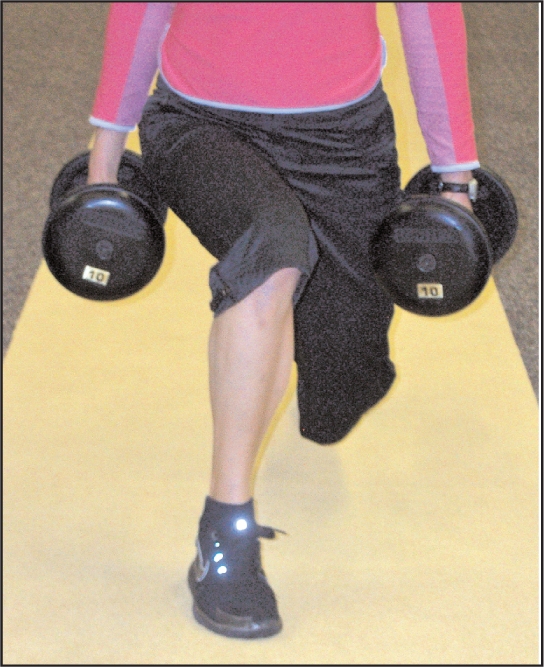
Dysfunctional lower extremity biomechanics during the lunge (demonstrating hip adduction and knee valgum)
The core muscular endurance tests (back extensor test, the flexor endurance test, and the lateral musculature test) should be conducted in order to assess trunk muscular endurance capacity (measured in seconds) and to compare times between the test positions.25 The back extensor test is performed with the athlete positioned in prone with his or her torso unsupported off of the edge of a treatment table with the lower extremities secured.25 Treatment tables utilizing a strap or a belt can be used to stabilize the lower extremities. Instruct the athlete to fold his or her arms across the chest with the hands resting on the opposite shoulders. Once the athlete assumes the testing position record in seconds how long he or she is able to hold the position. The test ends once athlete is unable to maintain this posture.
The flexor endurance test assesses the endurance capacity of the anterior musculature (rectus abdominis) of the core. To conduct the test, the athlete should be positioned supine with the hips and knees in a 90-90 position and the arms folded across the chest. Record how long in seconds, the athlete is able to maintain this position. The test is stopped when any part of the athlete's back comes in contact with a bolster placed 4 cm behind the person.25
The lateral musculature test is performed having the athlete assume the classic side plank pose. To perform the test correctly the athlete will assume the side plank position with the top leg placed in front of the lower leg. The athlete uses the forearm and his or her feet to support themselves.25 Again, record in seconds how long the athlete is able to maintain proper alignment in this position.
INTEGRATING SHOULDER AND CORE EXERCISES
McGill25 advocates increasing the endurance capacity of the torso musculature and improving the ratios between the core endurance tests. Core exercises designed to increase muscular endurance capacity include planks (side and front), crunches, bridges, and the bird dog.
The prescription of therapeutic exercises that address both shoulder weakness and core dysfunction simultaneously, may serve as a transitional program between the initial shoulder rehabilitation program and the terminal sport specific functional exercises. Published shoulder rehabilitation protocols highlight the inclusion of core training although there appears to be paucity in the literature as to the optimal point to initiate these exercises.1,3 The exercises presented in this article could be introduced as soon as the athlete can correctly perform each exercise without symptom reproduction. An athlete should be instructed to perform a high volume of repetitions utilizing light weights (Table 4). Employing this exercise strategy will allow the athlete to continue to increase shoulder strength while beginning to address core weakness. When progressing from a traditional shoulder therapeutic exercise (e.g. sidelying external rotation) to a combined core-shoulder exercise (e.g. side plank with shoulder external rotation), an athlete may need to actually decrease the amount of weight he or she lifts.
Table 4.
A Sample Integrated Core-Shoulder Exercise Routine
| Exercise | Reps/ Sets |
|---|---|
| Side plank with shoulder external rotation | 2-3 sets × 15 repetitions each side |
| Three point plank with shoulder horizontal abduction with external rotation | 2-3 sets × 15 repetitions each side |
| Three point plank with shoulder extension | 2-3 sets × 15 repetitions each side |
| Three point plank with shoulder row | 2-3 sets × 15 repetitions each side |
| Three point plank with diagonal arm raise | 2-3 sets × 15 repetitions each side |
COMBINING CORE AND SHOULDER EXERCISES
The following section describes several combined core-shoulder exercises and presents a sample rehabilitation program (Table 4). Whenever core training is conducted during clinical rehabilitation, the athlete should be instructed to perform an abdominal bracing maneuver with each exercise.25–27 The abdominal brace is an isometric contraction of the abdominal wall musculature with no inward or outward movement of the abdominal wall; or stated another way, an active muscular contraction of the abdomen without either “hollowing” or expanding the abdominal wall.25–28 Evidence exists to show that an abdominal brace is superior to the abdominal hollowing for promoting torso stability.25,26
Side Plank with Shoulder External Rotation
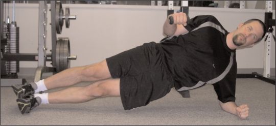
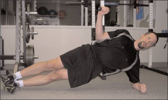
The side plank is an effective core exercise for training the obliques and the transversus abdo-minis muscles,25 when an athlete is able to assume the side plank pose, and incorporate shoulder external rotation (Figures 2A and 2B). If an athlete is initially unable to assume the traditional side plank position, consider having the athlete perform the basic or introductory side plank pose. The introductory side plank pose is similar to the traditional side plank except that the athlete supports his or her body with his or her knees on the ground (instead of his or her feet).
Figure 2A.
Side plank with shoulder external rotation (starting position)
Figure 2B.
Side plank with shoulder external rotation (demonstrating full shoulder external rotation)
Three Point Plank with Upper Extremity Movements
Several shoulder exercises can be performed in the 3-point plank position. Prior to initiating each shoulder component, ensure that the athlete is able to assume the 3-point position. Observe the athlete for any of the following technique errors: scapular winging, inability to maintain a neutral spine position, or hip/trunk flexion. Any deviations or compensation patterns should be corrected prior to proceeding with the combined exercises.
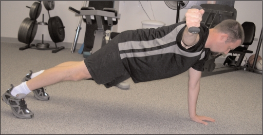
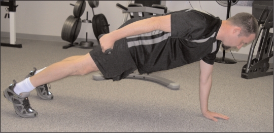
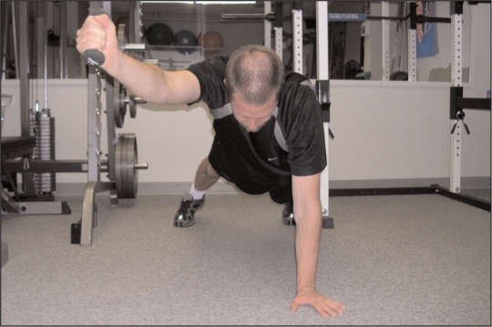
Prior to initiating each shoulder movement, instruct the athlete to retract the scapula and perform the abdominal brace. Horizontal abduction of the arm with the shoulder in external rotation (Figure 3) is an effective exercise for training the external rotators, the supraspinatus, the middle trapezius, and the rhomboid muscles.14–16 Extension of the shoulder (Figure 4) activates the middle trapezius and the posterior deltoid muscles.14,16 The shoulder row (Figure 5) activates the trapezius, the rhomboid, and the posterior deltoid muscles.14,16 A shoulder diagonal arm raise (in line with fiber orientation of the lower trapezius muscle) (Figure 6) also strengthens the middle and lower trapezius muscles.13,15 However, care must be followed when prescribing this exercise. The shoulder diagonal arm raise may minimize subacromial joint space which may potentially provoke symptoms in some athletes performing overhead activities.
Figure 3.
Three point plank with shoulder horizontal abduction with external rotation
Figure 4.
Three point plank with shoulder extension
Figure 5.
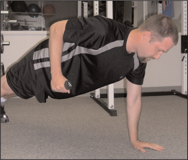
Three point plank with shoulder row
Figure 6.
Three point plank with diagonal arm raises
CONCLUSION
The inclusion of integrated core and shoulder exercises may help to bridge the gap between the initial rehabilitation exercises and later functional rehabilitation exercises. Future research should investigate the muscular activation levels of the rotator cuff and scapular muscles within these unique exercise postures.
REFERENCES
- 1.Wilk KE, Meister K, Andrews JR. Current concepts in the rehabilitation of the overhead throwing athlete. Am J Sports Med. 2002;30:136–151 [DOI] [PubMed] [Google Scholar]
- 2.Kibler WB, McMullen J, Uhl T. Shoulder rehabilitation strategies, guidelines, and practice. Orthop Clin North Am. 2001;32:527–538 [DOI] [PubMed] [Google Scholar]
- 3.Kibler WB, Press J, Sciascia A. The role of core stability in athletic function. Sports Med. 2006;36:189–198 [DOI] [PubMed] [Google Scholar]
- 4.Davies GJ, Dickoff-Hoffman S. Neuromuscular testing and rehabilitation of the shoulder complex. J Orthop Sports Phys Ther. 1993;18:449–458 [DOI] [PubMed] [Google Scholar]
- 5.Litchfield R, Hawkins R, Dillman CJ, et al. Rehabilitation for the overhead athlete. J Orthop Sports Phys Ther. 1993;18:433–441 [DOI] [PubMed] [Google Scholar]
- 6.Hodges PW, Gandevia SC, Richardson CA. Contractions of specific abdominal muscles in postural tasks are affected by respiratory maneuvers. J Appl Physiol. 1997;83:753–760 [DOI] [PubMed] [Google Scholar]
- 7.Tarnanen SP, Ylinen JJ, Siekkinen KM, et al. Effect of isometric upper-extremity exercises on the activation of core stabilizing muscles. Arch Phys Med Rehabil. 2008;89:513–521 [DOI] [PubMed] [Google Scholar]
- 8.Moreside JM, Vera-Garcia FJ, McGill SM. Trunk muscle activation patterns, lumbar compressive forces, and spine stability when using the bodyblade. Phys Ther. 2007;87:153–163 [DOI] [PubMed] [Google Scholar]
- 9.Crosbie J, Kilbreath SL, Hollmann L, York S. Scapulohumeral rhythm and associated spinal motion. Clin Biomech. 2008;23:184–192 [DOI] [PubMed] [Google Scholar]
- 10.Kibler WB. The role of the scapula in athletic shoulder function. Am J Sports Med. 1998;26:325–337 [DOI] [PubMed] [Google Scholar]
- 11.Ebaugh DD, McClure PW, Karduna AR. Scapulothoracic and glenohumeral kinematics following an external rotation fatigue protocol. J Orthop Sports Phys Ther. 2006;36:557–571 [DOI] [PubMed] [Google Scholar]
- 12.Tsai NT, McClure PW, Karduna AR. Effects of muscle fatigue on 3-dimensional scapular kinematics. Arch Phys Med Rehabil. 2003;84:1000–1005 [DOI] [PubMed] [Google Scholar]
- 13.Ekstrom RA, Donatelli RA, Soderberg GL. Surface electromyographic analysis of exercises for the trapezius and serratus anterior muscles. J Orthop Sports Phys Ther. 2003;33:247–258 [DOI] [PubMed] [Google Scholar]
- 14.Moseley JB, Jr., Jobe FW, Pink M, et al. EMG analysis of the scapular muscles during a shoulder rehabilitation program. Am J Sports Med. 1992;20:128–134 [DOI] [PubMed] [Google Scholar]
- 15.Reinold MM, Wilk KE, Fleisig GS, et al. Electromyographic analysis of the rotator cuff and deltoid musculature during common shoulder external rotation exercises. J Orthop Sports Phys Ther. 2004;34:385–394 [DOI] [PubMed] [Google Scholar]
- 16.Townsend H, Jobe FW, Pink M, Perry J. Electromyographic analysis of the glenohumeral muscles during a baseball rehabilitation program. Am J Sports Med. 1991;19:264–272 [DOI] [PubMed] [Google Scholar]
- 17.Hodges PW, Richardson CA. Relationship between limb movement speed and associated contraction of the trunk muscles. Ergonomics. 1997;40:1220–1230 [DOI] [PubMed] [Google Scholar]
- 18.Hodges PW, Richardson CA. Feedforward contraction of transversus abdominis is not influenced by the direction of arm movement. Exp Brain Res. 1997;114:362–370 [DOI] [PubMed] [Google Scholar]
- 19.Richardson CA, Hodges P, Hides J. Therapeutic Exercise for Lumbopelvic Stabilization. 2nd ed.Edinburgh: Churchill Livingstone; 2004 [Google Scholar]
- 20.Ellenbecker TS, Davies GJ. Closed Kinetic Chain Exercise: A Comprehensive Guide to Multiple-Joint Exercises. Champaign, IL: Human Kinetics; 2001 [Google Scholar]
- 21.Leetun DT, Ireland ML, Willson JD, Ballantyne BT, Davis IM. Core stability measures as risk factors for lower extremity injury in athletes. Med Sci Sports Exerc. 2004;36:926–934 [DOI] [PubMed] [Google Scholar]
- 22.Willson JD, Dougherty CP, Ireland ML, Davis IM. Core stability and its relationship to lower extremity function and injury. J Am Acad Orthop Surg. 2005;13:316–325 [DOI] [PubMed] [Google Scholar]
- 23.Powers CM. The influence of altered lower-extremity kinematics on patellofemoral joint dysfunction: A theoretical perspective. J Orthop Sports Phys Ther. 2003; 33:639–646 [DOI] [PubMed] [Google Scholar]
- 24.Willson JD, Ireland ML, Davis I. Core strength and lower extremity alignment during single leg squats. Med Sci Sports Exerc. 2006;38:945–952 [DOI] [PubMed] [Google Scholar]
- 25.McGill S. Low Back Disorders: Evidence-Based Prevention and Rehabilitation. Champaign, IL: Human Kinetics; 2002 [Google Scholar]
- 26.Grenier SG, McGill SM. Quantification of lumbar stability by using 2 different abdominal activation strategies. Arch Phys Med Rehabil. 2007;88:54–62 [DOI] [PubMed] [Google Scholar]
- 27.McGill SM, Grenier S, Kavcic N, Cholewicki J. Coordination of muscle activity to assure stability of the lumbar spine. J Electromyogr Kinesiol. 2003;13:353–359 [DOI] [PubMed] [Google Scholar]
- 28.McGill SM. Low back stability: From formal description to issues for performance and rehabilitation. Exerc Sport Sci Rev. 2001;29:26–31 [DOI] [PubMed] [Google Scholar]


