Abstract
Examination and evaluation of the patient a with multiple ligament injured knee is a complicated process and best done in a methodical, comprehensive fashion with a particular emphasis placed on assessment of supporting soft tissues. Tissues that can be damaged during this devastating injury include bones, ligaments, meniscus, articular cartilage, and neurovascular structures. Uses of diagnostic imaging and the physical examination process will be described in this article.
Keywords: knee dislocation, physical examination, diagnostic imaging
INTRODUCTION
Patients with multiple ligament knee injuries (MLKI) always have the risk of severe disability due to the resultant loss of stability from damage to passive and active knee stabilizers. Additionally concerning is the possible compromise of neurovascular structures surrounding the knee. Although rare, the average sports and orthopedic clinician will undoubtedly have a patient with an injury of this magnitude in their career. This injury may be due, in part, to improved physical examination testing, diagnostic technology, and enhanced awareness of these complex injuries. Therefore, it is paramount that clinical examination of this potentially disabling condition be performed in a thorough and expedient manner for optimal functional outcomes to occur following this injury. Failure to properly diagnose, evaluate, and treat this injury can lead to a whole host of complications, some of which require amputation.1 Although many times clinicians rely on various forms of advanced technology via imaging, the foundation of evaluation of this, or any musculoskeletal condition, is performance of an efficient hands-on clinical physical examination.2
HISTORY
The most common mechanism of injury causing knee dislocation is that of high- velocity motor vehicle or motorcycle accidents.3,4 Knee dislocations have occurred from low-velocity trauma in athletics also. Another common mechanism of injury is represented by trampoline accidents5 and spontaneous dislocations in the morbidly obese.6
OBSERVATION/INSPECTION
Due to the deformity, pain, and swelling, the dislocated knee is not hard to diagnose while not reduced. Although the severely deformed knee dislocation is usually obvious to the naked eye, spontaneous reduction of dislocations and subluxations have been reported between 20–50% of the time.7–11 In these knees that spontaneously reduce, it is hard to believe that absence of a deformity would allow such a high level injury. In these cases the extent of injury may be easily mis-diagnosed. Additionally, in the obese patient the deformity and swelling may be obscured by excess adipose tissue surrounding the knee.1 Because of the potential for disastrous complications, the evaluation and treatment of the knee that has acute, multiple ligament injuries should be considered a knee dislocation, until proven otherwise.1,8
Swelling can be helpful in defining the zone of injury of the knee. The knee may appear to have extensive soft tissue swelling proximally or surrounding the actual tibiofemoral joint. Swelling can be both intra-articular and extra-articular. Due to extravasations (leakage) of joint fluid into peri-articular soft tissues around the knee, intraarticular swelling may only be moderate if the synovial capsule is breeched. If the joint capsule is not ruptured, a significant joint effusion will most likely be present intracapsularly.
The patient should be examined for “dimple sign.” This finding is one of the hallmark signs of a posterolateral dislocation and occurs as a result of the infolding of the medial collateral ligament and capsule into the joint as the medial femoral condyle is forced posterolaterally.12–14
Discoloration is not always noted acutely, but generally within several days following insult can give the clinician an idea of the extent of soft tissue injury. Similarly, bogginess and extra-articular edema may suggest areas where capsular or collateral complexes have been disrupted.15
NEUROVASCULAR EXAMINATION
First and foremost during the clinical examination is the immediate assessment of the neurovascular status of the dislocated knee. Due to the high incidence of vascular insult, all knee dislocations should assume vascular compromise until proven otherwise. Early diagnosis and appropriate immediate treatment of vascular injury is required to save the extremity. Collateral circulation may initially be sufficient enough to provide a normal pulse, giving the examiner a false sense of security.16–18
Despite this fact, lower extremity pulses should be assessed. Distal pulses are normally assessed by manual palpation and compared to the contralateral, normal leg. The presence or absence of the dorsalis pedis and the posterior tibial arteries should always be documented. The absence of pulses is a true medical vascular emergency.19 If not assessed early, the posterior tibial artery is often-times very hard to assess due to the significant amount of soft tissue swelling seen in and around the knee following dislocations. It must be recognized that a strong palpable pulse does not rule out an arterial injury. Both a palpable pulse and a warm foot can exist in the face of popliteal artery injury.1 The danger in this injury is an intimal flap tear of the artery which can cause a delayed occlusion up to 24 to 72 hours after the injury.19
The popliteal artery always warrants assessment due to the high incidence of injury in the dislocated knee. The popliteal artery is transected with a posterior tibial dislocation, while it is stretched during the anterior tibial dislocation.20,21 Capillary refill alone is not accurate in evaluating the integrity of the popliteal artery.1 Tenseness in the popliteal region and the inability to “move the fossa” (defined as an inability to pinch the skin over the popliteal fossa because the fossa is so tight and filled with blood) are grave signs of vascular insufficiency.1
Peroneal nerve palsy is also common following MLKI. The peroneal nerve is even more important to assess in the posterior knee dislocation due to disruption of multiple posterolateral structures. Examination of peroneal nerve function can be assessed by determining the sensory function of the dorsum and lateral aspects of the foot and first web space on the affected extremity. This examination can be assessed by light touch and sharp/dull discrimination. Motor function is assessed by active contraction of foot and toe dorsiflexion. Loss of peroneal nerve function will result in loss of sensation, foot drop, and an altered gait pattern.
DIAGNOSTIC TESTING-RADIOGRAPHS
Radiographic examination is paramount to begin the assessment of the dislocated knee. Anterior, posterior, and lateral views are used to assess the direction of dislocation, integrity of the bones, and to look for other clues about the injury. Avulsion fractures from attachments of cruciate and collateral ligaments are sometimes visualized.
MAGNETIC RESONANCE IMAGING
Magnetic resonance imaging (MRI) is almost an essential tool to assist in the diagnosis of the MLKI and assists in the formulation of the treatment plan.22 The MRI helps to evaluate meniscal tears, intraosseous contusions, occult fractures, capsular tears, and muscle strains. The MRI additionally helps with the preoperative planning for specific ligament reconstructions or repairs and determining the amount of graft that is needed for reconstruction.
ARTERIOGRAPHY
The selective use of arteriography based on physical examination is a safe and prudent policy following knee dislocation rather than routine arteriography in all instances. Stannard et al23 developed an algorithm for indication of ordering arteriography. The algorithm was successfully used to diagnose all clinically important vascular injuries in a large series of patients. The first step in this algorithm is to perform a physical examination of the dorsalis pedis and posterior tibial arteries, as well as a gross evaluation of color and temperature. If any asymmetry between the two lower extremities is present, the patient should have an arteriogram made either in the angiography suite or on the operating-room table. If any history of an abnormal vascular examination in the pre-hospital setting exists, the patient should also undergo arteriography. In the absence of these findings, the patient should be admitted for careful observation. Neurovascular checks should be performed by the nursing staff every 2 to 4 hours for the first 48 hours. The vascular examination should be documented by a surgeon at the time of admission, 4 to 6 hours after admission, and again 24 and 48 hours after admission. If any clinical abnormalities are detected, an arteriogram should be made.23
DIAGNOSTIC ARTHROSCOPY
Diagnostic arthroscopy allows for further evaluation of the knee. This examination technique allows direct visualization of intra-articular anatomy and allows for clues as to what is damaged that is extra-articular. Capsular tears can be seen, as well as intra-articular capsular hemorrhaging. Articular surfaces, as well as the meniscus, can be evaluated for injury and a treatment plan can be instituted.
Caution must be employed with arthroscopy after a knee dislocation. If fluid extravasation through a ruptured capsule occurs, and if a pump is employed as part of the arthroscopy, a potential compartment syndrome could be created. A delay of 10–14 days from the injury can allow the capsule tear to seal and reduce the risk of a compartment syndrome. The use of a low pressure system including gravity and an outflow portal can reduce the potential risk of this serious complication.
INITIAL TREATMENT AND REDUCTION
The patient with the dislocated knee rarely presents with the knee dislocated. If the patient presents with a dislocated knee, evaluation should be performed with radiographs to evaluate for concomitant fractures. Once the patient has been evaluated with radiographs a reduction maneuver can be performed. Post-reduction radiographs are important to evaluate for a concentric reduction. These radiographs should be carefully studied for osteochondral fractures, as well. In addition, avulsion fractures can give clues to the soft tissue restraints that have been injured and help guide surgical technique in repair. If the knee is not reducible, then consideration should be given to soft tissue entrapment of the femoral condyle making the knee irreducible. Following reduction, the knee should be re-examined for neurovascular pathology.
CLASSIFICATION
Knee dislocations can be classified, according to the anatomic system proposed by Wascher.24 A KD-I is a dislocation associated with multiple-ligament injuries that did not include both cruciate ligaments, KD-II is a dislocation associated with a bi-cruciate ligament injury only, KD-III is a dislocation associated with a bi-cruciate ligament injury and a tear of either the posteromedial or posterolateral knee ligaments, KD-IV is a dislocation associated with tears of both cruciate ligaments and both posteromedial and posterolateral ligaments, or KD-V which is a dislocation associated with a periarticular fracture and multiple-ligament injuries.
LIGAMENT EXAMINATION
Following reduction, the ligament examination should be performed very carefully to avoid redislocation or further compromise to neurovascular structures. As with most examination procedures, stability examination should begin with an evaluation of the contralateral, non involved extremity.25 This examination allows the athlete to understand what will happen to the involved knee and, hopefully, decrease any anxiety associated with clinical examination procedures. It is easy to get a false first impression and note abnormal translational movement on an individual who demonstrates a significant amount of generalized ligamentous laxity, which may not actually be pathologic.
Anterior Cruciate Ligament
The “gold standard” test for anterior instability is the Lachman's test, which is performed at 30 degrees of knee flexion (Figure 1). Because the Lachman's test is for single plane anterior instability, it is imperative in the athlete with MLKI to control rotary motions while performing this test. Allowing excessive internal or external tibial rotation while performing the Lachman's test may result in false-positive results. Also remember that false-positives can occur in the athlete with a posterior cruciate tear because of the posterior tibial drop-back that occurs with that particular injury. In the face of combined ligamentous injuries the clinician may be misled by a knee that has an intact anterior cruciate ligament (ACL) but ruptured posterior cruciate ligament (PCL), medial collateral ligament (MCL), and lateral collateral ligament (LCL). To avoid this mis-interpretation, place the knee in the anatomic position prior to performing the examination techniques.26 Flexing the knee to 90 degrees and positioning the tibia so that an approximate 10-mm step-off between the femoral condyles and the edge of the tibial plateau exist.27 Another method to find the neutral position is to palpate the step-off of the femoral condyles to the tibial plateau in the uninvolved knee and compare to the involved knee in a similar position. Both the end-point (mushy or absent) and the length of excursion determine a positive test.28
Figure 1.
Lachman's test
The anterior drawer test can then be used to verify anterior single plane instability. This test may have decreased sensitivity and specificity in those with meniscal tears, acutely swollen knees, and protective hamstring muscle spasm. An advantage of this test is that the technique can be performed in a position of tibial internal and external rotation to assess for rotary instability of the posterolateral corner and posteromedial capsule, respectively.29 Grading of ligament laxity is always compared to the uninjured side. Grade I is defined as 0–5 mm, grade II from 6–10 mm, while a grade III is more than 10 mm of motion compared to that of the uninvolved side.
Posterior Cruciate Ligament
The PCL is examined via the posterior drawer test. This test is performed from a position of 45 degrees of hip flexion and 80 degrees of knee flexion with the feet flat on the examining table. The neutral position of the tibia must be found as described previously, before the test is performed to decrease the risk of obtaining false positive results. The clinician's thumbs palpate the anterior joint between the femoral condyles and the tibial plateau. The fingers wrap around the tibia to palpate the hamstring muscles to assess for spasm. A posterior directed force is placed upon the tibia while feeling for both the quality and quantity of the end feel. Excessive posterior tibial translation, a soft end feel, or combination of the two is indicative of a PCL tear.
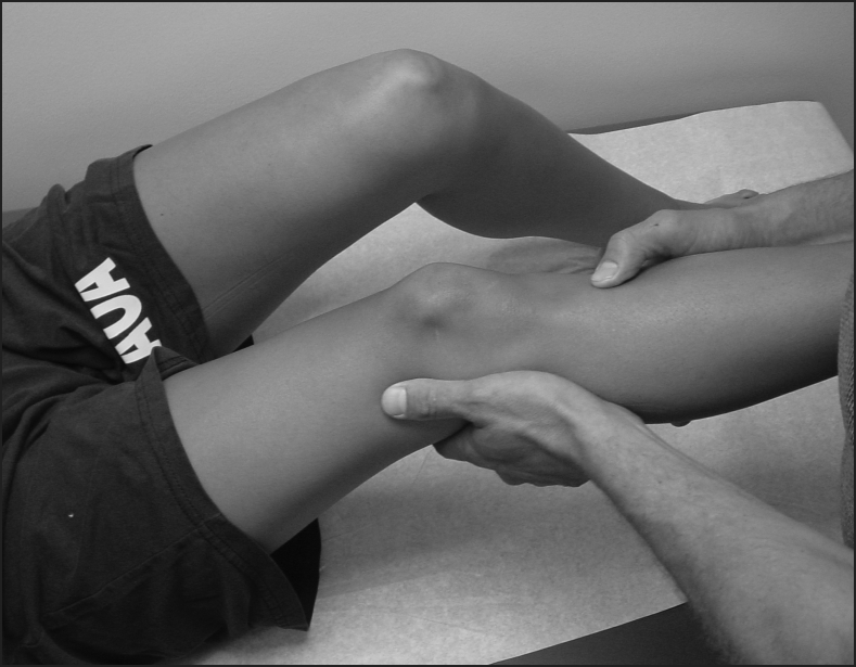
In this same position, the step-up test can be performed. In position above to start the posterior drawer test, the anterior portion of the tibial plateau is located approximately 10 mm anterior to the distal femoral condyles. Absence of this step-up is indicative of PCL injury (Figure 2). Lastly, the posterior sag test can be performed in which in the same position previously described, the patient is asked to completely relax and the affected knee is viewed from the side. A positive test occurs when the anterior aspect of the proximal tibia is found to “sag” behind the anterior aspect of the femoral condyles or in comparison to the contralateral knee.
Figure 2.

Absence of step-up
Collateral Ligaments
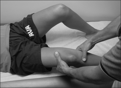
The valgus stress test is applied to the knee in both extension and at 30 degrees of flexion (Figure 3). When the MCL is disrupted in the unanesthetized patient, pain may decrease accuracy of this test due to guarding. This test is done with the hip in only slight flexion and full knee extension with or without recurvatum equal to that of the uninvolved limb. A significant amount of valgus motion in full extension is indicative of an ACL or PCL rupture, the posterior oblique ligament, and the medial portion of the posterior capsule. The ACL is a secondary restraint to medial joint opening.
Figure 3.
Valgus stress test (medial collateral ligament)
The test is then performed again in 30 degrees of knee flexion to place laxity on the posterior capsular structures. Medial laxity in only 30 degrees of flexion that is not present in full extension indicates injury to only the MCL.
The varus stress test is performed in a similar manner as the valgus stress test (Figure 4). The knee is tested at both full extension and 30 degrees of knee flexion. Lateral instability in full extension indicates rupture of LCL, lateral capsule, and PCL. Laxity at 30 degrees only indicates an isolated tear to the LCL.
Figure 4.

Varus stress test (lateral collateral ligament)
Posterolateral Corner
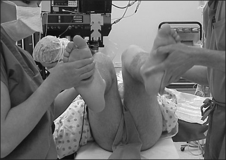
To examine for capsular sprain of the posterolateral corner of the capsule, the external rotation recurvatum test (ERRT) is performed. During the ERRT, the examiner grasps the great toe of each foot while the supine athlete's knees are allowed to fall toward full passive extension (Figure 5). This test is considered positive when the affected knee assumes a posture of varus angulation, hyperextension, and external rotation of the tibia as compared to the uninjured extremity.30 This test must be done very carefully in the patient with MLIK since disruption of the posterior capsule may allow excessive genu recurvatum, allowing the knee to re-dislocate.15
Figure 5.
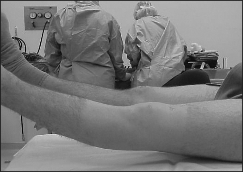
External rotation recurvatum test
The dial test assesses abnormal external tibial rotation to differentiate between isolated posterolateral corner injury and combined ACL/PCL injuries. This test is most commonly performed in the prone position with the knees flexed to 30 degrees (Figure 6).31 An external rotation force is applied to the athlete's heels by placing the fingers and thumb along side the talor-calcaneal bony contours. The foot thigh angle is measured and compared to the uninjured knee. The test is then performed in 90 degrees of flexion and the foot-thigh angle is re-measured (Figure 7). When comparing at either angle a difference of 10 degrees or more is significant. As the knee is flexed to 90 degrees a reduction in increased rotation may occur although the amount of motion remains greater than the uninjured side if the PCL is still intact. This increased rotation occurs because the PCL is the secondary stabilizer to external rotation and gains mechanical advantage when the knee is flexed.15 When the amount of external rotation is increased from 30 to 90 degrees, a combined PCL and posterolateral corner of the capsule injury is incurred.31,32
Figure 6.
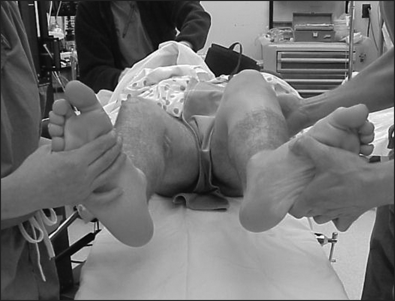
Dial test at 30° of knee flexion
Figure 7.
Dial test at 90° of knee flexion
Although not usually tolerated well in the athlete who is awake, the pivot shift test is best described as a reduction from a state of anterior tibial subluxation.33 The athlete will assume a supine position with the leg relaxed as much as possible. The examiner should hold the affected knee in full extension and internal rotation with the hip flexed about 45 degrees. The knee is flexed while the examiner applies a valgus stress. In an athlete with a deficient ACL, the lateral tibial plateau will be anteriorly subluxated at the beginning of the test. As the amount of flexion increases to 30–40 degrees, this anterior lateral position of the tibia will be abruptly reduced. This reduction can be both palpable and audible.34
SUMMARY
The acute examination and assessment of patients with the MLKI is crucial for multiple reasons including accurate knowledge of injury pattern and surgical or conservative treatment and decision making. The evaluation technique should be consistent, methodical, and comprehensive. As an adjunct to the physical examination, diagnostic imaging should be used when appropriate.
REFERENCES
- 1.Taft TN, Almekinders LC. The dislocated knee. In: Knee Surgery, Fu FH, Harner CD, Vince KG. (Eds). Williams and Wilkins, Baltimore, 1994 [Google Scholar]
- 2.Brautigan B, Johnson D. The epidemiology of knee dislocations. Clin Sports Med. 2000;19:387–397 [DOI] [PubMed] [Google Scholar]
- 3.Alberty R, Goodfried G, Boyden A. Popliteal artery injury with fractural dislocation of the knee. Am J Surg. 1981;142:36–38 [DOI] [PubMed] [Google Scholar]
- 4.Kendall R, Taylor D, Slavian A, et al. The role of arteriography in assessing vascular injuries associated with dislocations of the knee. J Trauma. 1993;35:875–878 [DOI] [PubMed] [Google Scholar]
- 5.Kwolek C, Sundaram S, Schwarcz T, et al. Popliteal thrombosis associated with trampoline injuries from anterior knee dislocations in children. Am J Surg. 1998;64:1183–1187 [PubMed] [Google Scholar]
- 6.Hagino R, DeCaprio J, Valentine R. Spontaneous popliteal vascular injury in the morbidly obese. J Vasc Surg. 1998;28:458–462 [DOI] [PubMed] [Google Scholar]
- 7.Walker DN, Hadison R, Schenck RC. A baker's dozen of knee dislocations. Am J Knee Surg.1994;7:117–124 [Google Scholar]
- 8.Wascher DC, Dvirnak PC, DeCoster TA. Knee dislocation: Initial assessment and implications for treatment. J Orthop Trauma.1997;11:525–529 [DOI] [PubMed] [Google Scholar]
- 9.Varnell RM, Coldwell DM, Sangeorzan BJ, et al. Arterial injury complicating knee dislocation. Am J Knee Surg. 1989;55:699–704 [PubMed] [Google Scholar]
- 10.Good L, Johnson RK. The dislocated knee. J Am Acad Orthop Surg.1995;3:284–292 [DOI] [PubMed] [Google Scholar]
- 11.Eastlack RK, Schenck RC, Guarducci C. The dislocated knee: Classification, treatment, and outcome. US Army Med Dept J.1997;11/12:1–9 [Google Scholar]
- 12.Haung F, Simonian P, Chansky H. Irreducible posterolateral dislocation of the knee. Arthroscopy.2000;16:323–327 [DOI] [PubMed] [Google Scholar]
- 13.Hill JA, Rana NA. Complications of posterolateral dislocation of the knee: Case report and literature review. Clin Orthop. 1981;154:212–215 [PubMed] [Google Scholar]
- 14.Quinlan AG, Sharrard WJ. Posterolateral dislocation of the knee with capsular interposition. J Bone Joint Surg.1958;40B:660–663 [DOI] [PubMed] [Google Scholar]
- 15.Romeyn RL, Davies GJ, Jennings J. The multiple ligament – injured knee: Evaluation, treatment and rehabilitation. In: Postsurgical Orthopedic Sports Rehabilitation: Knee and Shoulder. Manske RC. (Ed). Mosby, St. Louis, 2006 [Google Scholar]
- 16.Meyers MH, Harvey JP., Jr Traumatic dislocation of the knee joint. A study of eighteen cases. J Bone Joint Surg. 1971;53A:16–29 [PubMed] [Google Scholar]
- 17.DeBacke ME, Simeone FA. Battle injuries of the arteries in WWII: An analysis of 2,471 cases. Ann Surg. 1946;123:534–579 [PubMed] [Google Scholar]
- 18.Hoover NW. Injuries of the popliteal artery associated with fractures and dislocations. Surg Clin North Am.1961;41:1099–1116 [DOI] [PubMed] [Google Scholar]
- 19.Shapiro MS. Multiligament injuries. In: Sports Injuries of the Knee. Surgical Approaches. Simonian PT, Cole BJ, Bach BR., Jr (Eds). Thieme, New York, New York, 2006 [Google Scholar]
- 20.Green NE, Allen BL. Vascular injuries associated with dislocation of the knee. J Bone Joint Surg. 1977;59A:236–239 [PubMed] [Google Scholar]
- 21.O'Donnell JF, Jr, Brewster DC, Darling RC, et al. Arterial injuries associated with fractures and/or dislocations of the knee. J Trauma. 1997;17:775–784 [DOI] [PubMed] [Google Scholar]
- 22.LaPrade RF, Bollom TS, Gilbert TJ, et al. The MRI appearance of individual structures of the posterolateral knee: A prospective study of normal and surgically verified grade 3 injuries. Am J Sports Med. 2000;28:191–199 [DOI] [PubMed] [Google Scholar]
- 23.Stannard JP, Sheils TM, Lopez-Ben RR, et al. Vascular injuries in knee dislocations: The role of physical examination in determining the need for arteriography. J Bone Joint Surg. 2004;86A:910–915 [PubMed] [Google Scholar]
- 24.Wascher DC. High-velocity knee dislocation with vascular injury. Treatment principles. Clin Sports Med. 2000;19:457–77 [DOI] [PubMed] [Google Scholar]
- 25.Magee DJ. Orthopedic Physical Assessment. 5th ed.Saunders, St. Louis, 2008 [Google Scholar]
- 26.Alicea JA, Scuderi GR. Knee dislocations. In: Ligaments of the Knee. Tria EJ. (Ed.) Churchill Livingstone, New York: 1995 [Google Scholar]
- 27.Clancy WG. Repair and reconstruction of the posterior cruciate ligament. In: Operative Orthopaedics. Chapman M. (Ed). JB Lippincott, Philadelphia, 1988 [Google Scholar]
- 28.Brand JC, Johnson DL. Initial assessment: Physical examination and imaging studies. In: The Multiple Ligament Injured Knee. A Practical Guide to Management. Fanelli GC. (Ed). Springer, New York, 2004 [Google Scholar]
- 29.Ritchie J, Miller M, Harner C. History and physical examination. In: Fu F, Harner C, Vince K. (Eds). Knee Surgery. Vol 1 Baltimore, MD: Williams and Wilkins, 1994 [Google Scholar]
- 30.Hughston JC, Norwood LA. The posterolateral drawer test and external rotational recurvatum test for posterolateral rotatory instability of the knee. Clin Orthop. 1980;147:82–87 [PubMed] [Google Scholar]
- 31.Gollehon DL, Torzilli PA, Warren RF. The role of posterolateral and cruciate ligaments in the stability of the human knee: A biomechanical study. J Bone Joint Surg. 1987;69A:233–242 [PubMed] [Google Scholar]
- 32.Nielsen S, Ovesen J, Rasmussen O. The posterior cruciate ligament and rotatory knee instability: An experimental study. Arch Orthop Trauma Surg. 1985;104:53–56 [DOI] [PubMed] [Google Scholar]
- 33.Ellison AE, The pathogenesis and treatment of anterolateral rotator instability. Clin Orthop Relat Res. 1980;147:51–55 [PubMed] [Google Scholar]
- 34.Lubowitz JH, Bernardini BJ, Reid JB. Comprehensive physical examination for instability of the knee. Am J Sports Med. 2008;36:577–594 [DOI] [PubMed] [Google Scholar]


