

Abstract
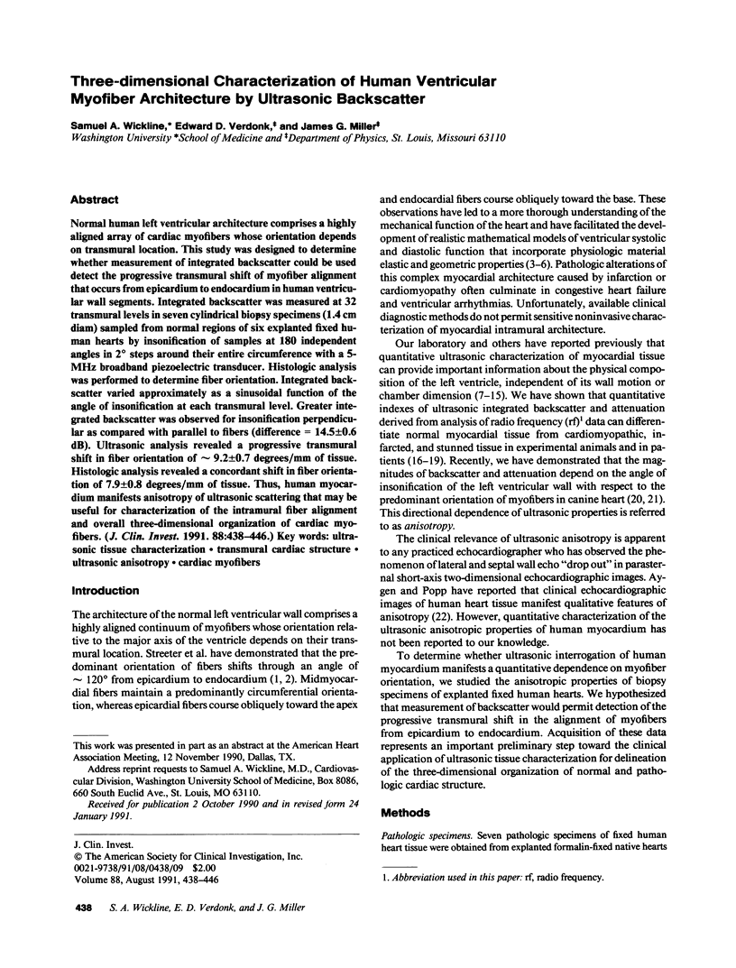
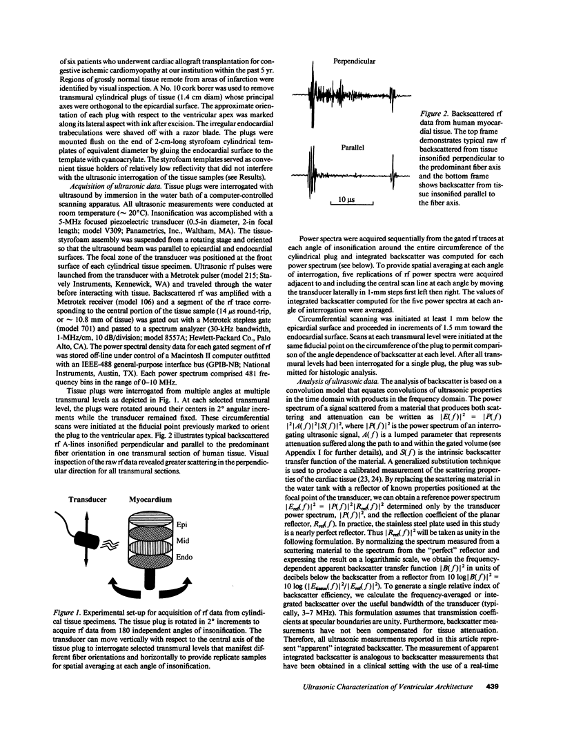
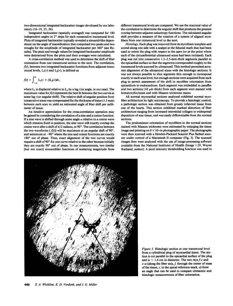
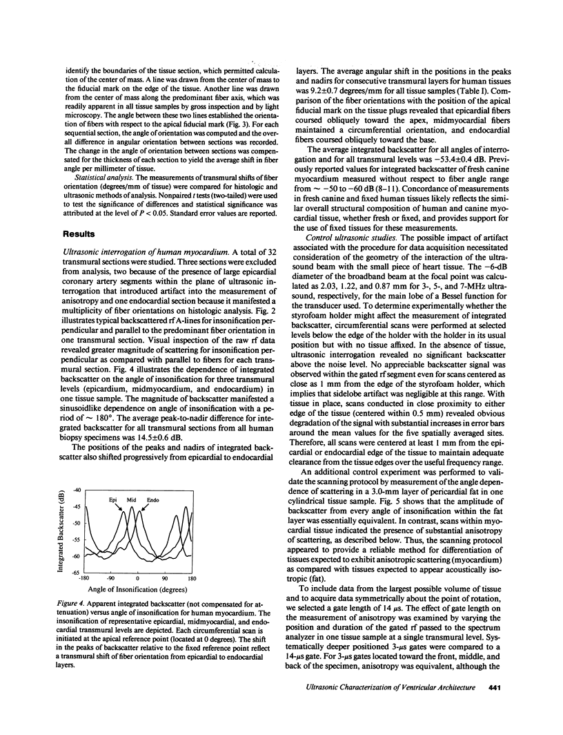
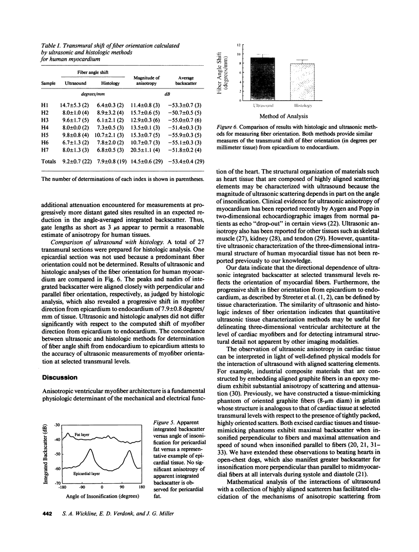
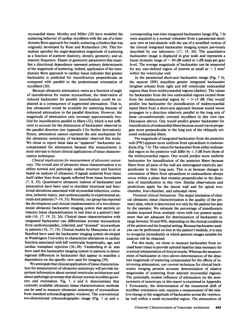
Normal human left ventricular architecture comprises a highly aligned array of cardiac myofibers whose orientation depends on transmural location. This study was designed to determine whether measurement of integrated backscatter could be used detect the progressive transmural shift of myofiber alignment that occurs from epicardium to endocardium in human ventricular wall segments. Integrated backscatter was measured at 32 transmural levels in seven cylindrical biopsy specimens (1.4 cm diam) sampled from normal regions of six explanted fixed human hearts by insonification of samples at 180 independent angles in 2 degrees steps around their entire circumference with a 5-MHz broadband piezoelectric transducer. Histologic analysis was performed to determine fiber orientation. Integrated backscatter varied approximately as a sinusoidal function of the angle of insonification at each transmural level. Greater integrated backscatter was observed for insonification perpendicular as compared with parallel to fibers (difference = 14.5 +/- 0.6 dB). Ultrasonic analysis revealed a progressive transmural shift in fiber orientation of approximately 9.2 +/- 0.7 degrees/mm of tissue. Histologic analysis revealed a concordant shift in fiber orientation of 7.9 +/- 0.8 degrees/mm of tissue. Thus, human myocardium manifests anisotropy of ultrasonic scattering that may be useful for characterization of the intramural fiber alignment and overall three-dimensional organization of cardiac myofibers.
Full text
PDF
Images in this article
Selected References
These references are in PubMed. This may not be the complete list of references from this article.
- Aygen M., Popp R. L. Influence of the orientation of myocardial fibers on echocardiographic images. Am J Cardiol. 1987 Jul 1;60(1):147–152. doi: 10.1016/0002-9149(87)91002-2. [DOI] [PubMed] [Google Scholar]
- Chadwick R. S. Mechanics of the left ventricle. Biophys J. 1982 Sep;39(3):279–288. doi: 10.1016/S0006-3495(82)84518-9. [DOI] [PMC free article] [PubMed] [Google Scholar]
- Chadwick R. S., Ohayon J., Lewkowicz M. Wall-thickness and midwall-radius variations in ventricular mechanics. Proc Natl Acad Sci U S A. 1989 May;86(9):2996–2999. doi: 10.1073/pnas.86.9.2996. [DOI] [PMC free article] [PubMed] [Google Scholar]
- Chandraratna P. A., Ulene R., Nimalasuriya A., Reid C. L., Kawanishi D., Rahimtoola S. H. Differentiation between acute and healed myocardial infarction by signal averaging and color encoding two-dimensional echocardiography. Am J Cardiol. 1985 Sep 1;56(7):381–384. doi: 10.1016/0002-9149(85)90870-7. [DOI] [PubMed] [Google Scholar]
- Fornage B. D. The hypoechoic normal tendon. A pitfall. J Ultrasound Med. 1987 Jan;6(1):19–22. doi: 10.7863/jum.1987.6.1.19. [DOI] [PubMed] [Google Scholar]
- Hoyt R. H., Collins S. M., Skorton D. J., Ericksen E. E., Conyers D. Assessment of fibrosis in infarcted human hearts by analysis of ultrasonic backscatter. Circulation. 1985 Apr;71(4):740–744. doi: 10.1161/01.cir.71.4.740. [DOI] [PubMed] [Google Scholar]
- Humphrey J. D., Yin F. C. Constitutive relations and finite deformations of passive cardiac tissue II: stress analysis in the left ventricle. Circ Res. 1989 Sep;65(3):805–817. doi: 10.1161/01.res.65.3.805. [DOI] [PubMed] [Google Scholar]
- Madaras E. I., Perez J., Sobel B. E., Mottley J. G., Miller J. G. Anisotropy of the ultrasonic backscatter of myocardial tissue: II. Measurements in vivo. J Acoust Soc Am. 1988 Feb;83(2):762–769. doi: 10.1121/1.396119. [DOI] [PubMed] [Google Scholar]
- Masuyama T., Nellessen U., Schnittger I., Tye T. L., Haskell W. L., Popp R. L. Ultrasonic tissue characterization with a real time integrated backscatter imaging system in normal and aging human hearts. J Am Coll Cardiol. 1989 Dec;14(7):1702–1708. doi: 10.1016/0735-1097(89)90019-3. [DOI] [PubMed] [Google Scholar]
- Masuyama T., St Goar F. G., Tye T. L., Oppenheim G., Schnittger I., Popp R. L. Ultrasonic tissue characterization of human hypertrophied hearts in vivo with cardiac cycle-dependent variation in integrated backscatter. Circulation. 1989 Oct;80(4):925–934. doi: 10.1161/01.cir.80.4.925. [DOI] [PubMed] [Google Scholar]
- Masuyama T., Valantine H. A., Gibbons R., Schnittger I., Popp R. L. Serial measurement of integrated ultrasonic backscatter in human cardiac allografts for the recognition of acute rejection. Circulation. 1990 Mar;81(3):829–839. doi: 10.1161/01.cir.81.3.829. [DOI] [PubMed] [Google Scholar]
- Miller J. G., Pérez J. E., Sobel B. E. Ultrasonic characterization of myocardium. Prog Cardiovasc Dis. 1985 Sep-Oct;28(2):85–110. doi: 10.1016/0033-0620(85)90020-9. [DOI] [PubMed] [Google Scholar]
- Milunski M. R., Mohr G. A., Pérez J. E., Vered Z., Wear K. A., Gessler C. J., Sobel B. E., Miller J. G., Wickline S. A. Ultrasonic tissue characterization with integrated backscatter. Acute myocardial ischemia, reperfusion, and stunned myocardium in patients. Circulation. 1989 Sep;80(3):491–503. doi: 10.1161/01.cir.80.3.491. [DOI] [PubMed] [Google Scholar]
- Milunski M. R., Mohr G. A., Wear K. A., Sobel B. E., Miller J. G., Wickline S. A. Early identification with ultrasonic integrated backscatter of viable but stunned myocardium in dogs. J Am Coll Cardiol. 1989 Aug;14(2):462–471. doi: 10.1016/0735-1097(89)90203-9. [DOI] [PubMed] [Google Scholar]
- Mol C. R., Breddels P. A. Ultrasound velocity in muscle. J Acoust Soc Am. 1982 Feb;71(2):455–461. doi: 10.1121/1.387467. [DOI] [PubMed] [Google Scholar]
- Mottley J. G., Miller J. G. Anisotropy of the ultrasonic attenuation in soft tissues: measurements in vitro. J Acoust Soc Am. 1990 Sep;88(3):1203–1210. doi: 10.1121/1.399751. [DOI] [PubMed] [Google Scholar]
- Mottley J. G., Miller J. G. Anisotropy of the ultrasonic backscatter of myocardial tissue: I. Theory and measurements in vitro. J Acoust Soc Am. 1988 Feb;83(2):755–761. doi: 10.1121/1.396118. [DOI] [PubMed] [Google Scholar]
- Pierce W. H. Body forces and pressures in elastic models of the myocardium. Biophys J. 1981 Apr;34(1):35–59. doi: 10.1016/S0006-3495(81)84836-9. [DOI] [PMC free article] [PubMed] [Google Scholar]
- Rubin J. M., Carson P. L., Meyer C. R. Anisotropic ultrasonic backscatter from the renal cortex. Ultrasound Med Biol. 1988;14(6):507–511. doi: 10.1016/0301-5629(88)90112-3. [DOI] [PubMed] [Google Scholar]
- Sagar K. B., Pelc L. E., Rhyne T. L., Wann L. S., Waltier D. C. Influence of heart rate, preload, afterload, and inotropic state on myocardial ultrasonic backscatter. Circulation. 1988 Feb;77(2):478–483. doi: 10.1161/01.cir.77.2.478. [DOI] [PubMed] [Google Scholar]
- Streeter D. D., Jr, Hanna W. T. Engineering mechanics for successive states in canine left ventricular myocardium. II. Fiber angle and sarcomere length. Circ Res. 1973 Dec;33(6):656–664. doi: 10.1161/01.res.33.6.656. [DOI] [PubMed] [Google Scholar]
- Streeter D. D., Jr, Vaishnav R. N., Patel D. J., Spotnitz H. M., Ross J., Jr, Sonnenblick E. H. Stress distribution in the canine left ventricle during diastole and systole. Biophys J. 1970 Apr;10(4):345–363. doi: 10.1016/S0006-3495(70)86306-8. [DOI] [PMC free article] [PubMed] [Google Scholar]
- Vandenberg B. F., Rath L., Shoup T. A., Kerber R. E., Collins S. M., Skorton D. J. Cyclic variation of ultrasound backscatter in normal myocardium is view dependent: clinical studies with a real-time backscatter imaging system. J Am Soc Echocardiogr. 1989 Sep-Oct;2(5):308–314. doi: 10.1016/s0894-7317(89)80005-7. [DOI] [PubMed] [Google Scholar]
- Vered Z., Barzilai B., Mohr G. A., Thomas L. J., 3rd, Genton R., Sobel B. E., Shoup T. A., Melton H. E., Miller J. G., Pérez J. E. Quantitative ultrasonic tissue characterization with real-time integrated backscatter imaging in normal human subjects and in patients with dilated cardiomyopathy. Circulation. 1987 Nov;76(5):1067–1073. doi: 10.1161/01.cir.76.5.1067. [DOI] [PubMed] [Google Scholar]
- Vered Z., Mohr G. A., Barzilai B., Gessler C. J., Jr, Wickline S. A., Wear K. A., Shoup T. A., Weiss A. N., Sobel B. E., Miller J. G. Ultrasound integrated backscatter tissue characterization of remote myocardial infarction in human subjects. J Am Coll Cardiol. 1989 Jan;13(1):84–91. doi: 10.1016/0735-1097(89)90553-6. [DOI] [PubMed] [Google Scholar]
- Wickline S. A., Sobel B. E. Ultrasonic tissue characterization: prospects for clinical cardiology. J Am Coll Cardiol. 1989 Dec;14(7):1709–1711. doi: 10.1016/0735-1097(89)90020-x. [DOI] [PubMed] [Google Scholar]
- Wickline S. A., Thomas L. J., 3rd, Miller J. G., Sobel B. E., Perez J. E. A relationship between ultrasonic integrated backscatter and myocardial contractile function. J Clin Invest. 1985 Dec;76(6):2151–2160. doi: 10.1172/JCI112221. [DOI] [PMC free article] [PubMed] [Google Scholar]
- Wickline S. A., Thomas L. J., 3rd, Miller J. G., Sobel B. E., Perez J. E. Sensitive detection of the effects of reperfusion on myocardium by ultrasonic tissue characterization with integrated backscatter. Circulation. 1986 Aug;74(2):389–400. doi: 10.1161/01.cir.74.2.389. [DOI] [PubMed] [Google Scholar]
- Wickline S. A., Thomas L. J., 3rd, Miller J. G., Sobel B. E., Pérez J. E. The dependence of myocardial ultrasonic integrated backscatter on contractile performance. Circulation. 1985 Jul;72(1):183–192. doi: 10.1161/01.cir.72.1.183. [DOI] [PubMed] [Google Scholar]