Abstract
Recent reports of mild to severe influenza-like illness in humans caused by a novel swine-origin 2009 A(H1N1) influenza virus underscore the need to better understand the pathogenesis and transmission of these viruses in mammals. Here, selected 2009 A(H1N1) isolates were assessed for their ability to cause disease in mice and ferrets, and compared with a contemporary seasonal H1N1 virus for their ability to transmit by respiratory droplets to naïve ferrets. In contrast to seasonal influenza H1N1 virus, 2009 A(H1N1) viruses caused increased morbidity, replicated to higher titers in lung tissue and were recovered from the intestinal tract of intranasally inoculated ferrets. The 2009 A(H1N1) viruses exhibited less efficient respiratory droplet transmission in ferrets in comparison to the high-transmissible phenotype of a seasonal H1N1 virus. Transmission of the 2009 A(H1N1) viruses was further corroborated by characterizing the binding specificity of the viral hemagglutinin to the sialylated glycan receptors (in the human host) using dose-dependent direct receptor binding and human lung tissue binding assays.
On June 11, 2009 the World Health Organization raised the global pandemic alert level to phase 6, the pandemic phase, in response to the emergence and global spread of a novel influenza A (H1N1) virus containing a unique combination of genes of swine origin (1). Leading up to this event were reports of increased numbers of patients with influenza-like illness and associated hospitalizations and deaths in several areas of Mexico during March and April (2). On April 15 and 17, 2009, two unrelated cases of febrile respiratory illness in children who resided in adjacent counties in southern California were confirmed to be caused by infection with a swine-origin influenza A (H1N1) virus (3,4), hereafter referred to as 2009 A(H1N1) viruses. In the period March-June 21, 2009, there have been over 44,000 laboratory-confirmed human cases of influenza 2009 A(H1N1) infections reported in 85 countries on 6 continents (5). Although most confirmed cases have occurred among individuals with uncomplicated, febrile, upper respiratory tract illnesses with symptoms similar to seasonal influenza, there have been over 180 deaths and approximately 40% of infected individuals have experienced symptoms that include gastrointestinal distress and vomiting, which is higher than that reported for seasonal influenza (6). The current case fatality rate of this global outbreak is uncertain as is the total number of persons infected with the 2009 A(H1N1) virus (7).
The factors that lead to the generation of pandemic viruses are complex and poorly understood, however the ability of a novel influenza virus to cause significant illness and transmit through the air are critical properties of pandemic influenza strains (8–10). Thus, knowing the inherent virulence and transmissibility of the 2009 A(H1N1) viruses, relative to seasonal influenza viruses, is important for executing appropriate public health responses.
We have therefore characterized the pathogenesis and transmissibility three 2009 A(H1N1) viruses (isolated from nasopharyngeal swabs) in the ferret (Mustela putorius furo) model, which appears to recapitulate not only human disease severity but also efficient transmission of seasonal (H1N1 and H3N2) influenza viruses and the poor transmission of avian (H5 and H7) influenza viruses (11–14). A/California/04/2009 (CA/04) virus was isolated from a pediatric patient with uncomplicated, upper respiratory tract illness; A/Mexico/4482/2009 (MX/4482) virus was isolated from a 29-year-old female patient with severe respiratory disease; and Texas/15/2009 (TX/15) virus was isolated from a pediatric patient with fatal respiratory illness. The three 2009 A(H1N1) viruses were compared with a representative seasonal H1N1 virus, A/Brisbane/59/2007 (Brisbane/07; H1N1) (14). To date, 2009 A(H1N1) viruses exhibit high genome sequence identity (99.9%) and lack previously identified molecular markers of influenza A virus virulence or transmissibility (1). Alignments of the deduced amino acid sequences between the three viruses we studied revealed a few differences. These were observed in the hemagglutinin (HA), neuraminidase (NA), polymerase (PA), nucleoprotein (NP), and non-structural proteins NS1 and NS2 (Supplementary Table 1). Viruses were propagated in Madin-Darby canine kidney cells (MDCK) or embroynated hens’ eggs (15).
For respiratory droplet transmission experiments, three ferrets were inoculated intranasally (i.n.) with 106 PFU (plaque forming units) of virus (15). Approximately 24 hours later, inoculated-contact animal pairs were established by placing a naïve ferret in each of three adjacent cages with perforated side walls, allowing exchange of respiratory droplets without direct or indirect contact (11). Direct contact transmission experiments were performed similarly except naïve ferrets were placed in the same cage as each of the inoculated ferrets where they shared a common food and water source. Inoculated and contact animals were monitored for clinical signs over a 14 day period. Transmission was assessed by titration of infectious virus in nasal washes and detection of virus specific antibodies in convalescent sera (11). Three additional inoculated ferrets from each virus-infected group were euthanatized on day 3 post-inoculation (p.i.) for assessment of infectious virus in tissues (15).
Ferrets inoculated with CA/04 virus showed no overt clinical signs but displayed mild signs of inactivity (relative inactivity index, RII = 1.0). TX/15 or MX/4482 virus infection resulted in more pronounced clinical features including a slight increase in RII (1.2). Significantly greater weight loss was observed with all of the 2009 A(H1N1) viruses than with the seasonal influenza virus, Brisbane/07 (P<0.05; Table 1). One ferret infected by direct contact transmission from a TX/15-inoculated ferret was euthanatized at 10 days p.i. due to excessive weight loss and three of six MX/4482-inoculated ferrets were euthanatized before the end of the experimental period due to severe lethargy or excessive weight loss (Table 1). Ferrets inoculated with any of the 2009 A(H1N1) virus isolates shed high peak mean titers of infectious virus in nasal washes as early as day 1 p.i (107.1–7.7 PFU/ml) (Supplementary Figures 1 & 2), that were sustained at titers of ≥104.4 PFU/ml for 5 days p.i. The 2009 A(H1N1) virus shedding showed similar kinetics to Brisbane/07 virus, which was also sustained for 5 days in ferrets at titers of ≥104.7 PFU/ml. In contrast to Brisbane/07, CA/04, TX/15 and MX/4482 viruses were detected in the lower respiratory tract at high titers (105.8–6.0 PFU/g lung tissue) and the intestinal tract. For the latter, viral titers were detected in rectal swabs or tissue samples collected throughout the intestinal tract (Table 1, Supplementary Figure 3). There was no evidence of viremia or infectious virus in the brain, kidney, liver and spleen tissues with any of the viruses tested (Supplementary Figure 3).
Table 1.
Replication and transmission of novel 2009 and seasonal H1N1 viruses in ferrets.
| Inoculated Animals | DC Contact Animalsa | RD Contact Animalsa | |||||||
|---|---|---|---|---|---|---|---|---|---|
| Virus | Clinical Signs | Virus Detection | Virus Detection |
Seroconversiong | Virus Detection |
Seroconversiong | |||
| Weight Loss (%)b |
Sneezingc | Lethalityd | Lung (peak titer)e |
Intestinal Tractf |
|||||
| A/California/04/2009 | 6/6 (10.3) | 2/6 | 0/6 | 3/3 (5.8) | 5/9 (2.3) | 3/3 | 3/3 | 2/3 | 2/3 |
| A/Texas/15/2009 | 6/6 (9.1) | 1/6 | 0/6 | 3/3 (6.0) | 1/9 (1.3) | 3/3 | 3/3 | 2/3 | 2/3 |
| A/Mexico/4482/2009 | 6/6 (17.5) | 3/6 | 3/6 | 2/3 (4.1) | 2/9 (2.7) | 3/3h | 3/3 | 2/3h | 2/3 |
| A/Brisbane/59/2007 | 6/6 (4.9) | 6/6 | 0/6 | 0/3 | 0/9 | 3/3 | 3/3 | 3/3 | 3/3 |
DC, direct contact; RD, respiratory droplet.
The percentage mean maximum weight loss observed during the first 10 days post-inoculation.
Number of animals in which sneezing was observed during the first 10 days post-inoculation.
Number of animals euthanatized before the end of the 14 day experimental period due to reaching a clinical end point.
Virus titers are expressed as mean log10 peak PFU/mL or PFU/gram of tissue.
Virus titers in intestinal tissue or rectal swabs are expressed as the mean log10 peak PFU/ml for the positive samples.
HI antibody titers ranged from 640 to 1280 and were determined using homologous virus and serum collected at least 17 days post contact.
Virus was detected in intestinal tract as well as nasal wash for 1 of 3 DC and 2 of 3 RD contact animals.
Consistent with the experimental transmission data obtained with contemporary human H1N1 and H3N2 viruses (11, 14, 17), the seasonal influenza H1N1 (Brisbane/07) virus efficiently transmitted via direct contact and respiratory droplets to all of the contact ferrets which shed virus as early as day 1 post-contact (p.c.) (Supplementary Figures 1 & 2). Direct contact transmission was observed between all animal pairs for CA/04, TX/15 and MX/4482 viruses; infectious virus was recovered from nasal washes and seroconversion was detected in all contact animals (Table 1, Supplementary Figure 1). However, the 2009 A(H1N1) viruses did not spread by respiratory droplet to every contact ferret and transmission was delayed by five or more days post exposure in two of six infected ferret pairs (Supplementary Figure 2). Respiratory droplet transmission of 2009 A(H1N1) viruses was significantly reduced compared to respiratory droplet transmission of the seasonal influenza virus (P≤0.01; Table 1). Sneezing was frequently observed in Brisbane/07-inoculated ferrets but was rarely observed in the CA/04-, TX/15- or MX4482-inoculated ferrets during the study period, similar to the infrequent sneezing observed in ferrets infected with avian influenza viruses (11). Collectively, these findings demonstrate that 2009 A(H1N1) viruses elicited elevated respiratory disease relative to seasonal H1N1 viruses in ferrets, yet despite efficient direct contact transmission, the viruses exhibited less efficient respiratory droplet transmission, compared with contemporary seasonal human influenza viruses (11, 14).
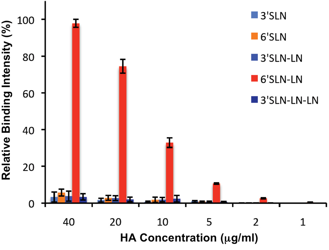
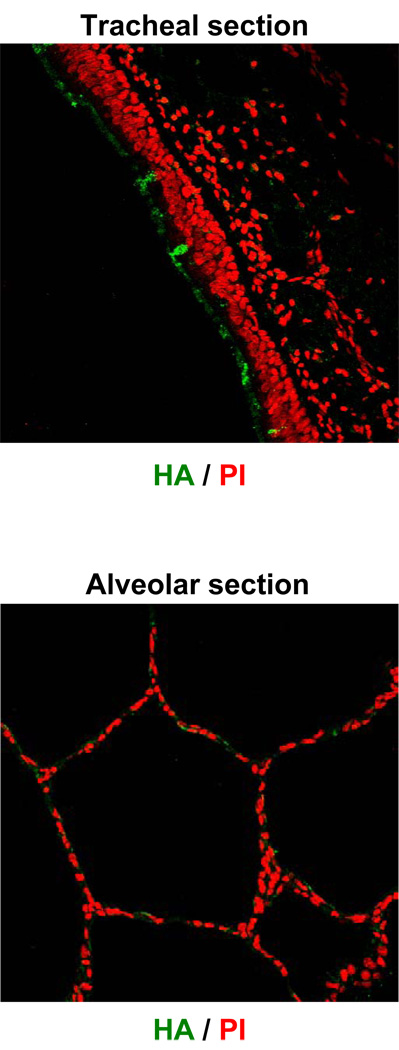
The binding of influenza viruses to their target cells is mediated by viral HA and binding preference to specific sialylated glycan receptors is a critical determinant of H1N1 virus transmission efficiency in ferrets (14, 17, 18). We examined the glycan binding properties of the 2009 A(H1N1) HA by dose-dependent direct receptor binding (18) and human lung tissue binding assays using recombinantly expressed soluble CA/04 HA (19). In the direct glycan receptor binding assay, CA/04 HA exhibited a dose-dependent binding to only a single α2–6 glycan (6’SLN-LN) and only minimal binding to α2–3 glycans (Figure 1). Although the binding pattern of CA/04 HA is similar to that of HA from the 1918 pandemic influenza A virus (A/South Carolina/1/1918 or SC18), the binding affinity of CA/04 HA is considerably lower than that of SC18 HA (Supplementary Figures 4 & 5). Examining the tissue binding of CA/04 HA indicates that it binds uniformly to the apical surface of the human tracheal (representative upper respiratory) tissue sections (Figure 2). This binding pattern correlates with the predominant distribution of α2–6 sialylated glycans on the apical surface of the tracheal tissue (19) and the α2–6 binding of CA/04 HA in the direct binding assay. Although CA/04 shows some binding to alveolus, it is not as extensive as the tracheal binding consistent with the minimal α2–3 binding observed in the direct binding assay.
Figure 1. Dose dependent direct receptor binding of CA/04 HA.
A streptavidin plate array comprising representative biotinylated α2–3 and α2–6 sialylated glycans were used for the assay. The biotinylated glycans include 3’SLN, 6’SLN, 3’SLN-LN, 6’SLN-LN and 3’SLN-LN-LN. LN corresponds to lactosamine (Galβ1–4GlcNAc) and 3’SLN and 6’SLN respectively correspond to Neu5Ac α2–3 and Neu5Ac α2–6 linked to LN. The assay was carried out as described previously (18) for an entire range of HA concentration from 0.01 – 40 µg/ml by pre-complexing HA: primary antibody: secondary antibody in the ratio 4:2:1 to enhance the multivalent presentation of HA. The binding signals for HA concentrations below 1 µg/ml were at background level and hence there concentrations are not shown on the x-axis. The y-axis shows the normalized binding signal as a percentage of the maximum value.
Figure 2. Human tissue binding of CA/04 HA.
Shown in the top is the binding of CA/04 HA at 20 µg/ml concentration to apical surface (white arrow) of human tracheal tissue sections (green as against propidium iodide staining in red). The binding of the recombinantly expressed HA to the human tissues was carried out as described previously (19) by precomplexing HA: primary antibody:secondary antibody in the ratio 4:2:1 to enhance multivalent presentation of HA. Note the binding of HA to the apical surface of tracheal tissue which is known to predominantly express α2–6 sialylated glycans (19). Shown in the bottom is the minimal binding of HA at 20 µg/ml concentration to the alveolar tissue section. The sialic acid specific binding of HA to the tracheal tissue section was confirmed by blocking of HA binding to the tissue section pre-treated with 0.2 U of Sialidase A (recombinantly expressed in Arthrobacter ureafaciens).
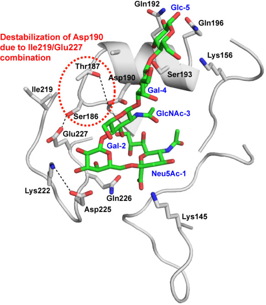
The receptor-binding site (RBS) of the 2009 A (H1N1) HAs (20), used in this study, were compared to those from SC18 and the recent seasonal influenza H1N1 viruses (Table 2). The similarity in the binding pattern between CA/04 HA and SC18 HA could potentially arise from the majority of the ”similar or analogous” RBS residues between these HAs including Asp190 and Asp225, which are ‘hallmark’ amino acids of human adapted H1N1 HAs that make optimal contacts with the α2–6 glycans. The main differences in RBS between SC18 and CA/04 HA are at positions 145, 186, 189, 219 and 227. The CA/04 HA has a unique Lys145 which provides an additional anchoring contact for the sialic acid (20). The residues at 186, 187, 189 are positioned to form an interaction network with Asp190 (18, 20). In the case of SC18 HA, this network involves oxygen atoms of Thr187, Thr189 and Asp190 (18). In a similar fashion, in the case of CA/04 HA, this network could potentially be formed by the oxygen atoms of Ser186, Thr187 and Asp190. The residues 219 and 227 in turn influence the orientation of residue 186. Comparison of residues 219 and 227 (Table 2) reveals that either both amino acids are hydrophobic such as Ala219 and Ala227 (as observed in SC18 HA) or they are charged residues such as Lys219 and Glu227 (as observed in the seasonal influenza HAs). Interestingly, the 2009 A(H1N1) HAs have a unique combination of Ile219 and Glu227 that results in a set of interactions that is neither fully hydrophobic nor fully charged. This combination could destabilize the hydrophobic or ionic network of residues at 186, 219 and 227 thereby disrupting optimal contacts with the α2–6 sialylated glycans (Figure 3). Analysis of the RBS of the 2009 A(H1N1) HA offers an explanation for the lower α2–6 binding affinity of CA/04 HA as compared to SC18 HA despite the similar binding pattern (Supplementary Figure 4). Taken together, the differences in the glycan binding property of the 2009 A(H1N1) HA when compared to that of SC18 and the recent seasonal influenza HAs correlate with the observed differences in the respiratory droplet transmission in ferrets.
Table 2.
Glycan binding residues of H1N1 HAs
| H1N1 Strains | Cluster 1 | Cluster 2 | Cluster 3 | Cluster 4 | Cluster 5 | |||||||||||||||||
|---|---|---|---|---|---|---|---|---|---|---|---|---|---|---|---|---|---|---|---|---|---|---|
| 136 | 138 | 226 | 137 | 153 | 155 | 194 | 183 | 145 | 222 | 225 | 190 | 189 | 187 | 186 | 219 | 227 | 192 | 193 | 156 | 159 | 196 | |
| SolIs_3_06 | S | S | Q | A | W | T | L | H | S | K | D | D | G | N | P | K | E | R | A | G | G | H |
| Bris_59_07 | S | S | Q | A | W | T | L | H | S | K | D | D | G | N | P | K | E | K | A | G | G | H |
| NewCal_20_ | S | S | Q | A | W | T | L | H | S | K | D | N | G | N | P | K | E | R | A | G | G | H |
| TX_36_91 | T | S | Q | T | W | T | I | H | S | K | D | D | R | N | S | K | E | R | A | E | G | H |
| SC18 | T | A | Q | A | W | T | L | H | S | K | D | D | T | T | P | A | A | Q | S | K | S | Q |
| TX/15 | T | A | Q | A | W | V | L | H | K | K | D | D | A | T | S | I | E | Q | S | K | N | Q |
| MX/4482 | T | A | Q | A | W | V | L | H | K | K | D | D | A | T | S | I | E | Q | S | K | N | Q |
| CA/04 | T | A | Q | A | W | V | L | H | K | K | D | D | A | T | S | I | E | Q | S | K | N | Q |
| Neu5Ac-1 | Gal-2 | GlcNAc-3 | Gal-4, Glc-5, … | |||||||||||||||||||
The residues are organized into network forming clusters. The sugar unit (numbered as shown in Figure 3), which makes contact with the clusters, is shown in the last row. The unique amino acids in 2009 H1N1 HAs are highlighted in red. The key for the virus strains SolIS_3_06 (A/Solomon Islands/3/06); Bris_59_07 (A/Brisbane/59/07); NewCal_20_99 (A/New Caledonia/20/99); TX_36_91 (A/Texas/36/91).
Figure 3. Structural model of CA/04 HA bound to α2–6 oligosaccharide.
The contacts of CA/04 HA with an α2–6 oligosaccharide (Neu5Ac α2–6Galβ1–4GlcNAcβ1–3Galβ1–4Glc) were analyzed by constructing a structural model as described previously (20). Shown in the figure is the cartoon representation of the glycan binding site of CA/04 HA where the side chains of the key amino acids are shown in stick representation (colored by atom carbon: gray; oxygen: red; nitrogen:blue). The α2–6 oligosaccharide is shown as a stick representation (colored by atom carbon: green; oxygen: red; nitrogen:blue) and labeled blue starting from non-reducing end Neu5Ac-1 to reducing end Glc-5. The potential destabilization of the interaction network due to the Ile219/Glu227 combination is highlighted in red dotted circle.
To evaluate the pathogenicity of the novel H1N1 viruses in another mammalian species, we inoculated BALB/c mice i.n. with three 2009 A(H1N1) isolates and then determined virus replication, morbidity (as measured by weight loss), 50% mouse infectious dose (MID50) and 50% lethal dose (LD50) titers. In addition to CA/04 and TX/15, A/Mexico/4108/2009 (Mex/4108; H1N1) virus, obtained from hospitalized case of non-fatal infection, was included in the mouse pathotyping experiments. The 2009 A(H1N1) viruses isolates did not kill mice (LD50 >106 PFU or EID50), which displayed only transient weight reduction (Table 3), however, all three 2009 A(H1N1) viruses replicated efficiently in mouse lungs without prior host adaptation (Table 3). Typically, human influenza A strains of the H1N1 subtype replicate efficiently in mice only after they are adapted to growth in these animals (21). The MID50 titers, determined by the detection of virus in the lungs of mice 3 days p.i., were markedly low (MID50 = 100.5–1.5 PFU or EID50), indicating high infectivity in this model. We next determined whether 2009 A(H1N1) viruses replicated systemically in the mouse after intranasal infection, a characteristic of virulent avian influenza (H5N1) viruses isolated from humans, but not 1918 (H1N1) virus (17, 22). All mice infected with CA/04, Tx/15, or Mex/4108 viruses had undetectable levels (<10 PFU/ml) of virus in whole spleen, thymus, brain and intestinal tissues, indicating that the 2009 A(H1N1) viruses did not spread to extrapulmonary organs in the mouse.
Table 3.
Pathogenicity of 2009 A(H1N1) viruses isolates in BALB/c mice
| Virus | PFU/mla | Weight lossb | Lung titersc | MID50d | LD50d |
|---|---|---|---|---|---|
| A/California/04/2009 | 7.4 | 5.3 | 5.8 ± 1.2 | 1.5 | >6 |
| A/Texas/15/2009 | 7.7 | 1.5 | 5.4± 0.9 | 0.5 | >6 |
| A/Mexico/4108/2009 | 7.8 | 3.5 | 6.8± 0.4 | 1.5 | >6 |
Titer of virus stocks prepared on Madin Darby canine kidney (MDCK) cells or eggs expressed as log10 PFU/ml.
Maximum percent weight loss (5 mice per group) following inoculation with 105 PFU or EID50.
Average lung titers of three mice on day 3 post-inoculation, expressed as PFU/ml ± SD.
Fifty percent mouse infectious dose (MID50) and 50% lethal dose (LD50) are expressed as the log10 PFU or EID50 required to give one MID50 or one LD50.
The full clinical spectrum of disease caused by 2009 A(H1N1) viruses and its transmissibility are not completely understood. The present study shows that overall morbidity and lung viral titers were higher in ferrets infected with 2009 A(H1N1) isolates compared with the seasonal H1N1 virus. Moreover, the detection of 2009 A(H1N1) viruses in the intestinal tissue of ferrets is consistent with gastro-intestinal involvement among some human 2009 A(H1N1) cases (6). Although the 2009 A(H1N1) viruses demonstrated similar replication kinetics as the seasonal H1N1 virus in the upper respiratory tract of inoculated ferrets, the 2009 A(H1N1) viruses did not spread to all naïve ferrets by respiratory droplets. This lack of efficient respiratory droplet transmission suggests that additional virus adaptation in mammals may be required to reach the high-transmissible phenotypes observed with seasonal H1N1 or the 1918 pandemic virus (14, 17).
It was demonstrated previously that the efficiency of respiratory droplet transmission in ferrets correlates with the α2–6 binding affinity of the viral HA (17). Only a single amino acid mutation in HA of the high-transmissible SC18 virus led to a virus (NY18) that transmitted inefficiently and the α2–6 binding affinity of NY18 HA was substantially lower than that of SC18 HA (Supplementary Figure 5). In a similar fashion the substantially lower α2–6 binding affinity of CA/04 HA to that of NY18 HA correlates with the less efficient 2009 A(H1N1) respiratory droplet transmission (Supplementary Figure 5).
Adaptation of the polymerase basic protein 2 (PB2) is also critical for efficient aerosolized respiratory transmission of an H1N1 influenza virus (14, 17, 23). A single amino acid substitution from glutamic acid to lysine at amino acid position 627 supports efficient influenza virus replication at the lower temperature (33°C) found in the mammalian airway, and contributes to efficient transmission in mammals (14, 23). All three of the 20th century influenza pandemics were caused by viruses containing human adapted PB2 genes and, in general, lysine is present at position 627 among the human influenza viruses whereas a glutamic acid is found in this position among the avian influenza isolates that fail to transmit efficiently among ferrets (14). In contrast to Brisbane/07 virus and other seasonal H1N1 viruses, all 2009 A(H1N1) viruses to date with an avian influenza lineage PB2 gene possess a glutamic acid at residue 627 (1). The phenotype of PB2 is determined by the amino acid at position 627, which can arise by mutant selection or reassortment, and along with adaptive changes in the RBS should be closely monitored as markers for enhanced virus transmission.
Supplementary Material
References and Notes
- 1.Garten RJ, et al. Science. 2009 [Google Scholar]
- 2.MMWR Morb Mortal Wkly Rep. 2009;58:453. [PubMed] [Google Scholar]
- 3.MMWR Morb Mortal Wkly Rep. 2009;58:400. [PubMed] [Google Scholar]
- 4.MMWR Morb Mortal Wkly Rep. 2009;58:467. [PubMed] [Google Scholar]
- 5. http://www.who.int/en/
- 6.Dawood FS, et al. N Engl J Med. 2009;360:2605. doi: 10.1056/NEJMoa0903810. [DOI] [PubMed] [Google Scholar]
- 7.Fraser C, et al. Science. 2009 [Google Scholar]
- 8.Tumpey TM, Belser JA. Annu Rev Microbiol. 2009 doi: 10.1146/annurev.micro.091208.073359. [DOI] [PubMed] [Google Scholar]
- 9.Webby RJ, Webster RG. Science. 2003;302:1519. doi: 10.1126/science.1090350. [DOI] [PubMed] [Google Scholar]
- 10.Viboud C, et al. Vaccine. 2006;24:6701. doi: 10.1016/j.vaccine.2006.05.067. [DOI] [PubMed] [Google Scholar]
- 11.Maines TR, et al. Proc Natl Acad Sci U S A. 2006;103:12121. doi: 10.1073/pnas.0605134103. [DOI] [PMC free article] [PubMed] [Google Scholar]
- 12.Belser JA, et al. Proc Natl Acad Sci U S A. 2008;105:7558. doi: 10.1073/pnas.0801259105. [DOI] [PMC free article] [PubMed] [Google Scholar]
- 13.Lowen AC, Palese P. Infect Disord Drug Targets. 2007;7:318. doi: 10.2174/187152607783018736. [DOI] [PubMed] [Google Scholar]
- 14.Van Hoeven N, et al. Proc Natl Acad Sci U S A. 2009;106:3366. doi: 10.1073/pnas.0813172106. [DOI] [PMC free article] [PubMed] [Google Scholar]
- 15.Material and Methods are available as supporting material on Science Online.
- 16.Yen HL, et al. J Virol. 2007;81:6890. doi: 10.1128/JVI.00170-07. [DOI] [PMC free article] [PubMed] [Google Scholar]
- 17.Tumpey TM, et al. Science. 2007;315:655. doi: 10.1126/science.1136212. [DOI] [PubMed] [Google Scholar]
- 18.Srinivasan A, et al. Proc Natl Acad Sci U S A. 2008;105:2800. doi: 10.1073/pnas.0711963105. [DOI] [PMC free article] [PubMed] [Google Scholar]
- 19.Chandrasekaran A, et al. Nat Biotechnol. 2008;26:107. doi: 10.1038/nbt1375. [DOI] [PubMed] [Google Scholar]
- 20.Soundararajan V, et al. Nat Biotechnol. 2009;27:510. doi: 10.1038/nbt0609-510. [DOI] [PubMed] [Google Scholar]
- 21.Hartley CA, Reading PC, Ward AC, Anders EM. Arch Virol. 1997;142:75. doi: 10.1007/s007050050060. [DOI] [PubMed] [Google Scholar]
- 22.Maines TR, et al. J Virol. 2005;79:11788. doi: 10.1128/JVI.79.18.11788-11800.2005. [DOI] [PMC free article] [PubMed] [Google Scholar]
- 23.Steel J, Lowen AC, Mubareka S, Palese P. PLoS Pathog. 2009;5 doi: 10.1371/journal.ppat.1000252. e1000252. [DOI] [PMC free article] [PubMed] [Google Scholar]
- 24.We thank Patrick Blair (Naval Health Research Center, San Diego), Gail J. Demmler (Texas Children’s Hospital, Houston), Celia Alpuche-Aranda (Instituto de Diagnóstico y Referencia Epidemiológicos (INDRE), Mexico), and WHO Collaborating Centre for Reference and Research on Influenza (Melbourne) for facilitating access to viruses. We also thank Xuihua Lu and Amanda Balish for preparation of viruses and Vic Veguilla for statistical analysis. RS would like to acknowledge the consortium for functional glycomics for providing glycan standards and support from the Singapore–Massachusetts Institute of Technology Alliance for Research and Technology and the National Institute of General Medical Sciences of the National Institutes of Health (GM 57073 andU54 GM62116). Confocal microscopy of the human lung tissue sections was performed utilizing the WM Keck Foundation Biological Imaging Facility at the Whitehead Institute The findings and conclusions in this report are those of the authors and do not necessarily reflect the views of the funding agency.
Associated Data
This section collects any data citations, data availability statements, or supplementary materials included in this article.


