Abstract
The Gram-negative bacterium Actinobacillus pleuropneumoniae is the etiologic agent of porcine contagious pleuropneumoniae, a lethal respiratory infectious disease causing great economic losses in the swine industry worldwide. In order to better interpret the genetic background of serotypic diversity, nine genomes of A. pleuropneumoniae reference strains of serovars 1, 2, 4, 6, 9, 10, 11, 12, and 13 were sequenced by using rapid high-throughput approach. Based on 12 genomes of corresponding serovar reference strains including three publicly available complete genomes (serovars 3, 5b, and 7) of this bacterium, we performed a comprehensive analysis of comparative genomics and first reported a global genomic characterization for this pathogen. Clustering of 26,012 predicted protein-coding genes showed that the pan genome of A. pleuropneumoniae consists of 3,303 gene clusters, which contain 1,709 core genome genes, 822 distributed genes, and 772 strain-specific genes. The genome components involved in the biogenesis of capsular polysaccharide and lipopolysaccharide O antigen relative to serovar diversity were compared, and their genetic diversity was depicted. Our findings shed more light on genomic features associated with serovar diversity of A. pleuropneumoniae and provide broader insight into both pathogenesis research and clinical/epidemiological application against the severe disease caused by this swine pathogen.
Actinobacillus pleuropneumoniae, a Gram-negative facultative anaerobic encapsulated coccobacillus, belongs to the Actinobacillus genus of the Pasteurellaceae family (19). A. pleuropneumoniae is a primary bacterial etiologic agent of porcine contagious pleuropneumonia, a severe respiratory disease leading to great economic losses to the global swine industry (7). The cases usually display pleuropneumonia and pulmonary lesions characterized by serious hemorrhage and necrosis. To date, several factors involved in the virulence of A. pleuropneumoniae have been described, including Apx exotoxins, capsular polysaccharides (CPS), lipopolysaccharides (LPS), outer membrane proteins, iron-acquisition proteins and adhesin factors (11, 19, 24). However, the genetic differences of pathogenesis remain poorly characterized and are worth interpreting from the perspective of comparative genomics for this bacterium.
Thus far, 15 serovars and two biotypes of A. pleuropneumoniae have been recognized, with great variations in virulence and interlocal distributions (6). The predominant serovar-specific antigens are composed of CPS, which could rigorously define serovars of A. pleuropneumoniae (6, 34). Antigenic differences in the LPS can further determine A. pleuropneumoniae subtypes within a same capsular serovar (13). The metabolic and virulent characteristics of this pathogen have been systematically described based on the prior knowledge and two complete genomes (18, 47), but the molecular basis and evolutionary mechanism of serotypic diversity are still not well explained due to the lack of sequence information. To investigate the associations of serovar diversity with the underlying genetic components, more serovar-related genomic islands involved in the biosynthesis of capsular and lipopolysaccharide antigens should be decoded at the pan-genome level of A. pleuropneumoniae. At present, through the next-generation of sequencing technique (454 GS FLX pyrosequencing platform), more and more bacterial species, subspecies or typical strains have been quickly sequenced, such as eight species in the Yersinia genus (9), 17 strains of Streptococcus pneumoniae (22), and 5 strains from different Francisella tularensis subspecies (8). Multiple genome sequences from different strains of a single species can offer comprehensive information to explore the relationship between genotypes and phenotypes and to further discover additional genetic markers for clinical purpose.
In the present study, we sequenced the A. pleuropneumoniae genomes of nine reference strains of serovars 1, 2, 4, 6, 9, 10, 11, 12, and 13. Together with three public complete genome sequences of A. pleuropneumoniae serovars 3, 5b, and 7, the analysis of comparative genomics was performed to report a global genomic characterization of this pathogenic bacterium. The acquisition and loss of genome compositions that contribute to virulence and serovar diversity were identified. The genetic loci involved in the biogenesis of capsule and O-specific polysaccharide were compared, and their vital roles in serotypic diversity were investigated.
MATERIALS AND METHODS
Bacterial strains.
Nine reference strains from different A. pleuropneumoniae serovars sequenced in the present study (Table 1) were kindly provided by Pat Blackall of Australasian Pig Institute, Australia. All strains were grown overnight in tryptic soy broth (TSB) medium at 37°C shaking on a rotary shaker (200 rpm), supplemented with 10 μg of NAD/ml and 10% bovine serum. Total genomic DNA was extracted by using the DNeasy tissue kit (Qiagen).
TABLE 1.
A. pleuropneumoniae strains and publicly available genomes used in this study
| Virulence | Serovar | Strain | GenBank accession no. | Source or reference |
|---|---|---|---|---|
| High | 1 | 4074 | This study | |
| Medium | 2 | S1536 | This study | |
| Low | 3 | JL03 | CP000687 | 47 |
| Medium | 4 | M62 | This study | |
| High | 5b | L20 | CP000569 | 18 |
| Medium | 6 | Femϕ | This study | |
| Medium | 7 | Ap76 | CP001091 | |
| High | 9 | CVJ13261 | This study | |
| Rarely isolated | 10 | D13039 | This study | |
| High | 11 | 56153 | This study | |
| Medium | 12 | 1096 | This study | |
| Rarely isolated | 13 | N273 | This study |
Sequencing and assembly.
Bacterial genomes were sequenced at the Chinese National Human Genome Center at Shanghai using the 454 GS FLX platform (Roche, Germany) (32). For each sample, a library containing fragments (500 to 800 bp) for sequencing was prepared from 4 μg of genomic DNA. On average, 59 contigs per genome were assembled by using the 454 Newbler de novo assembler (version 2.3).
Genome annotation.
Nine newly sequenced draft genomes and three publicly available complete genomes of different A. pleuropneumoniae serovars were annotated by using an automated bacterial annotation pipeline DIYA (40) and custom Perl scripts. Using DIYA, the genomic contigs were tiled against a complete genome (from strain L20) by the promer script in the MUMmer package (28). The order of the matching contigs was thus inferred, and all contigs for each sample were concatenated into a pseudogenome with random nonmatching contigs on the end. To identify the interrupt protein-coding genes at the terminals of a contig, a contig linker (NNNNNCATTCCATTCATTAATTAATTAATGAATGAATGNNNNN) that contains stop and start codons in all six reading frames was used (44). Each pseudogenome was then used for the identification of protein-coding sequences (CDSs), tRNAs, and rRNAs implemented by the programs Glimmer3 (37), tRNASCAN-SE v1.23 (30), and RNAmmer v1.2 (29), respectively. All protein-coding genes translated in all six reading frames were searched against the proteins from the UniRef50 database (which was updated in February 2010) (42) by using Blastx (10−10 cutoff E-value and 35% minimum identity) (2). Gene annotation was supplied with the cluster of orthologous groups (COG) database (43) by using rpsblast (10−10 cutoff E-value) (2). A combined data set including nine genomes sequenced in the present study and three reannotated intact genomes was generated for the following analysis of comparative genomics for A. pleuropneumoniae.
Accession numbers.
The draft genome assemblies for the nine A. pleuropneumoniae strains were deposited in the GenBank database (http://www.ncbi.nih.gov/GenBank/index.html). The accession numbers for these sequences, as well as the accession numbers for three publicly available A. pleuropneumoniae complete genomes, were as follows with corresponding strains: ADOD00000000 (4074), ADOE00000000 (S1536), ADOF00000000 (M62), ADOG00000000 (Femϕ), ADOI00000000 (CVJ13261), ADOJ00000000 (D13039), ADOK00000000 (56153), ADOL00000000 (1096), ADOM00000000 (N273), CP000569 (L20), CP000687 (JL03), and CP001091 (AP76).
Whole-genome alignment.
Based on the blastn hits (minimum identity of 95% and a cutoff E-value of 10−5) of a reference complete genome (from strain L20) searched against the other genomes of A. pleuropneumoniae, genome comparative circular maps were constructed by using CGview software (41). Multiple A. pleuropneumoniae genomes were aligned and visualized by using Mauve v2.3.1 with the default settings (12).
Gene clustering and alignment.
The CDSs were extracted from all annotated genomes of A. pleuropneumoniae. The small CDSs with length shorter than 40 amino acids were removed from the data set. The orthologous proteins were grouped by using the program Blastclust (1). An all-versus-all BLASTP of all proteomes was first performed to define the orthologous pairs satisfying the following criteria: a cutoff E-value of 10−6, over 70% length coverage, and at least 70% identity. Pairs of sequences that have statistically significant matches were then clustered into the same group by using single-linkage clustering. Multiple protein sequence alignment for each cluster was performed with the program MUSCLE 3.6 (14). The corresponding nucleotide sequence alignment was produced based on the aligned amino acid sequences from each gene cluster using custom Perl scripts.
Phylogeny of A. pleuropneumoniae serovars.
To infer the evolutionary relationships between 12 different serovar reference strains of A. pleuropneumoniae, two types of dendrograms were generated, respectively. According to a gene possession-based phylogenetic approach, we defined the genetic distance between a pair of genomes (i and k), to be Σn|gn,i − gn,k|, where gn,i is 1 if gene n is present in strain i and 0 otherwise (23). The distance matrix was then used to reconstruct species phylogeny with the unweighted pair group method with arithmetic mean (UPGMA) method implemented in the Phylip package (15). The second type of phylogenetic tree was reconstructed using sequence alignments of 1,287 single-copy core genes with nearly identical length and exactly one member in each of the 12 genomes. These gene alignments were concatenated into a large alignment of 1,211,061 nucleotides. A maximum-likelihood tree was built under the HKY85 substitution model with the estimated transition/transversion rate ratio (κ) and gamma distributed rate heterogeneity of four categories (Γ4) in PhyML (20).
RESULTS AND DISCUSSION
General features of sequenced genomes.
In the present study, nine genomes from different A. pleuropneumoniae serovar reference strains, 4074 of serovar 1, S1536 of serovar 2, M62 of serovar 4, Femϕ of serovar 6, CVJ13261 of serovar 9, D13039 of serovar 10, 56153 of serovar 11, 1096 of serovar 12, and N273 of serovar 13, were sequenced by using 454 GS FLX. The depth of each genomic sequencing is 14- to 26-fold, and the average number of assembled contigs for each genome is 59 with a range from 44 to 89 (see Table S1 in the supplemental material). All nine genome draft sequences have been submitted to GenBank.
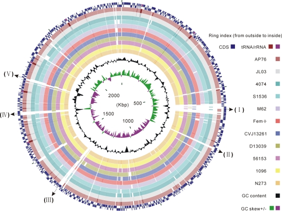
Although these genomes were extracted from the reference strains of different A. pleuropneumoniae serovars that can be assigned to three levels of virulence (Table 1) (10), the overall genomic characteristics were quite similar (Table 2). All of the genomes were comprised of a circular chromosome with ∼2.19 Mb ∼2.33 Mb in length. The average GC content of each genome was 41%, which was consistent with that of an entire A. pleuropneumoniae chromosome (47). The median number of CDSs per strain was 2,174, the largest number was 2,223 for serovar 4 strain M62, and the least was 2,096 for serovar 12 strain 1096. Pairwise nucleotide alignments using blastn (>95% identity) revealed high sequence conservation between each draft genome and the A. pleuropneumoniae serovar 5b strain L20 complete genome (Fig. 1). The percentage of total length of matched sequences accounting for the L20 genome (2,274,482 bp) was ranged from ∼90.8% (S1536 versus L20) to ∼92.7% (56153 versus L20). Meanwhile, the global pairwise genomic alignment also showed several large genetic differences that may be relative to bacterial virulence and serotypic diversity. Notably, the genomic regions bearing aberrant GC content may represent the occurrence of horizontal gene transfer events in different serovar reference strain, such as biosynthetic loci of the LPS O antigen and capsule (Fig. 1). Detailed analyses of these featured genomic regions using the local multiple sequence alignments among subsets of genomes are described below.
TABLE 2.
Summary of genome features and gene clusters of A. pleuropneumoniae
| Serovar | Strain | Genome size (Mb) | G+C (%) | CDSs | Total gene clusters | Distributed clusters | Unique clusters | Noncore clusters (%) |
|---|---|---|---|---|---|---|---|---|
| 1 | 4074 | 2.26 | 41.2 | 2,180 | 2,150 | 415 | 26 | 20.5 |
| 2 | S1536 | 2.22 | 41.1 | 2,137 | 2,111 | 351 | 51 | 19.0 |
| 3 | JL03 | 2.24 | 41.2 | 2,101 | 2,048 | 289 | 50 | 16.6 |
| 4 | M62 | 2.27 | 41.2 | 2,223 | 2,193 | 317 | 167 | 22.1 |
| 5b | L20 | 2.27 | 41.3 | 2,137 | 2,076 | 289 | 78 | 17.7 |
| 6 | Femϕ | 2.31 | 41.0 | 2,219 | 2,184 | 375 | 100 | 21.7 |
| 7 | AP76 | 2.33 | 41.2 | 2,203 | 2,134 | 377 | 48 | 19.9 |
| 9 | CVJ13261 | 2.26 | 41.2 | 2,204 | 2,178 | 432 | 37 | 21.5 |
| 10 | D13039 | 2.27 | 41.2 | 2,168 | 2,137 | 321 | 107 | 20.0 |
| 11 | 56153 | 2.27 | 41.2 | 2,195 | 2,164 | 438 | 17 | 21.0 |
| 12 | 1096 | 2.19 | 41.2 | 2,096 | 2,072 | 302 | 61 | 17.5 |
| 13 | N273 | 2.25 | 41.2 | 2,149 | 2,124 | 385 | 30 | 19.5 |
FIG. 1.
Circular representation of sequence conservation between A. pleuropneumoniae serovar 5b strain L20 and 11 strains belonging to different serovars. Circles are numbered from 1 (outermost circle) to 16 (innermost circle). The outermost two circles show CDSs, rRNAs and tRNAs in the L20 genome of A. pleuropneumoniae serovar 5b. The next 11 circles show the coordinates of BLAST hits detected through blastn comparisons (minimum sequence identity of 95% and expected threshold of 10−5) of the L20 reference genome against 11 A. pleuropneumoniae genomes, including two public complete genomes and nine contig sets of new genomes, and each circle is colored according to serovar reference strains: maroon for serovar 7 strain AP76, silver for serovar 3 strain JL03, teal for serovar 1 strain 4074, cyan for serovar 2 strain S1536, light purple for serovar 4 strain M62, red for serovar 6 strain Femϕ, blue for serovar 9 strain CVJ13261, olive for serovar 10 strain D13039, fuchsia for serovar 11 strain 56153, yellow for serovar 12 strain 1096, and orange for serovar 13 strain N273. Overlapping hits appear as darker arcs. The innermost two circles show GC content and GC skew plot of the L20 genome. Several known serovar-specific genomic regions with low sequence identity were numbered as follows: I, the ∼38-kb prophage region; II and III, the coding gene cluster involved in type I restriction-modification system; IV, the LPS O-antigen biosynthesis region; V, the CPS biosynthesis region.
Identification of gene clusters.
A multi-fasta file with 26,012 CDSs of all 12 A. pleuropneumoniae genomes used for clustering was available in Text S1 in the supplemental material. The total number of A. pleuropneumoniae orthologous gene clusters (designated APO, hereafter), also including unique genes that were exclusive by only a single strain, was 3,303. Of these, 52% were identified to be core gene clusters that were shared by all strains and accounts for 79% of the total number of CDSs, 25% were dispensable gene clusters that were found to be possessed by at least two strains but not all, and the remaining 23% were unique genes, only accounting for ca. 3% of the total CDSs (Table 3). A gene clustering table that contained the gene content of 12 A. pleuropneumoniae genomes was summarized (see Table S2 in the supplemental material) and the relative identifiers of CDSs were listed (see Table S3 in the supplemental material). Each genome contained some strain-specific protein coding genes, with a range of 167 for strain M62 of serovar 4 to 17 for strain 56153 of serovar 11 (Table 2). Among 1,594 noncore clusters, including distributed and unique genes, serovar 3 strain JL03 had the lowest percentage (16.6%), and serovar 4 strain M62 had the highest percentage (22.1%) (Table 2). The pairwise comparison of gene content demonstrated that the average number of genes associated with the gain or loss between any two strains was 429, with a standard deviation of 97. The maximum and minimum numbers of genic differences, 611 and 96, were identified in strain pairs M62 (serovar 4)/CVJ13261 (serovar 9) and CVJ13261 (serovar 9)/56153 (serovar 11), respectively.
TABLE 3.
Summary of gene clusters for 12 A. pleuropneumoniae strains
| Gene class | No. of orthologous clusters | No. of CDSs |
|---|---|---|
| Core | 1,709 | 20,597 |
| Distributed | 822 | 4,411 |
| Unique | 772 | 772 |
| Excluded by size | 232 | |
| Total | 3,303 | 26,012 |
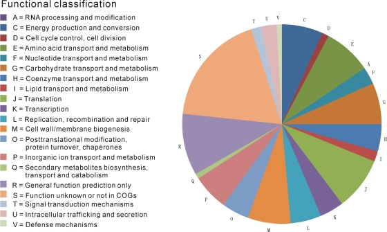
To some extent, the proteins encoded by the core genes present in all 12 genomes should participate in the fundamental metabolic activities of A. pleuropneumoniae and be essential for the growth and survival of this bacterium. The distribution of cellular functions of these core proteins indicated that protein-coding genes involved in translation were assigned into the largest category (8.54%) (Fig. 2; see also Table S4 in the supplemental material). As expected, there was no core protein involved in cell motility, which was coincident with the common phenotype of nonmotile. It was worth noting that a flagellin gene fliC reported previously in A. pleuropneumoniae (33) was absent in the genomic sequences of the 12 reference strains. The set of noncore proteins had relatively more elements involved in surface polysaccharide biogenesis and the bacterial pathogenic process compared to that of core proteins. These distributed or unique proteins that may play a potential role in differentiating serovars and virulence were assigned to the function categories of defense mechanism, replication and recombination, and the type I and III restriction-modification system, including diverse transposases, recombinases, integrases, and DNA helicases (see Table S5 in the supplemental material). Twenty-four genes encoding autotransporter adhesins involved in extracellular structures were found to be distributed or unique in 12 strains. In addition, ∼5.1% of the unique genes and ∼5.2% of distributed genes had annotations associated with phage, prophage, or bacteriophage; whereas very few phage protein coding genes of 0.2% was present in the core genes. Among 2,531 core and distributed orthologous gene clusters (Table 3), ca. 21.4% (542) were annotated as hypothetical or uncharacterized proteins (see Table S5 in the supplemental material), suggesting that a significant percentage of even the bacterial housekeeping genes remain unknown in A. pleuropneumoniae.
FIG. 2.
Distribution of cellular function categories of core orthologous protein clusters.
The differences of gene components between the high- and low-virulence strains may provide insight for identifying novel candidate virulence factors. The 54 distributed genes that were shared by the high-virulence strains from serovars 1, 5b, 9, and 11 but absent in the low-virulence strain JL03 of serovar 3 are summarized in Table 4. As expected, genes (APO_2026 and APO_2004) encoding a toxin activator ApxIC and a structural toxin ApxIA, respectively, were exclusively present in the reference strains of serovars 1, 5b, 9, 10, and 11, all of which secrete the strongly hemolytic and cytotoxic ApxI (19, 38). Compared to apxIC and apxIA, the genes apxIIIC (APO_2098) and apxIIIA (APO_2049) involved in expression of the nonhemolytic but strongly cytotoxic ApxIII were present only in the reference strains of serovars 2, 3, 4, and 6. The genetic compositions of apx genes in newly sequenced genomes conformed to the apx gene patterns of corresponding serovar reference strain previously reported (4, 25). Of 54 distributed genes, 17 were annotated as hypothetical proteins, and their roles in bacterial virulence need to be further investigated.
TABLE 4.
Gene clusters shared by the highly virulent serovars in A. pleuropneumoniae
| Cluster | Distributed serotypes | Annotation of predicted protein product |
|---|---|---|
| APO_1721 | 1, 2, 5b, 6, 7, 9, 10, 11, 12, 13, 4 | SNF2-related protein |
| APO_1723 | 1, 2, 7, 12, 13, 6, 5b, 9, 11, 4, 10 | Truncated transferrin-binding protein 1 |
| APO_1726 | 1, 2, 4, 5b, 6, 7, 9, 10, 11, 12, 13 | Homoserine dehydrogenase |
| APO_1729 | 1, 2, 4, 5b, 6, 7, 9, 10, 11, 12, 13 | GTP pyrophosphokinase |
| APO_1730 | 1, 2, 4, 5b, 6, 7, 9, 10, 11, 12, 13 | Elongation factor G |
| APO_1746 | 1, 2, 4, 5b, 6, 7, 9, 10, 11, 12, 13 | Exodeoxyribonuclease 7 large subunit |
| APO_1764 | 1, 2, 4, 5b, 6, 7, 9, 10, 11, 12, 13 | Malate dehydrogenase (oxaloacetate-decarboxylating) [NADP(+)], phosphate acetyltransferase |
| APO_1767 | 1, 2, 4, 5b, 6, 7, 9, 10, 11, 12, 13 | Molybdopterin biosynthesis protein moeA |
| APO_1779 | 1, 2, 4, 5b, 6, 7, 9, 10, 11, 12, 13 | Glycerophosphoryl diester phosphodiesterase |
| APO_1797 | 1, 2, 4, 5b, 6, 7, 9, 10, 11, 12, 13 | Uncharacterized periplasmic iron-binding protein |
| APO_1806 | 1, 2, 4, 6, 7, 9, 10, 11, 12, 13, 5b | DacC protein |
| APO_1811 | 1, 2, 4, 5b, 6, 7, 10, 11, 12, 13, 9 | Carbonic anhydrase |
| APO_1812 | 1, 2, 4, 5b, 6, 7, 9, 10, 11, 12, 13 | tRNA [adenine-N(6)-]-methyltransferase |
| APO_1817 | 1, 2, 4, 5b, 6, 7, 9, 10, 11, 12, 13 | FKBP-type peptidyl-prolyl cis-trans isomerase 1 |
| APO_1825 | 1, 2, 4, 5b, 6, 7, 9, 10, 11, 12, 13 | 50S ribosomal protein L10 |
| APO_1852 | 1, 2, 4, 5b, 6, 7, 9, 10, 11, 12, 13 | Hypothetical protein |
| APO_1866 | 1, 7, 9, 11, 13, 2, 4, 5b, 12, 6 | Acetyltransferase |
| APO_1874 | 1, 2, 5b, 6, 7, 9, 10, 11, 13, 12 | Threonine efflux protein |
| APO_1878 | 1, 2, 4, 5b, 7, 9, 10, 11, 12, 13 | Uncharacterized protein |
| APO_1879 | 1, 2, 5b, 6, 7, 9, 10, 11, 12, 13 | Endoribonuclease L-PSP |
| APO_1881 | 1, 2, 4, 5b, 6, 7, 9, 10, 11, 13 | Putative uncharacterized protein |
| APO_1895 | 1, 9, 11, 4, 5b, 7, 10, 12, 13 | ATP-dependent RNA helicase RhlB |
| APO_1896 | 1, 4, 5b, 7, 9, 11, 12, 13, 10 | Flp operon protein C, RcpC |
| APO_1897 | 1, 2, 4, 5b, 7, 9, 11, 12, 13 | Probable ABC transporter permease protein |
| APO_1901 | 7, 12, 1, 2, 5b, 6, 9, 11, 13 | Putative uncharacterized protein |
| APO_1905 | 1, 2, 5b, 6, 7, 9, 11, 12, 13 | Putative uncharacterized protein |
| APO_1916 | 1, 5b, 9, 11, 12, 6, 7, 13 | Hemolysin activation/secretion protein |
| APO_1920 | 1, 2, 5b, 7, 9, 11, 12, 13 | Transposase IS200-like protein |
| APO_1927 | 1, 2, 4, 5b, 9, 10, 11, 12 | Hypothetical protein |
| APO_1961 | 10, 5b, 7, 1, 9, 11 | Phage terminase large subunit |
| APO_1967 | 10, 5b, 7, 1, 9, 11 | Bacteriophage capsid protein |
| APO_1968 | 1, 9, 11, 12, 5b, 10 | Undecaprenyl-phosphate galactose phosphotransferase, WbaP |
| APO_1973 | 12, 10, 1, 5b, 9, 11 | Hypothetical protein |
| APO_1987 | 2, 5b, 6, 1, 9, 11 | Outer membrane receptor proteins, mostly Fe transport |
| APO_1988 | 10, 1, 5b, 7, 9, 11 | Uncharacterized protein |
| APO_2004 | 1, 5b, 9, 10, 11 | RTX-I toxin determinant A from serotypes 5/10, ApxIA |
| APO_2006 | 7, 1, 9, 11, 5b | ATP-dependent Clp protease proteolytic subunit |
| APO_2010 | 1, 5b, 9, 10, 11 | Putative uncharacterized protein |
| APO_2012 | 1, 5b, 6, 9, 11 | Predicted phage integrase family protein |
| APO_2016 | 1, 9, 11, 5b, 10 | ABC transporter ATP binding subunit, Wzm |
| APO_2018 | 1, 5b, 9, 10, 11 | Possible bacteriophage antirepressor |
| APO_2024 | 1, 5b, 7, 9, 11 | Putative phage tail component |
| APO_2025 | 1, 5b, 9, 11, 10 | Possible DNA methylase |
| APO_2026 | 5b, 10, 1, 9, 11 | Hemolysin-activating lysine-acyltransferase HlyC, ApxIC |
| APO_2027 | 5b, 1, 9, 11, 12 | Putative uncharacterized protein |
| APO_2030 | 5b, 10, 1, 9, 11 | Putative uncharacterized protein |
| APO_2031 | 1, 5b, 7, 9, 11 | Possible bacteriophage tail protein |
| APO_2033 | 1, 5b, 9, 10, 11 | Hypothetical protein |
| APO_2038 | 1, 5b, 9, 10, 11 | Hypothetical protein |
| APO_2039 | 10, 1, 5b, 9, 11 | Hypothetical protein |
| APO_2040 | 1, 9, 11, 5b, 10 | Hypothetical protein |
| APO_2043 | 1, 2, 5b, 9, 11 | Hypothetical protein |
| APO_2085 | 1, 5b, 9, 11 | Site-specific DNA methylase |
| APO_2140 | 5b, 9, 11, 1 | Hypothetical protein |
Phylogenetic relationships among serovar reference strains.
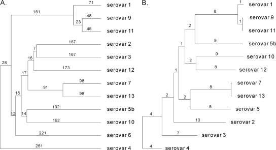
To understand the phylogenetic relationships among the 12 serovars of A. pleuropneumoniae, we used two approaches based on noncore genic differences and the concatenated sequences of 1,287 single-copy core genes among all serovar reference strains, respectively. Figure 3 A demonstrated phylogenetic differentiation among A. pleuropneumoniae serovars. The differentiation was represented by the total numbers of gene loss and gain between any two genomes of reference strains. Except for A. pleuropneumoniae strain L20 of serovar 5b, which originated in the United States, the other highly virulent strains from serovars 1, 9, and 11 belonged to a common clade and differed from each other by fewer than 157 genes, hinting that the strains from the three serovars probably derived from a recent common ancestor. As expected, serovar 9 strain CVJ13261 and serovar 11 strain D13039, which were both isolated from the same geographical location in the Netherlands, had a more close relationship (27). Notably, strains AP76 of serovar 7 and N273 of serovar 13 isolated in Canada and Hungary, respectively, were the second closely related pair and had the genic differentiation bearing 196 genes with gain or loss between them (3). Figure 3B showed a maximum-likelihood tree estimated by the large sequence alignment of 1,287 single-copy core genes. The partial topology types of the genic differences and multi-sequence alignments based trees were similar. Three A. pleuropneumoniae reference strains of serovars 1, 9, and 11 were also grouped into an individual clade, as well as serovars 7 and 13.
FIG. 3.
A. pleuropneumoniae whole-genome phylogeny. (A) Dendrogram showing the phylogenetic relationship based on differences in genetic gain or loss of noncore genes among the 12 strains of diverse A. pleuropneumoniae serovars. The numbers on the branch represent the number of genic differences that occurs from the previous bifurcation node. (B) Maximum-likelihood tree estimated from a data set of 1,287 concatenated, conserved genic sequences in 12 A. pleuropneumoniae genomes. The numbers on the branch stand for the number of nucleotide substitutions per kilobase that occur prior to the next level of separation.
Whole-genome alignment.
A global multiple genome alignment is shown in Fig. S1 in the supplemental material, and it demonstrates that A. pleuropneumoniae chromosomes had highly colinear arrangements without largely internal rearrangements among all genomes from 12 serovars. Comparative analysis between two complete genomes of strains JL03 and L20 has shown that the serotypic diversity of A. pleuropneumoniae is likely to associate with several serovar-specific genomic regions, which encode the gene clusters involved in the biosynthesis of CPS and LPS O antigen (47).
Genes involved in CPS biosynthesis.
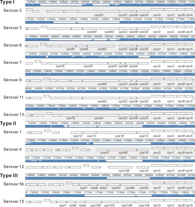
The genetic organization of the CPS biosynthesis and export locus is shown in Fig. 4, demonstrating that the genes cpxDCBA involved in CPS export are present and highly conserved in all of the serovar reference strains, whereas genes of the cps cluster involved in the capsule biosynthesis exhibited high genetic diversity in different sets of serovars. According to the results generated by Blastclust, 33 orthologous genes encoding CPS biosynthetic enzymes were identified in the reference strains of 12 A. pleuropneumoniae serovars (Table 5), 24 of which encode strain- or serovar-specific enzymes that are probably responsible for the dissimilarity of the CPS chemical structures. However, previous studies have pointed out that A. pleuropneumoniae serovars 1 to 13 can be divided into three groups according to differences of their chemical compositions and the structures of the capsule: type I of CPS consisted of teichoic acid polymers joined by phosphate diester bonds is present in serovars 2, 3, 6, 7, 8, 9, 11, and 13; type II consisted of oligosaccharide polymers joined through phosphate bonds includes serovars 1, 4, and 12; and type III solely containing repeats of oligosaccharide units includes serovars 5a, 5b, and 10 (26, 34).
FIG. 4.
Schematic comparison of the genetic organizations of the CPS biosynthesis and export gene clusters in the reference strains of 12 A. pleuropneumoniae serovars. Three types of CPS have been defined in A. pleuropneumoniae as follows: type 1, serovars 2, 3, 6, 7, 9, 11, and 13 are composed of teichoic acid polymers linked by phosphate diester bonds; type II, serovars 1, 4, and 12 are composed of oligosaccharide polymers linked by phosphate bonds; and type III, serovars 5b and 10 are composed of repeating oligosaccharide units (26, 34).
TABLE 5.
Genes encoding enzymes in the capsular polysaccharide biosynthesis locus of A. pleuropneumoniae
| ClusterID | Name | Putative function | CDS no. |
|---|---|---|---|
| APO_1934 | cps2C | Cps2C | appser2_15930 appser3_16160 appser6_17380 appser7_16930 appser13_16580 appser9_17130 appser11_17250 |
| APO_1935 | cps2A | Cps2A | appser2_15950 appser3_16180 appser6_17400 appser7_16950 appser9_17150 appser11_17270 appser13_16600 |
| APO_1947 | cps2B | Glycerol-3-phosphate cytidylyltransferase | appser2_15940 appser3_16170 appser6_17390 appser7_16940 appser9_17140 appser11_17260 appser13_16590 |
| APO_2170 | cps2D | Cps2D | appser2_15920 appser7_16920 appser13_16570 |
| APO_2202 | cps1A | Capsular polysaccharide phosphotransferase | appser12_16150 appser1_17000 appser4_16240 |
| APO_2331 | cps9D | Teichoic acid biosynthesis protein | appser9_17120 appser11_17240 |
| APO_2349 | cps7E | CapZD protein | appser7_16910 appser13_16560 |
| APO_2361 | Putative uncharacterized protein | appser11_17220 appser9_17090 | |
| APO_2380 | Putative uncharacterized protein | appser9_17110 appser11_17230 | |
| APO_2543 | cps1B | Glycosyl transferase family protein | appser1_16990 |
| APO_2554 | cps4B | Glycosyl transferase family protein | appser4_16230 |
| APO_2555 | cps6D | Cps6D | appser6_17370 |
| APO_2562 | cps12B | Cps12B | appser12_16140 |
| APO_2582 | cps9D' | Teichoic acid biosynthesis protein | appser3_16150 |
| APO_2601 | cps10B | Acetyltransferase (Isoleucine patch superfamily) protein | appser10_16450 |
| APO_2602 | cps5B | Region 2 capsular polysaccharide biosynthesis protein | appser5b_16510 |
| APO_2607 | cps10D | Putative uncharacterized protein | appser10_16430 |
| APO_2609 | Putative uncharacterized protein | appser3_16120 | |
| APO_2611 | cps6F | Glycosyl transferase group 1 | appser6_17350 |
| APO_2615 | cps10C | Cap29eA protein | appser10_16440 |
| APO_2625 | cps9D' | Putative uncharacterized protein | appser3_16140 |
| APO_2628 | cps10A | GDP-mannose pyrophosphorylase | appser10_16460 |
| APO_2632 | Putative uncharacterized protein | appser2_15900 | |
| APO_2644 | Putative uncharacterized protein | appser2_15910 | |
| APO_2651 | cps1C | Putative uncharacterized protein | appser1_16980 |
| APO_2663 | cps5C | Region 2 capsular polysaccharide biosynthesis protein | appser5b_16500 |
| APO_2684 | cps1D | Acetyltransferase (Isoleucine patch superfamily) protein | appser1_16970 |
| APO_2685 | cps4C | Possible lipopolysaccharide biosynthesis glycosyltransferase | appser4_16220 |
| APO_2686 | Putative uncharacterized protein | appser3_16130 | |
| APO_2688 | cps6E | Cps6E (fragment) | appser6_17360 |
| APO_2702 | cps5A | Putative glycosyltransferase | appser5b_16520 |
| APO_2724 | Hypothetical protein | appser6_17340 | |
| APO_3193 | Hypothetical protein | appser9_17100 |
The genetic organization of the cps biosynthetic loci of 12 serovar reference strains provided molecular evidence to further support the grouping of A. pleuropneumoniae serovars described above. First, the proteins encoded by cps2ABC were present only in serovars 2, 3, 6, 7, 9, 11, and 13 of type I. Cps2A and Cps2B encode teichoic acid glycerol transferase and glycerol-3-phosphate cytidylyltransferase, respectively, which are required for the sequential transfer of glycerol phosphate units (17). Teichoic acid synthases encoded by the genes cps2D (APO_2170), cps6D (APO_2555), and cps9D (APO_2331) were also identified in the serovars of type I. Although these synthases share low sequence identity, they all contain two conserved domains Glyphos_transf (PF04464) and Glycos_transf_1 (PF00534). It is worth mentioning that the teichoic acid biosynthetic enzyme encoded by cps9D (APO_2331) was present only in strains CVJ13261 and 56153 of serovars 9 and 11 but became a pseudogene in strain JL03 of serovar 3. Second, Cps1A encodes a capsular polysaccharide phosphotransferase and is 73% similar to the LcbA protein of Neisseria meningitidis, which may help pathogens to evade the host innate immune system (21, 39). This enzyme shared by serovars 1, 4, and 12 of type II may be involved in the chemical linkage of phosphate in the linear CPS backbone. Third, a KdsA homolog required for the synthesis of monosaccharide dOclA that is a structural component of the A. pleuropneumoniae serovars 5 and 10 CPSs (13, 46), was identified just in their reference strains L20 and D13039, respectively. It has been reported that the CPSs of A. pleuropneumoniae are the immunodominant antigens bearing greater serological specificities than the O antigens (35). Concordantly, the genetic organizations of the cps biosynthetic loci were found to be distinct from each other in all 12 serovar reference strains, potentially leading to differences in their CPS structures.
Genes involved in LPS biosynthesis.
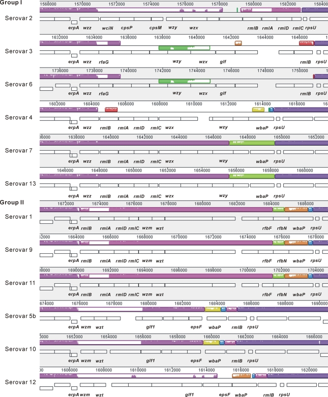
LPS, the major adhesin of A. pleuropneumoniae involved in adherence to porcine respiratory tract cells and mucus, plays an important role in virulence (5, 36). The structures of LPS O antigen (or O polysaccharide) were reported to be chemically identical or similar in different sets of A. pleuropneumoniae serovars: serovars 3, 6, 8, and 15; serovars 1, 9, and 11; and serovars 4, 7, and 13 (31, 35). In the present study, we identified 19 core genes involved in the synthetic pathways of lipid A and core oligosaccharide, the majority of which were dispersed throughout the chromosome and highly conserved among different organisms within the family Pasteurellaceae (see Table S6 in the supplemental material). On the other hand, a cluster of genes coding for enzymes that catalyze the biosynthesis of O antigen were identified between the conserved genes erpA (APO_1556) and rpsU (APO_0575), and these genes were transcribed in the same orientation in all A. pleuropneumoniae serovar reference strains. A total of 52 orthologous genes distributed in the 12 O-antigen chains were identified, 20 of which were strain- or serovar-specific genes probably involved in the structural diversity of O polysaccharide (Table 6). The genetic organizations of the LPS O-antigen biosynthesis are shown in Fig. 5. Three sets of serovars (serovars 1, 9, and 11; serovars 7 and 13; and serovars 3 and 6) were observed to have identical gene components; this finding was consistent with the characterization of the O-antigen structures described above. Two different O-antigen biosynthetic pathways were identified for the first time in A. pleuropneumoniae. The Wzy/Wzx-dependent pathway of O-antigen biosynthesis was possessed by the serovars 2, 3, 4, 6, 7, and 13 (group I), whereas the ABC-2 transporter-dependent pathway was shared by the serovars 1, 5b, 9, 10, 11, and 12 (group II) (45). The genes wzm and wzt encoding ABC-2 transporters that were the integral membrane subunit and the ATP-binding subunit, respectively, were identified within the O-antigen chains of group II. Wzm proteins in the corresponding A. pleuropneumoniae reference strains shared low amino acid sequence identity (58%) and were all predicted to have six transmembrane domains, like the Wzm homologue in Escherichia coli (16). We deduced that the diversified gene composition of O-antigen chains should also play a role in the serotypic diversity of A. pleuropneumoniae.
TABLE 6.
Genes encoding enzymes in the lipopolysaccharide O-antigen biosynthesis locus of A. pleuropneumoniae
| Cluster | Name | Putative function | CDS no. |
|---|---|---|---|
| APO_0575 | rmlB | dTDP-glucose 4,6-dehydratase | appser3_15020 appser5b_15380 appser6_16250 appser10_15330 appser12_15050 appser2_14830 appser4_15060 appser7_15190 appser13_15410 appser1_15770 appser9_15920 appser11_16040 |
| APO_1940 | rmlA | Glucose-1-phosphate thymidylyltransferase | appser1_15780 appser2_14840 appser4_15070 appser7_15200 appser9_15930 appser11_16050 appser13_15420 |
| APO_1941 | rmlD | dTDP-4-dehydrorhamnose reductase | appser1_15790 appser2_14850 appser4_15080 appser7_15210 appser9_15940 appser11_16060 appser13_15430 |
| APO_1942 | rmlC | dTDP-4-dehydrorhamnose 3,5-epimerase | appser1_15800 appser2_14860 appser4_15090 appser7_15220 appser9_15950 appser11_16070 appser13_15440 |
| APO_1968 | wbaP | Undecaprenyl-phosphate galactose phosphotransferase, WbaP | appser1_15880 appser9_16030 appser11_16150 appser12_15040 appser5b_15370 appser10_15320 |
| APO_1972 | wzz | Wzz-like protein | appser2_14740 appser3_14920 appser4_15050 appser6_16150 appser7_15180 appser13_15400 |
| APO_2016 | wzt | ABC transporter ATP binding subunit | appser1_15820 appser9_15970 appser11_16090 appser5b_15300 appser10_15260 |
| APO_2172 | Putative glycosyltransferase | appser1_15830 appser9_15980 appser11_16100 | |
| APO_2187 | wbaP | Undecaprenyl-phosphate galactose phosphotransferase, WbaP | appser4_15150 appser7_15280 appser13_15500 |
| APO_2191 | Serotype b-specific antigen synthesis gene cluster | appser1_15840 appser11_16110 appser9_15990 | |
| APO_2192 | wzx | Putative O-antigen transporter | appser4_15100 appser7_15230 appser13_15450 |
| APO_2194 | wzy | Putative membrane protein | appser7_15260 appser13_15480 appser4_15130 |
| APO_2198 | glf1 | Probable UDP-galactopyranose mutase | appser12_15010 appser10_15280 appser5b_15330 |
| APO_2209 | Glycosyl transferase, family 2 | appser1_15850 appser9_16000 appser11_16120 | |
| APO_2212 | Glycosyl transferase, family 2 | appser5b_15350 appser10_15300 appser12_15020 | |
| APO_2213 | rfbN | O antigen biosynthesis rhamnosyltransferase rfbN | appser1_15870 appser9_16020 appser11_16140 |
| APO_2215 | Glycosyl transferase family 2 | appser4_15140 appser7_15270 appser13_15490 | |
| APO_2216 | rfbF | Putative rhamnosyl transferase | appser1_15860 appser9_16010 appser11_16130 |
| APO_2228 | wzm | ABC transporter integral membrane subunit | appser1_15810 appser9_15960 appser11_16080 |
| APO_2230 | epsF | Exopolysaccharide biosynthesis protein | appser5b_15360 appser10_15310 appser12_15030 |
| APO_2352 | wzx | Flippase Wzx | appser3_14980 appser6_16210 |
| APO_2357 | wzy | Oligosaccharide repeat unit polymerase | appser3_14970 appser6_16200 |
| APO_2359 | Undecaprenyl-phosphate galactose phosphotransferase | appser3_15000 appser6_16230 | |
| APO_2373 | glf | UDP-galactopyranose mutase | appser3_14990 appser6_16220 |
| APO_2376 | Polysacchride biosynthesis protein | appser6_16240 appser3_15010 | |
| APO_2378 | Glycosyltransferase | appser3_14950 appser6_16180 | |
| APO_2379 | Glycosyltransferase | appser3_14960 appser6_16190 | |
| APO_2383 | Putative glycosyltransferase | appser7_15240 appser13_15460 | |
| APO_2398 | Glycosyltransferase | appser7_15250 appser13_15470 | |
| APO_2402 | rfaG | CpsF protein | appser3_14930 appser6_16160 |
| APO_2403 | wzm | ABC transporter integral membrane subunit | appser5b_15290 appser10_15250 |
| APO_2430 | Putative uncharacterized protein | appser5b_15340 appser10_15290 | |
| APO_2585 | Putative uncharacterized protein | appser10_15270 | |
| APO_2587 | Putative glycosyltransferase | appser12_15000 | |
| APO_2613 | wzx | Flippase Wzx | appser2_14800 |
| APO_2631 | Putative uncharacterized protein | appser12_14990 | |
| APO_2634 | Undecaprenyl-phosphate galactosephosphotransferase | appser2_14820 | |
| APO_2668 | wciN | WciN beta, glycosyltransferase | appser2_14750 |
| APO_2674 | VI polysaccharide biosynthesis protein | appser6_16170 | |
| APO_2678 | VI polysaccharide biosynthesis protein | appser3_14940 | |
| APO_2696 | Putative glycosyltransferase | appser4_15110 | |
| APO_2699 | Glycosyl transferase, group 2 family protein | appser2_14770 | |
| APO_2703 | Glycosyl transferase family 2 | appser12_14980 | |
| APO_2711 | wzy | Eps4N | appser2_14790 |
| APO_2725 | cpsP | CpsP, glycosyltransferase | appser2_14760 |
| APO_2755 | wzm | ABC-2 type transporter family protein | appser12_14960 |
| APO_2759 | Putative uncharacterized protein | appser5b_15310 | |
| APO_2791 | Chloramphenicol acetyltransferase | appser2_14810 | |
| APO_2796 | wzt | O-antigen export system ATP-binding protein RfbB | appser12_14970 |
| APO_2810 | cpsM | Eps7I | appser2_14780 |
| APO_2980 | Putative uncharacterized protein | appser5b_15320 | |
| APO_3164 | Glycosyltransferase | appser4_15120 |
FIG. 5.
Schematic comparison of the genetic organizations of the LPS O-antigen biosynthesis gene clusters in the reference strains of 12 A. pleuropneumoniae serovars. A. pleuropneumoniae serovars can be divided into two groups based on two different mechanisms for the assembly and translocation of O antigen: group I, the Wzy/Wzx-dependent pathway is present in serovars 2, 3, 4, 6, 7, and 13; and group II, the ABC-2 transporter-dependent pathway is present in serovars 1, 5b, 9, 10, 11, and 12.
In summary, comparative genomic analysis using genome sequences originated from 12 serovars showed that the pan genome of A. pleuropneumoniae consists of 3,303 gene clusters, which contain 1,709 core genome genes, 822 distributed genes, and 772 strain-specific unique genes. The genetic diversity of strain (serovar)-specific genomic islands related to the biogenesis of capsule and lipopolysaccharide O antigen should offer powerful molecular evidence explaining the mechanisms of the serotypic diversity of A. pleuropneumoniae. We believe that these findings will provide crucial clues for the development of genomic typing of A. pleuropneumoniae and new-style universal vaccines against the severe swine disease caused by this pathogen.
Supplementary Material
Acknowledgments
We thank Huajun Zheng and members in the Sequencing Division of the Chinese National Human Genome Center in Shanghai, China, for the genome sequencing and analysis work.
This study was supported by grants from the National Basic Research Program of China (973 Program; 2006CB504402), the National Natural Science Foundation of China (30771599, 30901075), the National Scientific and Technical Supporting Program of China (2006BAD06A01), and the Program for Changjiang Scholars and Innovative Research Team in University (IRT0726).
Footnotes
Published ahead of print on 27 August 2010.
Supplemental material for this article may be found at http://jb.asm.org/.
REFERENCES
- 1.Altschul, S. F., W. Gish, W. Miller, E. W. Myers, and D. J. Lipman. 1990. Basic local alignment search tool. J. Mol. Biol. 215:403-410. [DOI] [PubMed] [Google Scholar]
- 2.Altschul, S. F., T. L. Madden, A. A. Schäffer, J. Zhang, Z. Zhang, W. Miller, and D. J. Lipman. 1997. Gapped BLAST and PSI-BLAST: a new generation of protein database search programs. Nucleic Acids Res. 25:3389-3402. [DOI] [PMC free article] [PubMed] [Google Scholar]
- 3.Anderson, C., A. A. Potter, and G.-F. Gerlach. 1991. Isolation and molecular characterization of spontaneously occurring cytolysin-negative mutants of Actinobacillus pleuropneumoniae serotype 7. Infect. Immun. 59:4110-4116. [DOI] [PMC free article] [PubMed] [Google Scholar]
- 4.Beck, M., J. F. van den Bosch, I. M. Jongenelen, P. L. Loeffen, R. Nielsen, J. Nicolet, and J. Frey. 1994. RTX toxin genotypes and phenotypes in Actinobacillus pleuropneumoniae field strains. J. Clin. Microbiol. 32:2749-2754. [DOI] [PMC free article] [PubMed] [Google Scholar]
- 5.Bélanger, M., D. Dubreuil, J. Harel, C. Girard, and M. Jacques. 1990. Role of lipopolysaccharides in adherence of Actinobacillus pleuropneumoniae to porcine tracheal rings. Infect. Immun. 58:3523-3530. [DOI] [PMC free article] [PubMed] [Google Scholar]
- 6.Blackall, P. J., H. L. Klaasen, H. van den Bosch, P. Kuhnert, and J. Frey. 2002. Proposal of a new serovar of Actinobacillus pleuropneumoniae: serovar 15. Vet. Microbiol. 84:47-52. [DOI] [PubMed] [Google Scholar]
- 7.Bossé, J. T., H. Janson, B. J. Sheehan, A. J. Beddek, A. N. Rycroft, J. S. Kroll, and P. R. Langford. 2002. Actinobacillus pleuropneumoniae: pathobiology and pathogenesis of infection. Microbes Infect. 4:225-235. [DOI] [PubMed] [Google Scholar]
- 8.Champion, M. D., Q. Zeng, E. B. Nix, F. E. Nano, P. Keim, C. D. Kodira, M. Borowsky, S. Young, M. Koehrsen, R. Engels, M. Pearson, C. Howarth, L. Larson, J. White, L. Alvarado, M. Forsman, S. W. Bearden, A. Sjöstedt, R. Titball, S. L. Michell, B. Birren, and J. Galagan. 2009. Comparative genomic characterization of Francisella tularensis strains belonging to low and high virulence subspecies. PLoS Pathog. 5:e1000459. [DOI] [PMC free article] [PubMed] [Google Scholar]
- 9.Chen, P. E., C. Cook, A. C. Stewart, N. Nagarajan, D. D. Sommer, M. Pop, B. Thomason, M. P. Thomason, S. Lentz, N. Nolan, S. Sozhamannan, A. Sulakvelidze, A. Mateczun, L. Du, M. E. Zwick, and T. D. Read. 2010. Genomic characterization of the Yersinia genus. Genome Biol. 11:R1. [DOI] [PMC free article] [PubMed] [Google Scholar]
- 10.Christensen, H., and M. Bisgaard. 2004. Revised definition of Actinobacillus sensu stricto isolated from animals. A review with special emphasis on diagnosis. Vet. Microbiol. 99:13-30. [DOI] [PubMed] [Google Scholar]
- 11.Chung, J. W., C. Ng-Thow-Hing, L. I. Budman, B. F. Gibbs, J. H. Nash, M. Jacques, and J. W. Coulton. 2007. Outer membrane proteome of Actinobacillus pleuropneumoniae: LC-MS/MS analyses validate in silico predictions. Proteomics 7:1854-1865. [DOI] [PubMed] [Google Scholar]
- 12.Darling, A. C., B. Mau, F. R. Blattner, and N. T. Perna. 2004. Mauve: multiple alignment of conserved genomic sequence with rearrangements. Genome Res. 14:1394-1403. [DOI] [PMC free article] [PubMed] [Google Scholar]
- 13.Dubreuil, J. D., M. Jacques, K. R. Mittal, and M. Gottschalk. 2000. Actinobacillus pleuropneumoniae surface polysaccharides: their role in diagnosis and immunogenicity. Anim. Health Res. Rev. 1:73-93. [DOI] [PubMed] [Google Scholar]
- 14.Edgar, R. C. 2004. MUSCLE: multiple sequence alignment with high accuracy and high throughput. Nucleic Acids Res. 32:1792-1797. [DOI] [PMC free article] [PubMed] [Google Scholar]
- 15.Felsenstein, J. 1989. PHYLIP: Phylogeny Inference Package (version 3.2). Cladistics 5:164-166. [Google Scholar]
- 16.Feng, L., S. N. Senchenkova, J. Yang, A. S. Shashkov, J. Tao, H. Guo, J. Cheng, Y. Ren, Y. A. Knirel, P. R. Reeves, and L. Wang. 2004. Synthesis of the heteropolysaccharide O antigen of Escherichia coli O52 requires an ABC transporter: structural and genetic evidence. J. Bacteriol. 186:4510-4519. [DOI] [PMC free article] [PubMed] [Google Scholar]
- 17.Fitzgerald, S. N., and T. J. Foster. 2000. Molecular analysis of the tagF gene, encoding CDP-glycerol:poly(glycerophosphate) glycerophosphotransferase of Staphylococcus epidermidis ATCC 14990. J. Bacteriol. 182:1046-1052. [DOI] [PMC free article] [PubMed] [Google Scholar]
- 18.Foote, S. J., J. T. Bossé, A. B. Bouevitch, P. R. Langford, N. M. Young, and J. H. Nash. 2008. The complete genome sequence of Actinobacillus pleuropneumoniae L20 (serovar 5b). J. Bacteriol. 190:1495-1496. [DOI] [PMC free article] [PubMed] [Google Scholar]
- 19.Frey, J. 1995. Virulence in Actinobacillus pleuropneumoniae and RTX toxins. Trends Microbiol. 3:257-261. [DOI] [PubMed] [Google Scholar]
- 20.Guindon, S., and O. Gascuel. 2003. A simple, fast and accurate algorithm to estimate large phylogenies by maximum likelihood. Syst. Biol. 52:696-704. [DOI] [PubMed] [Google Scholar]
- 21.Hammerschmidt, S., C. Birkholz, U. Zähringer, B. D. Robertson, J. van Putten, O. Ebeling, and M. Frosch. 1994. Contribution of genes from the capsule gene complex (cps) to lipooligosaccharide biosynthesis and serum resistance in Neisseria meningitidis. Mol. Microbiol. 11:885-896. [DOI] [PubMed] [Google Scholar]
- 22.Hiller, N. L., B. Janto, J. S. Hogg, R. Boissy, S. Yu, E. Powell, R. Keefe, N. E. Ehrlich, K. Shen, J. Hayes, K. Barbadora, W. Klimke, D. Dernovoy, T. Tatusova, J. Parkhill, S. D. Bentley, J. C. Post, G. D. Ehrlich, and F. Z. Hu. 2007. Comparative genomic analyses of seventeen Streptococcus pneumoniae strains: insights into the pneumococcal supragenome. J. Bacteriol. 189:8186-8195. [DOI] [PMC free article] [PubMed] [Google Scholar]
- 23.Hogg, J. S., F. Z. Hu, B. Janto, R. Boissy, J. Hayes, R. Keefe, J. C. Post, and G. D. Ehrlich. 2007. Characterization and modeling of the Haemophilus influenzae core and supragenomes based on the complete genomic sequences of Rd and 12 clinical nontypeable strains. Genome Biol. 8:R103. [DOI] [PMC free article] [PubMed] [Google Scholar]
- 24.Jacques, M. 2004. Surface polysaccharides and iron-uptake systems of Actinobacillus pleuropneumoniae. Can. J. Vet. Res. 68:81-85. [PMC free article] [PubMed] [Google Scholar]
- 25.Jansen, R., J. Briaire, A. B. van Geel, E. M. Kamp, A. L. Gielkens, and M. A. Smits. 1994. Genetic map of the Actinobacillus pleuropneumoniae RTX-toxin (Apx) operons: characterization of the ApxIII operons. Infect. Immun. 62:4411-4418. [DOI] [PMC free article] [PubMed] [Google Scholar]
- 26.Jessing, S. G., P. Ahrens, T. J. Inzana, and Ø. Angen. 2008. The genetic organization of the capsule biosynthesis region of Actinobacillus pleuropneumoniae serotypes 1, 6, 7, and 12. Vet. Microbiol. 129:350-359. [DOI] [PubMed] [Google Scholar]
- 27.Kokotovic, B., and Ø. Angen. 2007. Genetic diversity of Actinobacillus pleuropneumoniae assessed by amplified fragment length polymorphism analysis. J. Clin. Microbiol. 45:3921-3929. [DOI] [PMC free article] [PubMed] [Google Scholar]
- 28.Kurtz, S., A. Phillippy, A. L. Delcher, M. Smoot, M. Shumway, C. Antonescu, and S. L. Salzberg. 2004. Versatile and open software for comparing large genomes. Genome Biol. 5:R12. [DOI] [PMC free article] [PubMed] [Google Scholar]
- 29.Lagesen, K., P. Hallin, E. A. Rødland, H. H. Staerfeldt, T. Rognes, and D. W. Ussery. 2007. RNAmmer: consistent and rapid annotation of rRNA genes. Nucleic Acids Res. 35:3100-3108. [DOI] [PMC free article] [PubMed] [Google Scholar]
- 30.Lowe, T. M., and S. R. Eddy. 1997. tRNAscan-SE: a program for improved detection of transfer RNA genes in genomic sequence. Nucleic Acids Res. 25:955-964. [DOI] [PMC free article] [PubMed] [Google Scholar]
- 31.MacLean, L. L., M. B. Perry, and E. Vinogradov. 2004. Characterization of the antigenic lipopolysaccharide O chain and the capsular polysaccharide produced by Actinobacillus pleuropneumoniae serotype 13. Infect. Immun. 72:5925-5930. [DOI] [PMC free article] [PubMed] [Google Scholar]
- 32.Margulies, M., M. Egholm, W. E. Altman, S. Attiya, J. S. Bader, L. A. Bemben, J. Berka, M. S. Braverman, Y. J. Chen, Z. Chen, S. B. Dewell, L. Du, J. M. Fierro, X. V. Gomes, B. C. Godwin, W. He, S. Helgesen, C. H. Ho, G. P. Irzyk, S. C. Jando, M. L. Alenquer, T. P. Jarvie, K. B. Jirage, J. B. Kim, J. R. Knight, J. R. Lanza, J. H. Leamon, S. M. Lefkowitz, M. Lei, J. Li, K. L. Lohman, H. Lu, V. B. Makhijani, K. E. McDade, M. P. McKenna, E. W. Myers, E. Nickerson, J. R. Nobile, R. Plant, B. P. Puc, M. T. Ronan, G. T. Roth, G. J. Sarkis, J. F. Simons, J. W. Simpson, M. Srinivasan, K. R. Tartaro, A. Tomasz, K. A. Vogt, G. A. Volkmer, S. H. Wang, Y. Wang, M. P. Weiner, P. Yu, R. F. Begley, and J. M. Rothberg. 2005. Genome sequencing in microfabricated high-density picolitre reactors. Nature 437:376-380. [DOI] [PMC free article] [PubMed] [Google Scholar]
- 33.Negrete-Abascal, E., M. E. Reyes, R. M. García, S. Vaca, J. A. Girón, O. García, E. Zenteno, and M. De La Garza. 2003. Flagella and motility in Actinobacillus pleuropneumoniae. J. Bacteriol. 185:664-668. [DOI] [PMC free article] [PubMed] [Google Scholar]
- 34.Perry, M. B., E. Altman, J.-R. Brisson, L. M. Beynon, and J. C. Richards. 1990. Structural characteristics of the antigenic capsular polysaccharides and lipopolysaccharides involved in the serological classification of Actinobacillus pleuropneumoniae strains. Serodiagn. Immuno. Infect. Dis. 4:299-308. [Google Scholar]
- 35.Perry, M. B., L. L. MacLean, and E. Vinogradov. 2005. Structural characterization of the antigenic capsular polysaccharide and lipopolysaccharide O-chain produced by Actinobacillus pleuropneumoniae serotype 15. Biochem. Cell Biol. 83:61-69. [DOI] [PubMed] [Google Scholar]
- 36.Rioux, S., C. Bégin, J. D. Dubreuil, and M. Jacques. 1997. Isolation and characterization of LPS mutants of Actinobacillus pleuropneumoniae serotype 1. Curr. Microbiol. 35:139-144. [DOI] [PubMed] [Google Scholar]
- 37.Salzberg, S. L., A. L. Delcher, S. Kasif, and O. White. 1998. Microbial gene identification using interpolated Markov models. Nucleic Acids Res. 26:544-548. [DOI] [PMC free article] [PubMed] [Google Scholar]
- 38.Schaller, A., S. P. Djordjevic, G. J. Eamens, W. A. Forbes, R. Kuhn, P. Kuhnert, M. Gottschalk, J. Nicolet, and J. Frey. 2001. Identification and detection of Actinobacillus pleuropneumoniae by PCR based on the gene apxIVA. Vet. Microbiol. 79:47-62. [DOI] [PubMed] [Google Scholar]
- 39.Sperisen, P., C. D. Schmid, P. Bucher, and O. Zilian. 2005. Stealth proteins: in silico identification of a novel protein family rendering bacterial pathogens invisible to host immune defense. PLoS Comput. Biol. 1:e63. [DOI] [PMC free article] [PubMed] [Google Scholar]
- 40.Stewart, A. C., B. Osborne, and T. D. Read. 2009. DIYA: a bacterial annotation pipeline for any genomics lab. Bioinformatics 25:962-963. [DOI] [PMC free article] [PubMed] [Google Scholar]
- 41.Stothard, P., and D. S. Wishart. 2005. Circular genome visualization and exploration using CGView. Bioinformatics 21:537-539. [DOI] [PubMed] [Google Scholar]
- 42.Suzek, B. E., H. Huang, P. McGarvey, R. Mazumder, and C. H. Wu. 2007. UniRef: comprehensive and non-redundant UniProt reference clusters. Bioinformatics 23:1282-1288. [DOI] [PubMed] [Google Scholar]
- 43.Tatusov, R. L., M. Y. Galperin, D. A. Natale, and E. V. Koonin. 2000. The COG database: a tool for genome-scale analysis of protein functions and evolution. Nucleic Acids Res. 28:33-36. [DOI] [PMC free article] [PubMed] [Google Scholar]
- 44.Tettelin, H., V. Masignani, M. J. Cieslewicz, C. Donati, D. Medini, N. L. Ward, S. V. Angiuoli, J. Crabtree, A. L. Jones, A. S. Durkin, R. T. Deboy, T. M. Davidsen, M. Mora, M. Scarselli, I. Margarit y Ros, J. D. Peterson, C. R. Hauser, J. P. Sundaram, W. C. Nelson, R. Madupu, L. M. Brinkac, R. J. Dodson, M. J. Rosovitz, S. A. Sullivan, S. C. Daugherty, D. H. Haft, J. Selengut, M. L. Gwinn, L. Zhou, N. Zafar, H. Khouri, D. Radune, G. Dimitrov, K. Watkins, K. J. O'Connor, S. Smith, T. R. Utterback, O. White, C. E. Rubens, G. Grandi, L. C. Madoff, D. L. Kasper, J. L. Telford, M. R. Wessels, R. Rappuoli, and C. M. Fraser. 2005. Genome analysis of multiple pathogenic isolates of Streptococcus agalactiae: implications for the microbial “pan-genome.” Proc. Natl. Acad. Sci. U. S. A. 102:13950-13955. [DOI] [PMC free article] [PubMed] [Google Scholar]
- 45.Valvano, M. A. 2003. Export of O-specific lipopolysaccharide. Front. Biosci. 8:s452-471. [DOI] [PubMed] [Google Scholar]
- 46.Ward, C. K., M. L. Lawrence, H. P. Veit, and T. J. Inzana. 1998. Cloning and mutagenesis of a serotype-specific DNA region involved in encapsulation and virulence of Actinobacillus pleuropneumoniae serotype 5a: concomitant expression of serotype 5a and 1 capsular polysaccharides in recombinant A. pleuropneumoniae serotype 1. Infect. Immun. 66:3326-3336. [DOI] [PMC free article] [PubMed] [Google Scholar]
- 47.Xu, Z., Y. Zhou, L. Li, R. Zhou, S. Xiao, Y. Wan, S. Zhang, K. Wang, W. Li, L. Li, H. Jin, M. Kang, B. Dalai, T. Li, L. Liu, Y. Cheng, L. Zhang, T. Xu, H. Zheng, S. Pu, B. Wang, W. Gu, X. L. Zhang, G. F. Zhu, S. Wang, G. P. Zhao, and H. Chen. 2008. Genome biology of Actinobacillus pleuropneumoniae JL03, an isolate of serovar 3 prevalent in China. PLoS One 3:e1450. [DOI] [PMC free article] [PubMed] [Google Scholar]
Associated Data
This section collects any data citations, data availability statements, or supplementary materials included in this article.


