Abstract
Fluorescence is widely used in biological research. Future advances in biology and medicine often depend on the advances in the capabilities of fluorescence measurements. In this overview paper we describe how a combination of fluorescence, and plasmonics, and nanofabrication can fundamentally change and increase the capabilities of fluorescence technology. This change will be based on the use of surface plasmons which are collective oscillations of free electrons in metallic surfaces and particles. Surface plasmon resonance is now used to measure bioaffinity reactions. However, the uses of surface plasmons in biology are not limited to their optical absorption or extinction. We have shown that fluorophores in the excited state can create plasmons which radiate into the far field; additionally fluorophores in the ground state can interact with and be excited by surface plasmons. These interactions suggest that the novel optical absorption and scattering properties of metallic nanostructures can be used to control the decay rates, location and direction of fluorophore emission. We refer to this technology as plasmon-controlled fluorescence. We predict that plasmon-controlled fluorescence (PCF) will result in a new generation of probes and devices. PCF is likely to allow design of structures which enhance emission at specific wavelengths and the creation of new devices which control and transport the energy from excited fluorophores in the form of plasmons, and then convert the plasmons back to light.
Keywords: Surface plasmons, plasmonics, metal enhanced fluorescence, fluorescence, surface plasmon coupled emission, plasmon controlled fluorescence, nanotechnology, lithography
1. INTRODUCTION
The applications of fluorescence have undergone extensive development since its introduction to biochemistry in the 1950s. Fluorescence is used for studies of biological macromolecules, cell imaging and many bioassays. Advanced methods such as multi-photon excitation and time-resolved microscopy are now routinely performed in many laboratories for cellular imaging. In our opinion fluorescence technology has reached a plateau in which the fundamental principles are mostly developed. However we are now poised for a paradigm shift which will change the ways we think about fluorescence and will expand its capabilities. Almost all uses of fluorescence depend on the spontaneous emission of photons occurring nearly isotropically in all directions (Figure 1). Information about the samples is obtained from changes in the non-radiative decay rates, such as collisions of fluorophores with quenchers and fluorescence resonance energy transfer (FRET). The rates of spontaneous emission are not significantly changed in such experiments.
Figure 1.
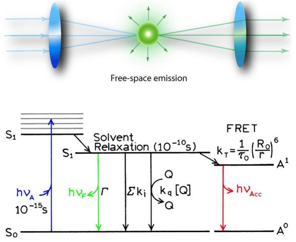
Free-space emission
We recently described methods to modify the radiative decay rates of fluorophores [1-2]. A review of the literature indicated that radiative decay rates could be modified by metallic particles such as silver colloids. By metal we mean conducting metallic surfaces and particles, not metal ions. The interactions of metals and fluorphores have been subject to prior theoretical reports [3-6]. However the practical usefulness of these effects was not explicitly recognized. We have described the use of the interactions of fluorophores with novel metal particles as radiative decay engineering (RDE) because the radiative decay rates of the fluorophores are modified in close proximity to the metal [7-8]. We observed several favorable effects due to metal particles, such as increased fluorescence intensities, increased photostability and increased distances for fluorescence resonance energy transfer (FRET). We refer to these favorable effects as metal-enhanced fluorescence (MEF).
We have also studied the interaction of fluorophores with planar metallic surfaces, similar to those used for surface plasmon resonance (SPR) - a continuous surface of silver or gold about 40 nm thick on a glass substrate. These thin metal films are optically dense. We found that excitation of fluorophores near these surfaces resulted in strong directional emission through the metal and into the substrate. The emission appeared as a cone with a half-angle consistent with the SPR angle for the emission wavelength, and there was little emission into the air away from the glass substrate [9-10]. The emission spectra were found to precisely match the emission spectra of the fluorophore, so it was not clear if the fluorophore or the plasmon was responsible for the observed emission. Other groups made similar observations [11-13] some dating back to 1975 [14], but in these papers it is often unclear if the emission is attributed to the fluorophore or scattered light. We refer to this unusual phenomenon as surface plasmon-coupled emission (SPCE) which is an appropriate name as the directional emission was subsequently shown to be due to plasmons created by the excited fluorophores, and not due to creation of plasmons by the incident light [15].
The observations of MEF [16] and SPCE led to our vision for plasmon-controlled fluorescence (PCF). As shown in Figure 2, the interaction of fluorophores with metals can affect both excitation and emission. Effects on excitation are possible because the electric fields from the incident light can be concentrated in small metallic colloids. Additionally, fluorophores near metal colloids can show increased rates of radiative decay than in the absence of metal particles. And finally, the emission can occur in defined directions by the use of metallic nanostructures. In our opinion, these effects will result in a new generation of devices in which the emission energy is controlled by metallic nanostructures.
Figure 2.
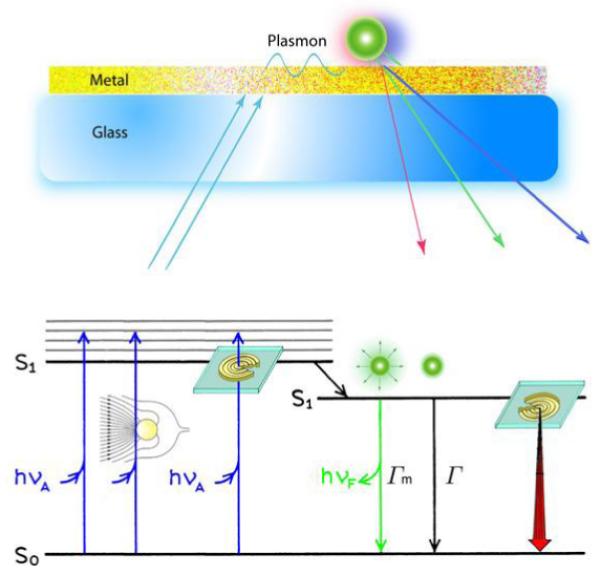
Plasmon controlled emission
1.1 Mechanism of Plasmon-Controlled Fluorescence
The phenomenon of SPCE showed that excited state fluorophores could create surface plasmons, which radiate into the substrate. The complete p-polarization of the SPCE indicated that the plasmons were responsible for the radiation propagating in the substrate. The ability of excited state fluorophores to create plasmons suggests the plasmonic structures to manipulate fluorescence. We believe that the scattering component of metal particle extinction is responsible for enhanced fluorescence because these plasmons can radiate, which occurs very rapidly. MEF is expected to occur when the absorption and/or emission bands of the fluorophore overlap with the scattering wavelength of the metal structure. We refer to this concept as the radiating plasmon (RP) model. This model is supported by some preliminary experiments which showed larger fluorescence enhancements with particles or particle aggregates displaying longer wavelength extinction [17-20], which is usually due to scattering more than absorption. The RP model has not been conclusively proven, but the reasonableness of the model has resulted in its general acceptance.
The effective use of PCF requires an understanding of the interaction mechanisms. The RP model provides a rational link between fluorescence and surface plasmons. Surface plasmons have become of great interest for nano-optics, nanolithography, and for the next generation of computer chips which utilize sub-wavelength size structures [21-23]. Surface plasmons can be engineered to either radiate or remain trapped at the metal surface, which suggest their use for the next generation of fluorescence technology.
1.2 Opportunities for Plasmonics in Biological Fluorescence
We see considerable potential for new capabilities in fluorescence technology. It is not possible to predict all the future developments in plasmon-controlled fluorescence. However, it seems valuable to suggest some of the future possibilities. Based on our current understanding we believe the rational use of fluorophore-plasmon interactions can result in a number of possibilities:
Ultrabright probes which are many-fold brighter than currently available fluorophores, and which can be monitored indefinitely at the single particle level without photobleaching.
The ability to measure distances between biomolecules in the presently inaccessible distance range from the 10 nm FRET limit up to 200 nm.
Probes which provide localized multi-photon excitation with low intensity wide-field illumination.
Metallic structures to selectively enhance excitation or emission at desired wavelengths.
New diagnosis devices based on plasmon-controlled fluorescence.
Wide-field sub-wavelength optical microscopy. One can already imagine schemes whereby 30 nm spatial resolution can be obtained using wide field methods. The availability of such microscopy would revolutionize cell biology and imaging.
In the following sections we will describe how these capabilities can be obtained based on information available in the plasmonics literature and the known ability of excited fluorophores to induce radiating plasmons.
1.3 Plasmonics and Fluorescence
Most applications of plasmonics to date have relied on the interaction of light with the metallic structures, followed by detection of the light at the same wavelength that is reflected or scattered from the structures [24-32]. These uses of plasmonics, while powerful, lack the advantage of a Stokes’ shift. We believe it will be possible to maintain some of the properties of plasmons (strong interactions with light) with the spectral characteristics of fluorescence (Stoke’s shift, time-delayed emission, etc.). This will be possible because the near fields created by plasmon resonances can interact with fluorophores and serve as a source of local fields and amplified excitation. Additionally, excited fluorophores can undergo near field interactions with metals, creating plasmons which then radiate light into free space.
The concept of using plasmonic structures to control fluorescence is shown in Figure 3. In such a device the fluorophores would be in close proximity to a silver surface and the silver surface will have a defined nanostructure. For example, the two regions on the left could contain nanohole arrays. The spacing of the nanoholes could be chosen for anomalous transmission of green (G) or red (R) wavelengths. The fluorophores with green or red emission will preferentially couple to plasmons in the respective structures. We expect the plasmons will then radiate light into the substrate for detection. Alternatively, the surface on the right can be patterned as a thin metal grating. In this case both the green and red fluorophores will couple to the surface, but the green and red emission will appear at different angles in the substrate.
Figure 3.
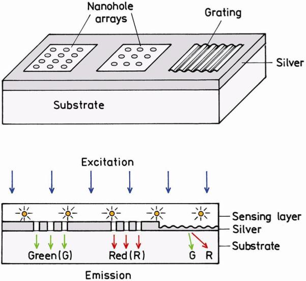
Schematic of using plasmon-controlled fluorescence for sensing. The periodicity of the nanohole array is chosen to couple to green (G) or red (R) emitting fluorophores. The periodicity of the grating is appropriate for separating red and green wavelengths.
1.4 Ultra-bright Fluorescent Particles
Plasmonic structures can also be used to obtain increased fluorescence intensities or to obtain ultra-bright particles. This application of PCF will depend on the use of metallic particles with varying sizes and shapes. For instance, consider a fluorophore contained within a metallic nanoshell, as compared with a fluorophore in solution (Figure 4). In the absence of a metal particle the fluorophore feels the incident light field (E) at the excitation wavelength (λex) and emits with the usual radiative decay rate at the emission wavelength λem. The extent of absorption is determined by the optical cross section of the fluorophore, which is usually a fraction of its physical cross section. Now consider a fluorophore within a silver shell and assume that light absorbed by the metal shell is transferred to the fluorophore. The effective cross section for absorption is now much larger than that of the fluorophore, and even larger than the colloid itself. This effect results in a higher effective incident field Em at the excitation wavelength and increased excitation of the fluorophore due to the amplified incident field. For the moment we are assuming the silver shell does not transform any of the energy into heat. As described in the introduction, fluorophores near metals can have higher radiative decay rates and higher quantum yields than in the absence of metals. Hence the emission intensity of the fluorophore in the metal shell can be increased further by the ability of the fluorophore to induce plasmons in the shell, which in turn radiate as light. These considerations suggest a fluorophore in a nanoshell or other suitable structures can have the useful Stokes’ shift of fluorescence with the high optical cross section of a PRP. There are several factors which result in large fluorescence enhancements inside nanoshells. The shorter lifetime of the fluorophore results in less time for photochemistry while in the excited state, and thus more excitation-emission cycles prior to photobleaching. The metal shell may be impermeable to oxygen and other species that react with the fluorophore and thus protect the fluorophore from photochemical reactions. It is known that the electric fields within these shells is uniform [33], suggesting that all the fluorophores may be equally enhanced irrespective of location within the nanoshell. These considerations suggest that fluorophores encapsulated in metal nanoshells can be bright and photostable particles for single molecule detection and imaging.
Figure 4.
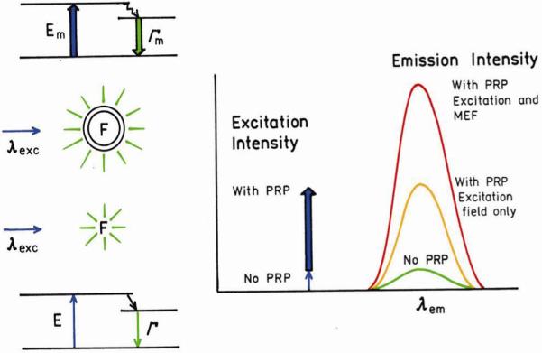
Comparison of fluorescence with no PRPs, with the effect PRPs on the enhanced excitation field, and with the PRPs enhancing both excitation and emission.
It is informative to compare a fluorophore in a nanoshell, which we will call a plasmonic probe, with semiconductor nanoparticles or Qdots. Since Qdots are already in use for cellular and whole-animal imaging [34-38], one can expect the plasmonic fluorophores to be used in similar applications. Qdots are frequently targeted to internal or external regions of cells using specific antibodies or peptides. Similar surface chemistries can be used to attach proteins to the surfaces of metal particles. This surface labeling of metal particles may be even easier with silver and gold particles because of the intrinsic affinity of these metals for amino and sulfhydryl groups. The Qdots have been found very useful even though their brightness is comparable to a single fluorophore or a few fluorophores. The Qdots contain materials such as CdS or CdSe, which can be toxic to cells under some conditions [39]. Impermeable surface coatings are often required to prevent leakage of the toxic materials. In contrast to Qdots, silver and gold are essentially non-toxic and if needed can also be coated with silica or biocompatible materials. The Qdots are claimed to be highly photostable. This is a result of the surface coating since uncoated semiconductor Qdots are sensitive to the local environment and undergo photobleaching. Similarly, the silver coating over the fluorophore is likely to result in increased photostability. We expect the plasmonic probes to be easily 100-fold brighter than a single fluorophore and thus as useful as Qdots.
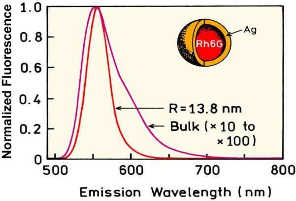
An advantage of Qdots is their ability to be excited at any wavelength and their narrow emission spectra [40-41]. These same advantages are expected for plasmonic probes. Gold and silver nanoshells are known to display broad absorption [42-43]. We expect the excitation spectra of plasmonic probes to be comparable to broad absorption of the nanoshells. Quantum dots are known to display narrow emission spectra. Fluorophores within metal shells are also expected to display more narrow emission spectra without the long wavelength tails [44-45]. In fact, the calculated emission spectrum for rhodamine 6G in a silver shell is about 2-fold more narrow than in solution (Figure 5). Hence plasmonic probes are potentially brighter, more photostable and less toxic than Qdots.
Figure 5.
Calculated intensities and emission spectra for Rh6G in a silver shell. From [44-45]
1.5 Nanostructures for Enhanced Wavelength-Selective Detection
Let us now consider the case where it is desirable to detect or count single fluorophores not coupled to metals, in the presence of background emission. Suppose the emission spectrum of the desired fluorophore occurs at 600 nm and that the sample also displays autofluorescence from 400-800 nm. The fluorophore emission overlaps with the background and would be difficult to detect without some enhancement (Figure 6). Now suppose a plasmonic structure flow cell can be created which has a strong resonance at 600 nm. As described below, one such structure is an array of metal nanowires. Such a structure will increase the emission intensity of the 600 nm emission, without increasing the intensity of the autofluorescence at other wavelengths. Such wavelength-selective enhancement over a narrow range of wavelengths requires a plasmonic structure which is strong but narrow resonance. Of course the autofluorescence at 600 nm would also be enhanced, but few natural substances have narrow emission spectra like modern fluorophores.
Figure 6.
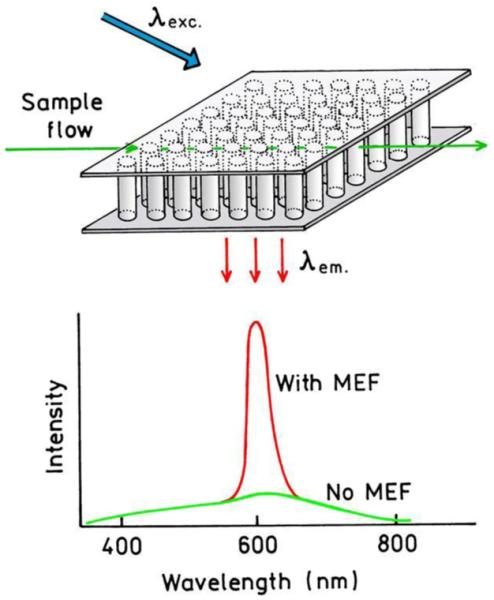
Schematic of wavelength-selective enhanced fluorescence in the presence of background. The top structure represents an array of metal nanowires spanning the top and bottom of a flow cell.
In recent years several groups have calculated the optical properties of metallic nanorods and chains of particles [46-51]. Some of the calculations predict very narrow plasmon resonances. Figure 7 shows the calculated spectra for a chain of spherical silver nanoparticles, 200 nm in diameter, with the center-to-center spacing shown on the figure. The peak wavelength and width of the resonance depends on both particle size (not shown) and the spacing between the particles. The resonances can be very narrow. For example, the resonance at 800 nm is below 4 nm in width. The resonances are very strong as seen by extinction up to 20-fold larger than the physical cross sections of the particles. As the resonances become sharper the peak intensity of the resonance increases [52]. Many organic fluorophores in current use have relatively narrow emission spectra and their emission is likely to be enhanced by such structures. Metallic structures of the type shown in Figures 6 and 7 may be used with QDots and lanthanides, both of which have emission spectra more narrow than typical fluorophores.
Figure 7.
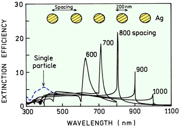
Extinction spectra for a one dimensional chain of silver nanoparticles. Revised from [51].
Various nanofabrication methods have been used to produce a variety of metal shapes bound to substrates [53-55]. Figure 8 shows an electron micrograph of nano-triangles on a glass substrate [56]. These triangles are formed by nanosphere lithographs (NSL) in which a regular array of polymeric beads is coated with a metal film. When the polymeric beads are removed the metal deposited between the beads remains on the substrate, typically with a triangular shape. Metallic shapes prepared by NSL have been shown to display a wide variety of extinction spectra. Depending on particle size and shape the resonances can be wide or narrow, can be dominated by a single resonance, or show multiple resonances at different wavelengths. Even a relatively simple fabrication method like NSL can be used to prepare particles which display resonances over a wide range of wavelengths (Figure 9). Shaped particles on substrates could provide plasmonic structures which enhance emission over a small range of wavelengths. If the particle spacing is varied gradually or stepwise, different wavelengths may be enhanced at different locations in the device, thereby providing spectral information about the sample from the spatial distribution of the emission.
Figure 8.
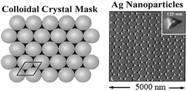
Schematic of a colloidal mask and electron micrograph of nano-triangles prepared by NSL. From [53]
Figure 9.
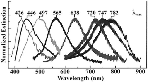
Extinction spectra of silver particles prepared by NSL. The oscillations in the spectra are due to the apparatus, not the particles. From [55].
So far we described wavelength-selective enhancements using particles fixed to a substrate. It may also be of interest to obtain wavelength-selective enhancements in solution. Such particles may be useful in multiplex assays. Assays of this type require an ability to create solid particles or shells with a narrow resonance at the desired wavelength. Fortunately, a variety of such particles have been made using batch chemistries suitable for preparation of reasonable quantities of particles [57-58]. Figure 10 shows calculated scattering spectra of silver or gold nanoshells on a silica core [59]. The peak scattering wavelength, and presumably the peak wavelength for fluorescence enhancement, can be varied throughout the visible and NIR wavelengths. Changes in the peak scattering wavelengths are obtained by variation of the size and thickness of the core and shell, while keeping the overall diameter constant. We note that the configurations shown in the preceding figures are only meant to show that a variety of geometries are possible for wavelength-selective enhancements. It is likely that the eventual devices will have very different geometries.
Figure 10.
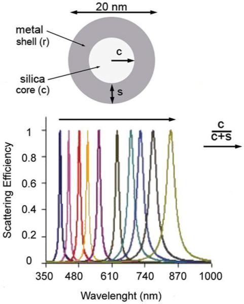
Normalized scattering efficiency calculated for metal nanoshells on silica cores. Revised from [59].
In suggesting structures for enhanced fluorescence, we make an assumption that the fluorescence enhancement was in some way proportional to the strength of the plasmon resonance and to the extent of spectral overlap between this resonance and the emission spectrum of the fluorophore. This assumption seems reasonable, and results show that structures for enhanced fluorescence can be designed using the assumption that a strong plasmon resonance is needed for maximum enhancements. However, this need not always be the case if the plasmon extinction is due to absorption rather than scattering.
1.6 Plasmon Engineering
The phenomenon of surface plasmon-coupled emission (SPCE) shows that excited state fluorophores can undergo a near-field non-radiative interaction with the metal to create propagating far-field radiation. This statement is supported by the observation of SPCE using silver, gold and aluminum, with wavelengths ranging from the UV for tryptophan through the visible and extending to NIR wavelengths [60-64]. These diverse results suggest that near-field coupling of excited state fluorophores with continuous metal surfaces is a general phenomenon that can be reliably predicted and utilized.
Surface plasmons are two-dimensional waves with optical frequencies which are trapped at an interface. This is an unusual phenomenon because light usually propagates freely in three dimensions. We are all aware of the remarkably complex electronic circuits which can be created because electrical currents can be manipulated on a flat surface. In contrast, optical devices such as waveguides and optical filter waveguides have the dimensions of light in three dimensions, and there are strict rules of reflection and refraction to change the direction of the propagation wave. The different ways we manipulate light and electrical current are ingrained in our thinking so that we rarely consider controlling light in the same way we control electrical currents.
Surface plasmons are, in a sense, light transformed into current. It is known that surface plasmons can migrate considerable distances in thin metal films with modest attenuation, and that plasmons can propagate in sub-wavelength size metallic structures [65]. This confinement property of plasmons is of interest for the design of components for optical computing. The ability to control the flow of optical energy in two-dimensions would have a profound impact on the design of devices for fluorescence sensing. Instead of using optical components to manipulate light, the fluorescence could be trapped in metallic nanostructures in the form of plasmons and moved to a different location on the surface. This capability would be useful in lab-on-a-chip or DNA array applications, where the chemistry is already on a surface, which can easily be a metal surface.
What is the evidence that plasmons can migrate usefully long distances without being dissipated by heat or by radiation into the substrate? How can the migrating plasmons be controlled? A relatively recent experiment has shown that plasmons with frequencies for wavelengths of 633 to 785 nm can migrate 50 to 150 μm with 1/e attenuation [66]. The detector was shielded from the incident light by the 50 nm aluminum film (Figure 11). It was found that the plasmons migrated further than expected if one accounted for attenuation due to radiation into the substrate. This radiation may have been prevented by the aluminum. The important point is that using available thin-film technology it should be possible to create structures which allow plasmons to propagate or to radiate, depending on the local metal structure.
Figure 11.
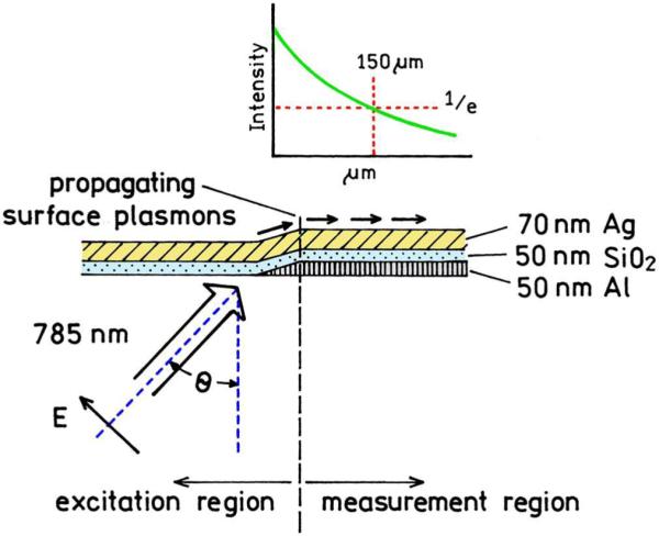
Experimental setup to measure the distance of plasmon propagation in a metal film. Revised from [66].
The use of plasmonic circuits requires ways to create directional plasmons and to reflect, diffract or focus the energy. These manipulations of plasmons are now becoming possible [67]. Figure 12 shows how directional surface plasmons can be created by laser illumination of a silver wire on a silver film. The plasmons are seen to be strongly directional and to migrate distances greater than 10μm. The next step for plasmon circuits is the fabrication of optical elements on a metal surface to manipulate the plasmons. This can now be accomplished using dielectric or metallic features on a metal film [68-70]. Two examples are shown in Figure 13. The plasmons are launched by irradiation of linear feature seen as a vertical line, similar to Figure 12. The lower panels in Figure 13 show a plasmon mirror created by five rows of dots. This mirror has a reflectivity of 90%. The upper panels show a beamsplitter which divides the plasmons reflected from the mirror.
Figure 12.
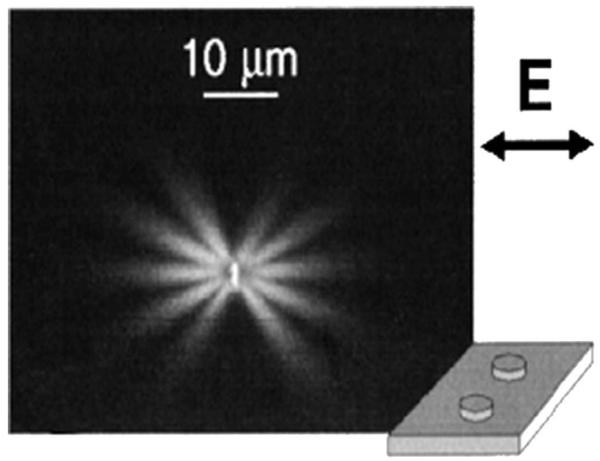
Fluorescence image of plasmons created by illumination of a silver wire on silver film. The two metal surfaces were separated by 10 nm of silica. From [70].
Figure 13.
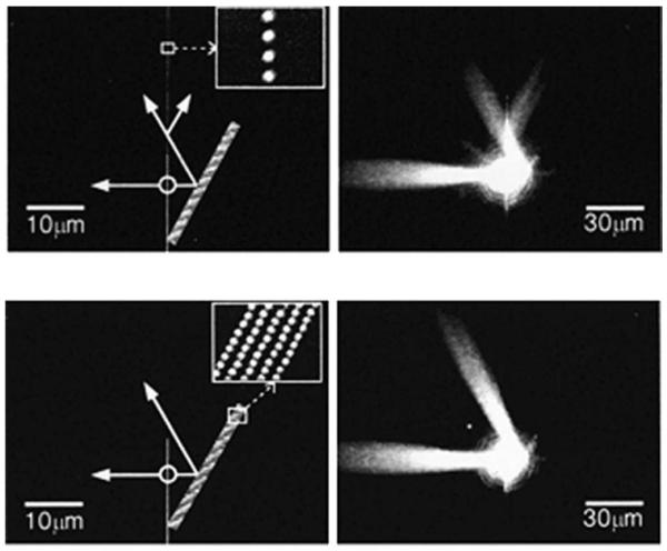
Optical elements for surface plasmons using bumps on a silver film. The left panel shows SEM images of the mirror (bottom) and mirror with beamsplitters (top). The right panels show the surface plasmons. From [69].
Another example of plasmon engineering is the use of two-dimensional lenses to focus the plasmons. In one such example a plasmon lens was made from a semi-circle of nanoholes in a metal film [71]. The intensities of the plasmon fields were measured using NSOM (Figure 14). The plasmons were found to form a focal point, which was explained by interference at the plasmon wavelength. The focal point could be formed or eliminated by rotation of the incident polarization. The results in Figures 11-14 show that optical frequency energy can be transported and manipulated in a two-dimensional surface.
Figure 14.
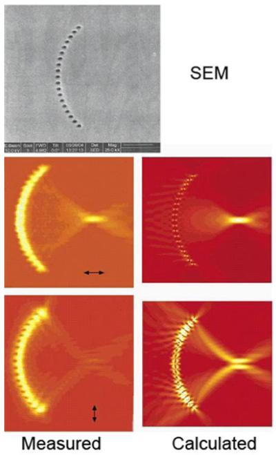
Top, scanning electron micrograph of a semi-circle of 200 nm holes in a 50 nm thick film. Bottom, NSOM images (left) and calculated intensities (right) for the plasmon lines illuminated at 532 nm. From [71].
So far we have shown that it is possible to couple the energy of excited fluorophores into plasmons (via SPCE), transport the plasmons across a surface, and to manipulate the direction of the plasmons (Figures 11-14). In order to detect the energy it may be necessary to convert the plasmons back into propagating light. This conversion can be accomplished with simple 2D structures [68]. Figure 15 (top) shows a SEM image of two nanohole arrays in a smooth gold film separated by 30 μm. The smaller array on the right, which is difficult to see in the figure, is illuminated at 815 nm, creating plasmons which migrate to the left. The lower panel shows a near-field optical image of the sample. The plasmons migrate from right to left between the arrays, and are converted back to visible light when they encounter the nanohole array on the left. Hence plasmons can be created in one region of a device, propagate a reasonable distance, and then be converted back into light. The ability to interconvert light and plasmons, and to manipulate the plasmons, can result in a new generation of devices for diagnostics, biotechnology, and other applications. Using currently available knowledge about plasmonics and fluorescence, one can propose a variety of configurations for nano devices designed to couple in the incident light or couple back to the emission frequency from surface-bound fluorophores. Such devices would be inexpensive and easy to manufacture with available lithographic methods.
Figure 15.
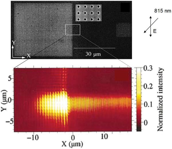
Top: SEM image of the nanohole arrays on 150 nm thick gold film. The array on the left is 41 × 41, and the array on the right is 11 × 11. Bottom: Near-field image of the central region of the top panel. Revised from [68].
The strong plasmon resonance in metal particles provides good opportunity for localized plasmon enhanced fluorescence applications near the particle. It is known that the incident fields can be dramatically amplified near non-spherical particles [72-73]. Calculations for a silver ellipsoid show an enhanced field near the tips (Figure 16. The peak enhancement for the ellipsoid is a factor of 4700, which is above the range of the color scale in this figure. For a triangle the enhanced fields are also located near the tips, with a peak enhancement of 3500-fold. Even more dramatic field enhancements are expected near clusters of particles [73]. For example, the field between two closely spaced silver particles can be enhanced by a factor of 104 (Figure 17). An appropriately designed metal structure can act like an antenna and show resonance enhancements of the fields between the particles [74], which suggest another approach for implementing localized plasmon enhanced fluorescence applications.
Figure 16.
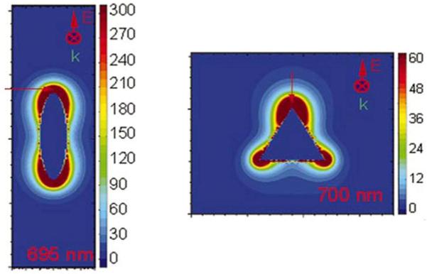
Field enhancements around the silver particles calculated using the discrete dipole approximations. The maximum enhancement for the ellipsoid is 4700 and for the triangle is 3500. From [74].
Figure 17.
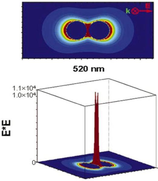
Calculated field enhancement bear two closely spaced silver particles. The particles are separated by 2nm. From [74].
1.7 Sub-Wavelength Microscopy
Optical microscopy is an important tool for cellular imaging. Optical imaging is accomplished using a wide variety of methods including wide field imaging, multi-photon excitation and/or laser scanning confocal microscopy. Based on the Rayleigh criteria the spatial resolution is limited to about λ/2. This resolution can be increased using complex methods such as 4Pi, stimulated emission depletion or near-field scanning microscopy [75], but the complexity and limitations of these methods have prevented their widespread use for cellular imaging. The use of plasmonics offers the opportunity for sub-wavelength resolution microscopy, with a spatial resolution as small as 25 nm. It is known that the transmission efficiency of light decreases dramatically as the size of the aperture becomes smaller than the wavelength. Unexpectedly high transmission efficiencies were observed for apparently opaque silver films which contained arrays of holes with sizes ranging from about 50 to 150 nm. This observation resulted in additional studies with metal films containing various types of periodic structures [76-79]. The possibility of sub-wavelength resolution is of great interest for nanotechnology. It is now known that the near-fields from surface plasmons can have dimensions much smaller than free-space radiation at the same frequency [80-84]. The concepts of plasmon-coupled transmission and optical near-fields are merging into proposals for wide-field optical microscopy with sub-wavelength resolution [85-87]. Such a microscope could be based on excitation of the specimen by the near-field near the nanohole array. It is now thought that the near-fields are tightly confined, or at least not diffracted into half-space, as will occur without plasmon-coupled transport (Figure 18). In order to construct a microscope, it would be necessary to measure the intensities from fluorophores excited by the near-fields with sub-wavelength dimensions. The emission from these regions could be imaged if the nanoholes were about 1 micron apart (Figure 19). The fluorescence excited near each nanohole could be imaged onto an array detector. The specimen or nanohole array would be moved and the multiple images used to reconstruct the entire image.
Figure 18.
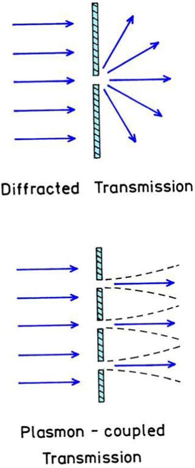
Transmission through nanoholes in a metal film.
Figure 19.
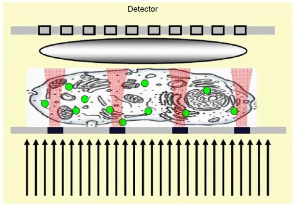
Possible schematic for a sub-wavelength plasmonic microscope. Revised from [87].
Construction of such a microscope faces several technological challenges, such as positioning and moving the specimen and array relative to each other, and obtaining near fields with sub-wavelength dimensions at reasonable distances from the metal. The extensive interest in nanolithography is already resulting in methods to control and focus the near fields. While there are certain to be many technical challenges, we are not aware of any physical limits that would prevent creation of a sub-wavelength plasmonic microscope.
2. CONCLUSION
The combination of fluorescence with plasmonics will result in a paradigm shift for both technologies. Fluorescence instrumentation, devices and sensors will begin to use plasmons for creation of excited states and manipulation of the emission energy. The field of plasmonics will expand to include studies of the factors which govern fluorophore-metal interactions and how to optimize the desired effects. These new approaches to fluorescence and plasmonics will be facilitated by the rapid growth of nanofabrication methods, resulting in new approaches to high throughput biology, diagnostics and molecular imaging.
ACKNOWLEDGEMENTS
Supported by the NIH NCRR, RR08119, NHGRI HG 002655 and NBIB EB.
REFERENCES
- 1.Lakowicz JR. Radiative decay engineering: Biophysical and biomedical applications. Anal. Biochem. 2001;298:1–24. doi: 10.1006/abio.2001.5377. [DOI] [PMC free article] [PubMed] [Google Scholar]
- 2.Lakowicz JR, Shen Y, D’Auria S, Malicka J, Fang J, Gryczynski Z. Gryczynski, IRadiative decay engineering. 2. Effects of silver island films on fluorescence intensity, lifetimes, and resonance energy transfer. Anal. Biochem. 2002;301:261–277. doi: 10.1006/abio.2001.5503. [DOI] [PMC free article] [PubMed] [Google Scholar]
- 3.Chance RR, Prock A, Silbey R. Molecular fluorescence and energy transfer near interfaces. Adv. Chem. Phys. 1973;37:1–65. [Google Scholar]
- 4.Ford GW, Weber WH. Electromagnetic interactions of molecules with metal surfaces. Phys. Reports. 1984;113(4):195–287. [Google Scholar]
- 5.Gersten J, Nitzan A. Spectroscopic properties of molecules interacting with small dielectric particles. J. Chem. Phys. 1981;75(3):1139–1152. [Google Scholar]
- 6.Gersten J. ITheory of fluorophore-metallic surface interactions. In: Geddes CD, Lakowicz JR, editors. Topics in Fluorescence Spectroscopy, Vol. 8: Radiative Decay Engineering. Springer Science+Business Media, Inc; New York: 2005. pp. 197–221. [Google Scholar]
- 7.Geddes CD, Aslan K, Gryczynski I, Malicka J, Lakowicz JR. Radiative decay engineering. In: Geddes CD, Lakowicz JR, editors. Topics in Fluorescent Spectroscopy, Vol. 8: Radiative Decay Engineering. Springer Science+Business Media, Inc; New York: 2005. pp. 405–448. [Google Scholar]
- 8.Geddes CD, Aslan K, Gryczynski I, Malicka J, Lakowicz JR. Noble-metal surfaces for metal enhanced fluorescence. In: Geddes CD, Lakowicz JR, editors. Topics in Fluorescent Spectroscopy, Vol. 8: Radiative Decay Engineering. Springer Science+Business Media, Inc; New York: 2005. pp. 365–401. [Google Scholar]
- 9.Lakowicz JR. Radiative decay engineering 3. Surface plasmon-coupled directional emission. Anal. Biochem. 2004;324:153–169. doi: 10.1016/j.ab.2003.09.039. [DOI] [PMC free article] [PubMed] [Google Scholar]
- 10.Gryczynski I, Malicka J, Gryczynski Z, Lakowicz JR. Radiative decay engineering 4. Experimental studies of surface plasmon coupled directional emission. Anal. Biochem. 2004;324:270–182. doi: 10.1016/j.ab.2003.09.036. [DOI] [PMC free article] [PubMed] [Google Scholar]
- 11.Benner RE, Dornhaus R, Chang RK. Angular emission profiles of dye molecules excited by surface plasmon waves at a metal surface. Opt. Commun. 1979;30(2):145–149. [Google Scholar]
- 12.Pockhand I, Brilliante A, Mobius D. Nonradiative decay of molecular excitation at a metal interface. Nuovo Cimento. 1980;63:350–357. [Google Scholar]
- 13.Hayashi S. Spectroscopy of gap modes in metal particle-surface systems. Topics Appl. Phys. 81:71–95. [Google Scholar]
- 14.Gerbshtein YM, Merkulov IA, Mirlin DN. Transfer of luminescence-center energy to surface plasmons. JETP Letts. 2001;22:35–36. 1975. [Google Scholar]
- 15.Zhang J, Gryczynski Z, Lakowicz JR. First observation of surface plasmon-coupled electro chemiluminescence. Chem. Phys. Letts. 2004;393:483–487. doi: 10.1016/j.cplett.2004.06.050. [DOI] [PMC free article] [PubMed] [Google Scholar]
- 16.Lakowicz JR. Radiative decay engineering 5: metal-enhanced fluorescence and plasmon emission. Anal. Biochem. 2005;337:171–194. doi: 10.1016/j.ab.2004.11.026. [DOI] [PMC free article] [PubMed] [Google Scholar]
- 17.Aslan K, Leonenko Z, Lakowicz JR, Geddes CD. Fast and slow deposition of silver nanorods on planar surfaces: Application to metal-enhanced fluorescence. J. Phys. Chem. B. 2005;109:3157–3162. doi: 10.1021/jp045186t. [DOI] [PMC free article] [PubMed] [Google Scholar]
- 18.Zhang J, Lakowicz JR. Enhanced luminescence of Phenyl-phenenthridine dye on aggregated small silver nanoparticles. J. Phys. Chem. B. 2005;109:8701–8706. doi: 10.1021/jp046016j. [DOI] [PMC free article] [PubMed] [Google Scholar]
- 19.Zhang J, Malicka J, Gryczynski I, Lakowicz JR. Surface enhanced fluorescence of fluorescein-labeled oligonucleotides capped on silver nanoparticles. J. Phys. Chem. B. 2005;109:7643–7648. doi: 10.1021/jp0490103. [DOI] [PMC free article] [PubMed] [Google Scholar]
- 20.Zhang J, Malicka J, Gryczynski I, Lakowicz JR. Oligonucleotide-displaced organic monolayer-protected silver nanoparticles and enhanced luminescence of their salted aggregates. Anal. Biochem. 2004;330:81–86. doi: 10.1016/j.ab.2004.04.001. [DOI] [PMC free article] [PubMed] [Google Scholar]
- 21.Tominaga J. The manipulation of surface and local plasmons. In: Tsai DP, editor. Optical Nanotechnologies. Springer; New York: 2003. p. 212. [Google Scholar]
- 22.Ohtsu M, Kobayashi K. Optical Near Fields. Springer; New York: 2004. Introduction to classical and quantum theories of electromagnetic phenomena at the nanoscale; p. 205. [Google Scholar]
- 23.Kawata S, Ohtsu M, Irie M, editors. Nano-Optics. Springer; New York: 2002. p. 321. [Google Scholar]
- 24.Yguerabide J, Yguerabide EE. Light scattering submicroscopic particles as highly fluorescent analogs and their use as tracer labels in clinical and biological applications. Anal. Biochem. 1998;262:137–156. doi: 10.1006/abio.1998.2759. [DOI] [PubMed] [Google Scholar]
- 25.Yguerabide J, Yguerabide EE. Light scattering submicroscopic particles as highly fluorescent analogs and their use as tracer labels in clinical and biological applications. Anal. Biochem. 1998;262:137–156. doi: 10.1006/abio.1998.2759. [DOI] [PubMed] [Google Scholar]
- 26.Cao YW, Jin R, Mirkin C. ADNA-modified core-shell Ag/Au nanoparticles. J. Am. Chem. Soc. 2001;123:7961–7962. doi: 10.1021/ja011342n. [DOI] [PubMed] [Google Scholar]
- 27.Jin R, Wu G, Li Z, Mirkin CA, Schatz GC. What controls the melting properties of DNA-linked gold nanopartcle assemblies? J. Am. Chem. Soc. 2003;125:1643–1654. doi: 10.1021/ja021096v. [DOI] [PubMed] [Google Scholar]
- 28.Enderlein J, Zander C. Theoretical foundations of single molecule detection in solutions. In: Zander Ch., Endelein J, Keller RA., editors. Single Molecule Detection in Solution. Wiley-VCH; Germany: 2002. p. 371. [Google Scholar]
- 29.Schultz S, Smith SR, Mock JJ, Schulz DA. Single target molecule detection with nonbleaching multicolor optical immunolabels. PNAS. 2000;97:996–1001. doi: 10.1073/pnas.97.3.996. [DOI] [PMC free article] [PubMed] [Google Scholar]
- 30.Mitchell JS, Wu Y, Cook CJ, Main L. Sensitivity enhancement of surface plasmon resonance biosensing of small molecules. Anal. Biochem. 2005;343:125–135. doi: 10.1016/j.ab.2005.05.001. [DOI] [PubMed] [Google Scholar]
- 31.He L, Smith EA, Natan MJ, Keating DD. The distacnce dependence of colloidal Au-Amplified surface plasmon resonance. J. Phys. Chem. B. 2004;108:10973–10980. [Google Scholar]
- 32.He L, Musick MD, Nicewarner SR, Salinas FG, Benkovic SJ, Natan MJ, Keating CD. Colloidal Au-enhanced surface plasmon resonance for ultrasensitive detection of DNA hybridization. J. Am. Chem. Soc. 2000;122:9071–9077. [Google Scholar]
- 33.Scheim S, Smith GB. Internal electrical field densities of metal nanoshells. J. Phys. Chem. B. 2005;109:1689–1694. doi: 10.1021/jp0450686. [DOI] [PubMed] [Google Scholar]
- 34.Jaiswal JK, Mattoussi H, Mauro JM, Simon SM. Long-term multiple color imaging of live cells using quatum dot bioconjugates. Nature Biotech. 2003;21:47–51. doi: 10.1038/nbt767. [DOI] [PubMed] [Google Scholar]
- 35.Osaki F, Kanamori H, Sando S, Sera T, Aoyama Y. A quantum dot conjugated sugar ball and its cellular uptake. On the side effects of endocytosis in the subviral region. J. Am. Chem. Soc. 2004;126:6520–6521. doi: 10.1021/ja048792a. [DOI] [PubMed] [Google Scholar]
- 36.Smith AM, Gao X, Nie S. Quatum dot nanocrystals for In Vivo molecular and cellular imaging. Photochem. Photobiol. 2004;80:377–385. doi: 10.1562/0031-8655(2004)080<0377:QDNFIV>2.0.CO;2. [DOI] [PubMed] [Google Scholar]
- 37.Michalet X, Pinaud FF, Bentolila LA, Tsay JM, Doose S, Li JJ, Sundaresan G, Wu AM, Gambhir SS, Weiss S. Quantum dots for live cells in Vivo imaging and diagnostics. Science. 2005;307:538–544. doi: 10.1126/science.1104274. [DOI] [PMC free article] [PubMed] [Google Scholar]
- 38.Jiang W, Papa E, Fischer H, Mardyani S, Chan WC. Semiconductor quantum dots as contrast agents for whole animal imaging. Trends in Biotech. 2004;22(12):604–609. doi: 10.1016/j.tibtech.2004.10.012. [DOI] [PubMed] [Google Scholar]
- 39.Derfus AM, Chan WC, Bhatia SN. Probing the cytotoxicity of semiconductor quantum dots. Nano Letts. 2004;4(1):11–18. doi: 10.1021/nl0347334. [DOI] [PMC free article] [PubMed] [Google Scholar]
- 40.Murphy CJ, Coffer JL. Quantum dots: A Primer. Appl. Spec. 2002;56:16A–27A. [Google Scholar]
- 41.Rosenthal S. JBarcoding biomolecules with fluorescent nanocrystals. Nature Biotech. 2001;19:621–622. doi: 10.1038/90213. [DOI] [PubMed] [Google Scholar]
- 42.Wang H, Goodrich GP, Tam F, Oubre C, Nordlander P, Halas NJ. Controlled texturing modifies the surface topography and plasmonic properties of Au nanoshells. J. Phys. Chem. B. 2005;109:11083–11087. doi: 10.1021/jp051466c. [DOI] [PubMed] [Google Scholar]
- 43.Shi W, Sahoo Y, Swihart MT, Prasad PN. Gold nanoshells on polystyrene cores for control of surface plasmon resonance. Langmuir. 2005;21:1610–1617. doi: 10.1021/la047628y. [DOI] [PubMed] [Google Scholar]
- 44.Enderlein J. Spectral properties of a fluorescing molecule within a spherical metallic cavity. Phys. Chem. Chem. Phys. 2002;4:2780–2786. [Google Scholar]
- 45.Enderlien J. Theoretical study of single molecule fluorescence in a metallic nanocavity. Appl. Phys. Letts. 2002;80:315–317. [Google Scholar]
- 46.Brioude A, Jiang XC, Pileni MP. Optical properties of gold nanorods: DDA simulations supported by experiments. J. Phys. Chem. B. 2005;109:13138–13142. doi: 10.1021/jp0507288. [DOI] [PubMed] [Google Scholar]
- 47.Park SY, Stroud D. Surface- plasmon dispersion realtions in chains of metallic nanoparticles: An exact quasistatic calculation. Phys. Rev. B. 2004;69:125418-1–125418-7. [Google Scholar]
- 48.Girard C, Quidant R. Near-field optical transmittance of metal particle chain wavelengths. Optics Express. 2004;12:6141–6146. doi: 10.1364/opex.12.006141. [DOI] [PubMed] [Google Scholar]
- 49.Panoiu NC, Osgood RM. Subwavelength nonlinear plasmonic nanowire. Nano Letts. 2004;4:2427–2430. [Google Scholar]
- 50.Hicks EM, Zou S, Schatz GC, Spears KG, van Duyne R, Gunnarson L, Rindzevicius T, Kasemo B, Kall M. Controlling plasmon lineshapes through Diffractive coupling linear arrays of cylindrical nanoparticles fabricated by electron beam lithography. Nano Letts. 2005;5:1065–1070. doi: 10.1021/nl0505492. [DOI] [PubMed] [Google Scholar]
- 51.Zou S, Janel N, Schatz GC. Silver nanoparticle array structures that produce remarkably narrow plasmon lineshapes. J. Chem. Phys. 2004;120:10871–10875. doi: 10.1063/1.1760740. [DOI] [PubMed] [Google Scholar]
- 52.Mulvaney P. Surface plasmon spectroscopy of nanosized metal particles. Langmuir. 1996;12:788–800. [Google Scholar]
- 53.Zhang H, Mirkin CA. DPN-Generated nanostructures made of gold, silver, and palladium. Chem. Mater. 2004;16:1480–1484. [Google Scholar]
- 54.Childs WR, Nuzzo RG. Large area patterning of coinage-metal thin films using decal transfer lithography. Langmuir. 2005;21:195–202. doi: 10.1021/la047884a. [DOI] [PubMed] [Google Scholar]
- 55.Haynes CL, Van Duyne RP. Nanosphere Lithography: A versatile nanofabrication tool for studies of size-dependent nanoparticle optics. J. Phys. Chem. B. 2001;105:5599–5611. [Google Scholar]
- 56.Liang HP, Wan LJ, Bai CL, Jiang L. Gold hollow nanospheres: Tunable surface plasmon resonance controlled by interior cavity sizes. J. Chem. Phys. B. 2005;109:7795–7800. doi: 10.1021/jp045006f. [DOI] [PubMed] [Google Scholar]
- 57.Chen MMY, Katz A. Synthesis and characterization of gold-silica nanoparticles incorporating a mercaptosilane core-shell interface. Langmuir. 2002;18:8566–8572. [Google Scholar]
- 58.Oldenburgh SJ, Westscott SL, Averitt RD, Halas NJ. Surface enhanced Raman scattering in the near infrared using metal nanoshell substrates. J. Chem. Phys. 1999;111(10):4729–4735. [Google Scholar]
- 59.Chen K, Lui Y, Ameer G, Backman V. Optimal design if structured nanospberes for ultrashap light-scattering resonances as molecular imaging mutilabels. J. Biomed. Optics. 2005;10(2):024005–024110. doi: 10.1117/1.1899684. [DOI] [PubMed] [Google Scholar]
- 60.Grycynski I, Mailicka J, Lukomska J, Grycynski Z, Lakowicz JR. Surface plasmon-coupled polarized enission of N-Acetyl-L-Tryptophanamide. Photochem, Photobiol. 2004;80:482–485. doi: 10.1562/0031-8655(2004)080<0482:SPPEON>2.0.CO;2. [DOI] [PMC free article] [PubMed] [Google Scholar]
- 61.Malicka J, Gryczynski I, Gryczynski Z, Lakowicz JR. Surface plasmon-coupled emission of 2,5-Diphenyl-1,3,4-oxadiazole. J. Phys. Chem. B. 2004;108:19114–19118. doi: 10.1021/jp047136u. [DOI] [PMC free article] [PubMed] [Google Scholar]
- 62.Gryczynski I, Malicka J, Gryczynski Z, Lakowicz JR. Surface plasmon-coupled emission with gold films. J. Phys. Chem. B. 2004;108:12568–12574. doi: 10.1021/jp040221h. [DOI] [PMC free article] [PubMed] [Google Scholar]
- 63.Grycynski I, Malicka J, Grycynski Z, Nowaczyk K, Lakowicz JR. Ultraviolet surface-plasmon-coupled emission using thin aluminum films. Anal. Chem. 2004;76:4076–4081. doi: 10.1021/ac040004c. [DOI] [PMC free article] [PubMed] [Google Scholar]
- 64.Geddes CD, Grycynski I, Malicka J, Grycynski Z, Lakowicz JR. Directional surface plasmon coupled emission. J. Fluores. 2004;14(1):119–123. doi: 10.1023/b:jofl.0000014809.64610.a8. [DOI] [PMC free article] [PubMed] [Google Scholar]
- 65.Barnes WL, Dereux A, Ebbesen TW. Surface plasmon subwavelength optics. Nature. 2003;424:824–830. doi: 10.1038/nature01937. [DOI] [PubMed] [Google Scholar]
- 66.Lamprecht B, Krenn JR, Schider G, Ditlbacher H, Felidj N, Leitner A, Aussenberg FR. Surface plasmon propagation in microsale metal stripes. Appl. Phys. Letts. 2001;79:51–53. [Google Scholar]
- 67.Ditlbacher H, Krenn JR, Felidj N, Lamprecht B, Schider G, Salerno M, Leitner A, Aussenberg FR. Fluorescence imaging of surface plasmon fields. Appl. Phys. Letts. 2002;80:404–406. [Google Scholar]
- 68.Devaux E, Ebbesen TW, Weeber JC, Dereux A. Launching and decoulping surface plasmons via micro-gratings. Appl. Phys. Letts. 2003;83:4936–4938. [Google Scholar]
- 69.Krenn JR, Ditlbacher H, Schider G, Hohenau A, Leitner A, Aussenberg FR. Surface plasmon micro-and nano optics. J. Micros. 2003;209:167–172. doi: 10.1046/j.1365-2818.2003.01088.x. [DOI] [PubMed] [Google Scholar]
- 70.Hohenau A, Krenn JR, Stepanov AL, Drezert A, Ditlbacher H, Steinberger B, Leitner A, Aussenberg FR. Dielectric optical elements for surface plasmons. Optics Letters. 2005;30:893. doi: 10.1364/ol.30.000893. [DOI] [PubMed] [Google Scholar]
- 71.Yin L, Vlasko-Vlasov VK, Pearson J, Hiller JM, Hua J, Welp U, Brown DE, Kimball CW. Subwavelength focusing and guiding of surface plasmons. Nano Letts. 2005;5(7):1399–1402. doi: 10.1021/nl050723m. [DOI] [PubMed] [Google Scholar]
- 72.Calander N, Willander M. Theory of surface-plasmon resonance optical field enhancement at prolate spheroids. J. Appl. Phys. 2002;92:4878–4884. [Google Scholar]
- 73.Hao E, Schatz GC. Electromagnetic fields around silver nanoparticles and dimers. J. Chem. Phys. 2004;120:357–366. doi: 10.1063/1.1629280. [DOI] [PubMed] [Google Scholar]
- 74.Muhlschlegel P, Eisler HJ, Martin OJF, Hecht B, Pohl DW. Resonant Optical Antennas. Science. 2005;308:1607–1609. doi: 10.1126/science.1111886. [DOI] [PubMed] [Google Scholar]
- 75.Garini Y, Vermolen BJ, Young IT. From micro to nano: recent advances in high resolution microscopy. Curr. Opin. in Biotech. 2005;16:3–12. doi: 10.1016/j.copbio.2005.01.003. [DOI] [PubMed] [Google Scholar]
- 76.Thomas DA, Hughes HP. Enhanced optical transmission through a subwavelength 1D aperture. Solid State Commun. 2004;129:519–524. [Google Scholar]
- 77.Martin-Moreno L, Garcia-Vidal FJ, Lezec HJ, Degiron A, Ebbesen TW. Theory of highly directional emission from a single subwavelength aperture surrounded by surface corrugations. Phys. Rev. Lett. 2003;90:167401–167404. doi: 10.1103/PhysRevLett.90.167401. [DOI] [PubMed] [Google Scholar]
- 78.Luo X, Shi J, Wang H, Yu G. Surface plasmon polariton radiation from metallic photonic crystal slabs breaking the diffraction limit: nano-storage and nano-fabrication. Modern Phys. Letts. B. 2004;18:945–953. [Google Scholar]
- 79.Sun Z, Kim HK. Refractive transmission of light and beam shaping with metallic nano-optic lenses. Appl. Phys. Letts. 2004;85:642–644. [Google Scholar]
- 80.Hohng SC, Yoon YC, Kim DS, Malyachuk V, Muller R, Lienau Ch., Park JW, Yoo KH, Kim J, Ryu HY, Park QH. Light emission from the shadows: Surface plasmon nano-optics at near light and far fields. Appl. Phys. Letts. 2002;81:3239–3241. [Google Scholar]
- 81.Luo X, Ishihara T. Surface plasmon resonant interference nanolithography technique. Appl. Phys. Letts. 2004;84:4780–4782. [Google Scholar]
- 82.Dragnea B, Szarko JM, Kowarik S, Weimann T, Feldmann J, Leone SR. Near-field surface plasmon excitation on structured gold films. Nano Letts. 2003;3(1):3–7. [Google Scholar]
- 83.Srituravanich W, Fang N, Sun C, Luo Q, Zhang X. Plasmonic nanolithography. Nano Letts. 2004;4(6):1085–1088. [Google Scholar]
- 84.Liu Z-W, Wei Q-H, Zhang X. Surface plasmon interference nanolithography. Nano Letts. 2005;5(5):957–961. doi: 10.1021/nl0506094. [DOI] [PubMed] [Google Scholar]
- 85.Smolyaninov II, Elliott J, Zayats AV, Davis CC. Far-field optical microscopy with a nanometer-scale resolution based on the in-plane image magnification by surface plasmon polaritons. Phys. Rev. Letts. 2005;94:057401–057401-4. doi: 10.1103/PhysRevLett.94.057401. [DOI] [PubMed] [Google Scholar]
- 86.Garini Y, Kutchoukov VG, Bossche A, Alkemade PFA, Docter M, Verbeek PW, van Vliet LJ, Young IT. Toward the development of a three-dimensional mid-field microscope. Proc. SPIE. 2004;5327:115–122. [Google Scholar]
- 87.Docter MW, Young IT, Kutchoukov VG, Bossche A, Alkemade PFA, Garini Y. A novel concept for a mid-field microscope. Proc. SPIE. 2005;5703:118–126. [Google Scholar]


