Abstract
p53 limits the proliferation of primary diploid fibroblasts by inducing a state of growth arrest named replicative senescence — a process which protects against oncogenic transformation and requires integrity of the p53 tumour suppressor pathway1–3. However, little is known about the downstream target genes of p53 in this growth-limiting response. Here, we report that suppression of the p53 target gene encoding plasminogen activator inhibitor-1 (PAI-1) by RNA interference (RNAi) leads to escape from replicative senescence both in primary mouse embryo fibroblasts and primary human BJ fibroblasts. PAI-1 knockdown results in sustained activation of the PI(3)K–PKB–GSK3β pathway and nuclear retention of cyclin D1, consistent with a role for PAI-1 in regulating growth factor signalling. In agreement with this, we find that the PI(3)K–PKB–GSK3β–cyclin D1 pathway is also causally involved in cellular senescence. Conversely, ectopic expression of PAI-1 in proliferating p53-deficient murine or human fibroblasts induces a phenotype displaying all the hallmarks of replicative senescence. Our data indicate that PAI-1 is not merely a marker of senescence, but is both necessary and sufficient for the induction of replicative senescence downstream of p53.
Primary murine fibroblasts activate the p19ARF–p53 tumour suppressor pathway during prolonged culturing in vitro, which induces a post-mitotic state referred to as replicative senescence. Senescence can be overcome by loss of either p19ARF, p53 or the combined loss of all three retinoblastoma family proteins1,2. Proliferation of fibroblasts is induced by growth factors which activate cyclin-dependent kinases (CDKs), and in turn inactivate pRb's growth-limiting ability2, a G1 cell-cycle checkpoint that is often deregulated in cancer3. It is unclear which of the many downstream p53-target genes is responsible for the p53-dependent induction of replicative senescence. An attractive candidate is the CDK inhibitor p21CIP1. However, mouse embryo fibroblasts (MEFs) knocked out for p21CIP1 are not immortal4.
Here, we identify an unexpected causal role for the urokinase type plasminogen activator (uPA)–PAI-1 system in the induction of replicative senescence. The serpin and extra-cellular matrix (ECM)-associated protein PAI-1 is a direct target of p53 (ref. 5, 6), is upregulated in ageing fibroblasts in vivo and in vitro, and is considered a marker of replicative senescence7–9. PAI-1 inhibits the activity of the secreted protease uPA by forming a stable complex. uPA expression can cause cells to progress through G1 into S phase10, most likely through activating a mitogenic signalling cascade by increasing the bioavailability of growth factors.
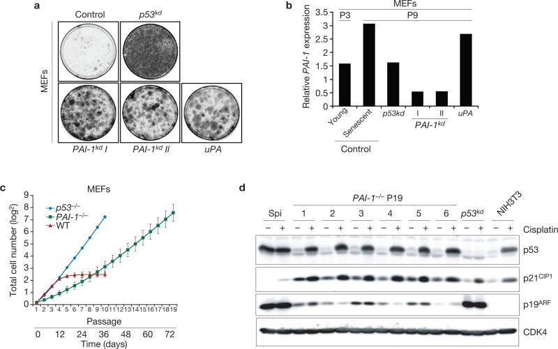
To investigate the role of PAI-1 in replicative senescence, two independent retroviral vectors were generated that targeted murine PAI-1 for suppression through RNAi11. When primary MEFs were infected with either PAI-1 knockdown (kd) construct (PAI-1kdI or PAI-1kdII), bypass of senescence was observed in a colony formation assay (Fig. 1a). As inhibition of PAI-1 expression leads to activation of uPA12,13, we asked whether overexpression of uPA also caused immortalisation. Retrovirus-mediated over-expression of pro-uPA was as efficient as PAI-1 knockdown in causing immortalisation of MEFs (Fig. 1a). To assess PAI-1 levels in infected MEFs, p53kd, PAI-1kd or uPA overexpressing cells were serially passaged until they had become post-senescent at passage 9 (P9) (that is, when control MEFs were senescent) and observed a significant reduction in PAI-1 mRNA expression in PAI-1kd MEFs by quantitative RT–PCR (QRT–PCR; Fig. 1b). As PAI-1 is a transcriptional p53 target5,6 and p53 is activated during replicative senescence, PAI-1 is highly expressed in senescent MEFs9. Accordingly, P9 MEFs expressed more PAI-1 than P3 or p53kd cells, which had comparable PAI-1 mRNA levels (Fig. 1b). uPA activity is downstream of PAI-1 and p53, and consequently immortal cells overexpressing uPA have PAI-1 expression levels similar to those observed in P9 MEFs (Fig. 1b). When examined in a long-term cell proliferation assay, PAI-1kd also extended the proliferative capacity of MEFs far beyond that of wild-type cells (see Supplementary Information, Fig. S1a). Spontaneous immortalisation of MEFs can be caused either by mutation of p53 or by loss of p19ARF expression14−16, which was not observed in PAI-1kd cells as judged by their normal p53-dependent DNA-damage response (see Supplementary Information, Fig. S1b). As p19ARF levels in PAI-1kd cells are comparable with those observed in wild-type senescent cells, we concluded that p19ARF expression is not lost after knockdown of PAI-1. Consistent with these observations, PAI-1 knockout (PAI-1–/–) MEFs proliferated well beyond the senescence checkpoint, albeit at a slower rate than p53–/– MEFs (Fig. 1c). Importantly, six independent immortal PAI-1–/– MEF cell lines showed normal p53 function after DNA-damage exposure (Fig. 1d).
Figure 1.
PAI-1 loss induces senescence-bypass in primary mouse fibroblasts. (a) Colony formation assay in primary MEFs overexpressing the indicated constructs. A knockdown vector for p53 was used as a positive control. (b) Relative PAI-1 expression analysed by QRT–PCR on extracts from the indicated post-senescent polyclonal cell lines. P3 are young passage 3 and P9 are senescent passage 9 MEFs. (c) Growth curves of PAI-1–/–, p53–/– and wild-type (WT) MEFs. For each genotype the mean ± s.d. of six independent cultures is shown. (d) Western blot analysis of a spontaneously immortal line (spi), six independent PAI-1–/– passage 19 (P19), p53kd and p16INK4A–p19ARF-deficient immortal NIH3T3 cell lines showing status of p53 and its targets p19ARF and p21CIP1 after cisplatin-induced DNA damage. CDK4 is a loading control. Uncropped full scans are shown in the Supplementary Information.
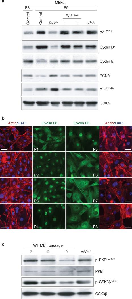
Next, we sought to understand the molecular pathway(s) involved in the immortalisation of MEFs after PAI-1 knockdown or uPA overexpression. The tumour suppressors p16INK4A and p21CIP1 were induced in P9 MEFs and immortal PAI-1kd or uPA overexpressing cells, indicating that the senescence-stress pathway is activated in these MEFs (Fig. 2a). Interestingly, a sharp increase in cyclin D1 levels were observed in both non-proliferating P9 and proliferating PAI-1kd or uPA overexpressing MEFs, which may neutralize the high levels of p21CIP1 (Fig. 2a). We asked whether signalling through PI(3)K, PKB (also known as AKT) and GSK3β was involved. Activation of PI(3)K and subsequent full activation of PKB by phosphorylation on Ser 473 leads to an inhibitory phosphorylation of GSK3β on Ser 9 by PKB17,18. GSK3β controls cyclin D1 localization and degradation through an inhibitory phosphorylation on Thr 286 and loss of this inhibitory phosphorylation protects cyclin D1 from nuclear exclusion and degradation19. Therefore, mitogenic signalling through PI(3)K–PKB–GSK3β influences cyclin D1 stability and its nuclear activity, leading to cell-cycle progression2. uPA induces growth factor-related PI(3)K–PKB signalling20 and, as a result, uPA activity may result in nuclear retention of cyclin D1 by inducing loss of GSK3β activity through phosphorylation on Ser 9. When cyclin D1 localization was determined in ageing MEFs, a striking but gradual nuclear exclusion of cyclin D1 was observed (Fig. 2b), which correlated with the decline in growth rate (note that wild-type MEFs became fully senescent at passage 8; see Supplementary Information, Fig. S2a, as confirmed by staining for senescence-associated acidic β-galactosidase21, SA-β-Gal; data not shown). Furthermore, during serial passaging, wild-type MEFs show a progressive decline in PKB activation by loss of phosphorylation and a gradually increased GSK3β activation by loss of inhibitory phosphorylation (Fig. 2c). In contrast, immortal PAI-1kd or uPA overexpressing MEFs had sustained PKB–GSK3β signalling (see Supplementary Information, Fig. S2b). We therefore concluded that during replicative senescence cyclin D1 is excluded from the nucleus and stabilized in the cytosol, correlating with decreased growth-rate and downregulation of PKB–GSK3β signalling. The high levels of cyclin D1 found in senescent cells (Fig. 2a) were unexpected because GSK3β activity induces not only nuclear exclusion, but also the turnover of cyclin D1 (ref. 19). Significantly, high levels of cyclin D1 were also observed in senescent human BJ fibroblasts (Fig. 5c). Apparently, the turnover of cyclin D1 is different in senescent cells as compared with cycling NIH3T3 cells19.
Figure 2.
MEFs reduce PKB activation and exclude cyclin D1 from the nucleus during replicative senescence. (a) Western blot analysis of P3, P9, p53kd, PAI-1kd or uPA overexpressing MEFs for cell-cycle related proteins. PCNA and CDK4 are proliferation and loading controls, respectively. (b) Qualitative immunofluorescence microscopy analysis of serially passaged MEFs, (P1–P8), for cyclin D1 expression. The scale bar represents 50 μm. (c) Expression of phosphorylated PKB or GSK3β related to unphosphorylated fraction of the same proteins in P3, P6 and P9 MEFs and post-senescent p53kd cells as analysed by western blot.
Figure 5.
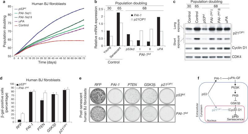
PAI-1 is necessary and sufficient for senescence in human BJ fibroblasts. (a) Growth curves of primary human BJ fibroblasts overexpressing the indicated constructs. The mean ± s.d. of three independent cultures per genotype is shown. (b) Relative PAI-1 and p21CIP1 expression analysed by QRT–PCR on the indicated post-senescent BJ cell lines and control-infected young or senescent BJ fibroblasts. (c) Western blot analysis of cells from a for p53-target p21CIP1 and cell-cycle related protein cyclin D1. CDK4 is a loading control. (d) Quantification of SA-β-Gal-positive post-senescent PAI-1kd or p53kd BJ fibroblasts after retroviral overexpression of PAI-1, PTEN, GSK3β, p21CIP1 or an RFP control. The mean ± s.d. of three independent plates is shown. (e) Colony formation assay in post-senescent p53kd or PAI-1kd BJ fibroblasts infected with the indicated retroviral overexpression constructs. RFP is a negative control. (f) Schematic representation of a proposed model for senescence in fibroblasts. p53 induces PAI-1 and p21CIP1 during ageing in culture. PAI-1 antagonizes uPA–GF (growth factor) signalling to cyclin D1 through PI(3)K–PKB–GSK3β and p21CIP1 blocks cyclin D1 activity directly. The PAI-1–cyclin D1 connection is dominant over p21CIP1 activity and controls induction of the senescence response downstream of p53 and upstream of pRb.
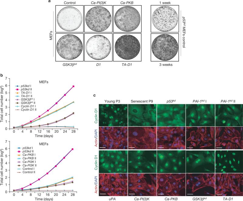
We next examined whether immortalisation of MEFs by PAI-1 knockdown may be the result of activated PI(3)K–PKB–GSK3β signalling. Primary MEFs were retrovirally transduced with constitutively active mutants of PI(3)K (p110αCAAX; Ca-PI(3)K) or PKB (Myr-PKB; Ca-PKB), a retroviral knockdown short hairpin RNA (shRNA) construct for GSK3β, or retroviral expression constructs for wild-type cyclin D1 (D1) or non-degradable cyclin D1 (T286A-cyclin D1; TA-D1), and determined their immortalizing potential in a colony-formation assay. The TA-D1 mutant is refractory to phosphorylation by GSK3β and is therefore constitutively nuclear19. We found that Ca-PI(3)K, Ca-PKB, GSK3βkd or TA-D1 (but not wild type D1) are individually capable of immortalizing wild-type MEFs (Fig. 3a). Furthermore, no loss of p19ARF expression or evidence for mutation of p53 was found in any of the immortalised MEFs (data not shown). When tested in a long-term proliferation assay, overexpression of Ca-PI(3)K, Ca-PKB, GSK3βkd or TA-D1 (but not wild type D1) also induced a bypass of senescence in MEFs (Fig. 3b). Again, immortalisation was not accompanied by loss of p19ARF or p53 function (data not shown). As previously observed in PAI-1kd or uPA overexpressing MEFs (Fig. 2a), induction of p21CIP1 protein levels in the post-senescent polyclonal cell lines expressing a Ca-PI(3)K, Ca-PKB, GSK3βkd, or TA-D1 construct was noted (see Supplementary Information, Fig. S2c). Furthermore, high PKB-GSK3β signalling was observed in Ca-PI(3)K or Ca-PKB overexpressing cells and reduced GSK3β expression in GSK3βkd cells (see Supplementary Information, Fig. S2d). Our results suggest that enforced constitutive activation of PI(3)K–PKB signalling, reduction of GSK3β activity or nuclear retention of cyclin D1, is sufficient to bypass senescence in MEFs downstream of p53. Accordingly, immunofluorescence microscopy analysis of post-senescent polyclonal cell lines of MEFs immortalised with the various constructs used in this study revealed nuclear localization of endogenous cyclin D1 when compared with senescent wild-type or HA-tagged cyclin D1 expressing MEFs (Fig. 3c and see Supplementary Information, Fig. S2e).
Figure 3.
Sustained PI(3)K–PKB signalling or nuclear retention of cyclin D1 induces bypass of senescence. (a) Colony formation assay of primary MEFs infected with the indicated retroviral constructs. Immortalizing efficiency controls are p53kd MEFs stained after 1 or 3 weeks. (b) Growth curves of various depicted immortalizing constructs versus wild-type cyclin D1 infected MEFs. Overexpression of p53kd or control vector are positive and negative controls, respectively. The results shown are of two independent infections per construct (I, II). (c) Qualitative immunofluorescence microscopy analysis for cyclin D1 of post-senescent MEFs immortalised with the indicated constructs and control P3 and P9 MEFs. The scale bar represents 50 μm.
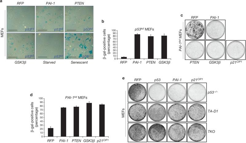
To examine whether reduction of PI(3)K–PKB–GSK3β signalling is sufficient for the induction of senescence, antagonists of this mitogenic signalling route were overexpressed in p53-depleted cells. Retroviral cDNA expression constructs were generated for human PAI-1 (which is not targeted by the mouse PAI-1kd vectors), mouse PTEN and GSK3β. PTEN is a potent tumour suppressor often deleted in cancer that blocks the activation of PI(3)K22and exerts its effect in part by regulating nuclear availability of cyclin D1 (ref. 23). When overexpressed in p53kd or PAI-1kd cells, PAI-1, PTEN and GSK3β were all individually able to induce senescence, as observed by a flat-cell morphology and positive staining for SA-β-Gal. (Fig. 4a–d). To test whether cyclin D1 is an essential target downstream of PAI-1, PAI-1 was overexpressed in p53–/– MEFs, MEFs overexpressing TA-D1, or pocket-protein deficient triple knockout cells (pRb–/––p107–/––p130–/–; TKO). The immortal TKO controls lack retinoblastoma family function and therefore have sustained E2F activity24,25. In normal fibroblasts pRb (family)-E2F-mediated repression is required for cell-cycle exit in response to p19ARF–p53 activation26. Overexpression of TA-D1 in MEFs may therefore resemble the phenotype seen in TKO cells by blocking retinoblastoma family function. Figure 4e shows that PAI-1 induced an arrest in p53–/– cells, but not when these cells also expressed TA-D1 or were pocket-protein deficient. These results suggest that in the presence of a wild-type cyclin D1 protein, PAI-1 is able to induce an arrest, but not when cyclin D1 is constitutively nuclear and insensitive to GSK3β.
Figure 4.
PAI-1 expression is sufficient for the induction of replicative senescence. (a) PAI-1, PTEN or GSK3β overexpression induces senescence in p53kd MEFs as indicated by staining for senescence-associated β-galactosidase (SA-β-Gal). Control cells are mock-infected, serum depleted (starved) and wild-type senescent MEFs. The scale bar represents 400 μm. (b) Quantification of SA-β-Gal-positive p53kd cells after retroviral overexpression of constructs as indicated in a. The mean ± s.d. of three independent plates is shown. (c) Colony formation assay in immortal PAI-1kd MEFs after retroviral overexpression of the indicated constructs. p21CIP1 and red fluorescent protein (RFP) are positive and negative controls, respectively. (d) Quantification of SA-β-Gal-positive PAI-1kd MEFs after retroviral overexpression of PAI-1, PTEN, GSK3β, p21CIP1 or an RFP control. Shown is the mean ± s.d. per plate per infection. (e) Colony formation assay in immortal p53–/–, TA-D1 or TKO (retinoblastoma family triple knockout; pRb–/––p107–/––p130–/–) fibroblasts infected with indicated retroviral overexpression constructs.
The senescence response of human fibroblasts is, in the first instance, primarily dependent on p53 (M1 checkpoint) and, as in MEFs, PAI-1 is upregulated during ageing and is a marker of senescence in these cells7,27. When primary human BJ fibroblasts were infected with either one of two independent human-specific PAI-1 knockdown constructs (PAI-1kd I or PAI-1kd II) an M1 senescence-bypass was observed as judged in a long-term growth assay (Fig. 5a). As in MEFs, overexpression of uPA also induced a bypass of senescence in primary BJ cells, albeit with lower efficiency (Fig. 5a). When the post-senescent and proliferating population doubling (PD) 68 PAI-1kd were assayed for PAI-1 levels, reduction to levels even below those observed in young PD 30 BJ fibroblasts was observed (Fig. 5b). PAI-1 levels in PD 68 uPA overexpressing BJ cells were similar to those seen in senescent cells, in agreement with the notion that uPA acts downstream of p53. PAI-1kd or uPA overexpressing fibroblasts have notably higher levels of p21CIP1 than p53kd cells (Fig. 5b, c), consistent with the notion that PAI-1kd mediates senescence-bypass downstream of p53. That p53 is wild type in the PAI-1kd or uPA over-expressing BJ fibroblast is also supported by their normal response to DNA damage (data not shown). As observed in MEFs, induction of cyclin D1 in PAI-1kd and uPA overexpressing post-senescent BJ cells, as well as in non-proliferating senescent BJ cells, was observed (Fig. 5c). Furthermore, an increase in cytoplasmic cyclin D1 in ageing BJ fibroblasts was noticed, which was associated with p21CIP1 (see Supplementary Information, Fig. S4a–c), although the nuclear-to-cytoplasmic transition of cyclin D1 seems to be not as pronounced as in ageing MEFs. Importantly, overexpression of murine PAI-1, PTEN, or GSK3β in immortal human p53kd or PAI-1kd cells induced an arrest and SA-β-Gal. staining (Fig. 5d, e), indicating that PAI-1 expression and downregulation of the PI(3)K–PKB–GSK3β signalling route are also sufficient for induction of senescence in human fibroblasts downstream of p53 and PAI-1. Taken together, we conclude that PAI-1 is necessary and sufficient for senescence in human BJ fibroblasts. As p21CIP1 is an essential p53 target in the senescence response of human fibroblasts28,29, we asked if simultaneous knockdown of both p21CIP1 and PAI-1 would induce a more efficient bypass of senescence than either one alone. Knockdown of the expression of either PAI-1 or p21CIP1 resulted in a less efficient senescence-bypass than observed with knockdown of p53 (see Supplementary Information, Fig. 5a, b). Interestingly, simultaneous knockdown of PAI-1 and p21CIP1 resulted in a more efficient bypass of the arrest than knockdown of p53 itself (see Supplementary Information, Fig. S5a−c). We conclude that PAI-1 and p21CIP1 are both relevant downstream targets of p53 in the induction of senescence in human fibroblasts, as evident from the effects of their combined knockdown.
Here, we show that PAI-1 is a critical downstream target of p53 in the senescence response of both ageing mouse and human diploid fibroblasts. Our data indicate that p53 controls growth factor-dependent proliferation by upregulating PAI-1, leading to down-regulation of PI(3)K–PKB signalling and nuclear exclusion of cyclin D1. Conversely, we found that loss of PAI-1 expression or uPA overexpression in MEFs conferred resistance to the anti-proliferative activity of p53 by inducing sustained PI(3)K–PKB signalling and cyclin D1 nuclear retention. Our data are consistent with a model in which PAI-1 acts to limit cyclin–CDK activity during the induction of replicative senescence and suggest a role for PAI-1 as a secreted gatekeeper of fibroblast proliferative capacity (Fig. 5f).
We found that the mitogen-stimulated PI(3)K–PKB pathway is causally involved in the senescence-bypass of fibroblasts, and that overexpression of its antagonist, PTEN22, can reverse this process. It has been reported that the levels of PTEN are crucial in determining the senescence response of fibroblasts — partial loss of PTEN confers a proliferative advantage, whereas acute loss of all PTEN induces senescence30. Consistent with this, we found that somatic knockdown of PTEN in wild-type MEFs resulted in bypass of senescence (data not shown) and constitutive activation of the PI(3)K–PKB signalling route, albeit not to the degree seen in Ca-PI(3)K or Ca-PKB overexpressing cells (see Supplementary Information, Fig. S3b). In apparent conflict with our data, which show senescence bypass by active PKB, it has been reported that overexpression of an active PKB resulted in induction of senescence. However, when these cells were followed over 6 days, proliferation was not entirely lost in the infected population30. We have selected PKB-infected wild type MEFs over a longer period of time, and consequently enriched for proliferating cells with potentially only moderately enhanced PKB activity. Taken together, these data support the notion that slightly elevated levels of PKB or partial loss of PTEN results in enhanced proliferation, whereas highly elevated PKB or complete loss of PTEN induces senescence. This is reminiscent of what has been observed in RAS signalling, where overexpression of an activated RAS oncogene induces senescence, whereas activated RAS expressed at physiological levels confers a growth advantage31.
PAI-1 is induced by a variety of growth factors and is a target of c-Myc7,32. uPA transcription and its extracellular activity are regulated by growth-factor signalling and proteases. Induction of PAI-1 may therefore be part of a growth factor-stimulated negative feedback loop that becomes constitutively activated by p53 in ageing fibroblasts. As a consequence, senescent fibroblasts may induce a state of growth-factor unresponsiveness by secreting PAI-1. It is therefore possible that induction of PAI-1 by p53 or disturbance of the uPA or PAI-1 levels influences intra-tumour heterotypic signalling and the local tumour microenvironment. This is particularly noteworthy as uPA and PAI-1 are causally involved in wound healing, angiogenesis and metastasis12,33 — processes that are dominantly regulated by cell–cell signalling34. Furthermore, uPA is secreted by stromal fibroblasts and myofibroblasts at the invasive front in breast and prostate cancer35 and loss of uPA can result in reduced metastasis in mouse models36,37. As it is becoming increasingly clear that stromal tissue is an indispensable player in neoplastic transformation and metastasis38,39, our observations may lead to a better understanding of the role of fibroblasts in cancer.
METHODS
Antibodies and Vectors
For western blotting, antibodies against p16INK4A (M156), p21CIP1 (F5, C19), SP1 (PEP2), cyclin D1 (H295, M20), cyclin E (M20), PCNA (PC-10), PKB–Akt1 (C20), p53 (DO-1), HA (Y11) and CDK4 (C22) were purchased from Santa Cruz Biotechnology (Santa Cruz, CA); anti p-PKB (Ser473; #9271), anti p-GSK3β (Ser9; #9336) and HSP90 (#4874) from Cell Signalling (Beverly, MA); anti p19ARF (Ab 80-100) from Abcam (Cambridge, UK); anti-GSK3β (610201) from BD Pharmingen (San Jose, CA); anti phosphotyrosine (PY20) from Calbiochem (San Diego, CA); and anti p53 (Ab7) from Oncogene Research Products (Boston, MA). Flag-tagged mouse cDNAs for PAI-1, GSK3β, PTEN or human PAI-1 were generated by PCR amplification and cloned into pLZRS–IRES–zeocin. Mouse cDNA for uPA was generated by PCR amplification of pro-uPA and cloned into pBABEpuro. The production of siRNAs in MEFs, BJ or tsLT–hTERT–BJ fibroblasts was achieved using the pRETRO–SUPER vector11. For the generation of mouse PAI-1 knockdown constructs the following 19-mer sequences were used: PAI-1 I, 5′-GAACAAGAATGAGATCAGT-3′; PAI-1 II, 5′-GTTGGGCATGCCTGACATG-3′. For the generation of human PAI-1 knockdown constructs the following 19-mer sequences were used: PAI-1 I, 5′-CTGACTTCACGAGTCTTTC-3′; PAI-1 II, 5′-CCTGGGAATGACCGACATG-3′. The 19-mer sequences used for knockdown of mouse p53 or GSK3β have been described elsewhere40,41, as have the 19-mer sequences used for knockdown of human p53 or p21CIP1 (ref. 29). Control infections were performed with nonfunctional hairpin or red fluorescent protein (RFP) vectors.
Cell culture, transfection and retroviral infection
MEFs, primary and tsLT hTERT human BJ fibroblasts and Phoenix cells were cultured in DMEM (Gibco, Carlsbad, CA) supplemented with 8% heat-inactivated fetal bovine serum (Perbo; PAA, Pasching, Austria), 2 mM l-glutamine and penicillin–streptomycin (Gibco). Transfections were performed with the calcium-phosphate precipitation technique. Retroviral supernatants were produced by transfection of Phoenix packaging cells. Viral supernatants were filtered through a 45 μm Millex HA filter (Millipore, Carrigtwohill, Ireland) and infections were performed in the presence of 4 μg ml–1 polybrene (Sigma, St Louis, MO). Drug selections in MEFs or BJs were performed with 1 μg ml–1 puromycin, 50 μg ml–1 hygromycin or 100 μg ml–1 zeocin.
Colony-formation assays
Wild-type MEFs were infected with shRNA or cDNA constructs at P3, selected and at P5 5 × 10⁴ cells were seeded onto 10-cm plates and stained after 3 weeks. p53kd control MEFs (5 × 10⁴) were seeded onto 10-cm plates and stained after 1, 2 or 3 weeks. Immortal PAI-1kd MEFs were infected, after 72 h were plated under low density (5 × 10⁴ cells in a 10-cm plate) and 2 weeks were later stained. p53–/–, TA-D1 or TKO MEFs were infected, 48 h after infection 5 × 10⁴ cells were seeded onto 10-cm plates and were stained after 1 week. Human post-senescent p53kd or PAI-1kd BJ fibroblasts were infected with cDNA constructs, plated under low density (1 × 10⁵ cells in a 10-cm plate) and were stained after 2 weeks. Human tsLT BJ fibroblasts were infected at 32 °C and after 48 h, 1 × 10⁵ cells were seeded per 10-cm plate, shifted to 39 °C and stained after 2 weeks. For all colony formations representative examples of at least three independent experiments are shown.
Growth curves
MEFs were infected with retroviral shRNA or cDNA expression constructs at P3, selected and at P5 1.5 × 10⁵ cells were plated in a 6-cm dish (time = 0 days). Every 4 days, cells were counted and 1.5 × 10⁵ cells were replated. A MEF passage, as defined for this paper, represents 4 days in culture. PAI-1–/–, p53–/– or wild-type MEFs (1.5 × 10⁵) were plated in a 6-cm dish at P1, and every 4 days cells were counted and 1.5 × 10⁵ cells were replated. Human primary BJ fibroblasts at population-doubling 53 were infected, selected and 1.5 × 10⁵ cells were plated in a 6-cm dish (time = 0 days). Every 4 days, cells were counted and 1.5 × 10⁵ cells were replated. Human tsLT BJ fibroblasts were infected, shifted to 39 °C after 48 h and 1.5 × 10⁵ cells were plated in a 6-cm dish (time = 0 days). Every 6 days, cells were counted and 1.5 × 10⁵ cells were replated. The p53 status of control, senescent or post-senescent primary MEFs, BJ fibroblasts or tsLT BJ fibroblasts was checked by DNA-damage induced p53 activation by overnight addition of 0.5 mM cisplatin and western blotting for p53 and its targets (p19ARF and p21CIP1 in MEFs, or p21CIP1 and Bax in BJ fibroblasts). Total cell amounts in all growth curves were displayed as cumulative over time. For all growth-curves representative examples of at least two independent experiments are shown.
Quantitative RT–PCR
Total RNA from immortal post-senescent MEF cell lines plus P3 and 9 control MEFs, control or immortalized human BJ fibroblasts and immortalised tsLT BJ fibroblasts at 39 °C or controls at 32 °C or 39 °C, was isolated with TRI-Zol (Invitrogen, Carlsbad, CA) according to manufacturers’ instructions. QRT–PCR was performed on an ABI Prism 7700 with Assays-on-Demand (Applied Biosystems, Foster City, CA) for mouse PAI-1 and TBP as a control housekeeping gene, or for human PAI-1 or p21CIP1 with GAPDH as a control housekeeping gene. QRT–PCR results in tsLT BJ fibroblasts are the mean ± s.d. of three independent cell lines per genotype. For all QRT–PCRs, representative examples of at least two independent experiments are shown.
Immunofluorescence microscopy
Cells were plated on 8-well chamber slides (Nutacon; Leimuiden, The Netherlands) and cultured overnight after which they were fixed with 4% formaldehyde in PBS for 15 min, permeabilized with 0.2% Triton X (Sigma), blocked and incubated with anti-cyclin D1 (M20) from Santa Cruz Biotechnology. Actin was stained with rhodamine-conjugated phalloidin (Invitrogen) and the nucleus with DAPI (Roche, Basel, Switzerland). Images were obtained using a Modified Zeiss Axiovert 100M (SP LSM 5) cooled CCD fluorescence microscope with a Plan-APOCHROMAT, 1.0 NA, 40× oil immersion objective plus a Zeiss single excitation-triple emission filter set 40 with a KP650 red blocking filter on a Photometrics MAC 200A camera and SmartCapture V2.0 software. For all immunostainings, representative examples of at least three independent experiments are shown.
Acidic β-galactosidase staining
p53kd or PAI-1kd MEFs were infected, after 4 days plated under low density (5 × 10⁴ cells in a 10-cm plate) and 24 h later stained overnight for senescence-associated acidic β-galactosidase as previously described21. Human post-senescent immortal p53kd or PAI-1kd BJ fibroblasts were infected, after 5 days plated under low density (1 × 10⁵ cells in a 10-cm plate), and 48 h later stained overnight for acidic β-galactosidase as previously described21. Per plate, three independent groups of 300 cells were counted for SA-β-Gal staining. For all SA-β-Gal stainings, representative examples of at least two independent experiments are shown. Images were obtained using a Zeiss Axiovert 25 microscope with A-Plan 10× or LD A-plan 20× objectives on a Canon Powershot G3 14× zoom camera.
Western blotting
Selected cells were lysed in RIPA buffer (50 mM Tris at pH 8, 150 mM NaCl, 1% NP40, 0.5% deoxycholate, 0.1% SDS). 20, 40 or 80 micrograms of Proteins (20, 40 or 80 μg) were separated by 8–12% SDS–PAGE and transferred to polyvinylidine difluoride membranes (Millipore, Billerica, MA). Western blots were probed with the indicated antibodies. For all western blots representative examples of at least two independent experiments are shown.
Coimmunoprecipitations
Total cell lysates were isolated with ELB buffer (0.25 M NaCl, 0.1% NP40, 50 mM HEPES at pH 7.3) supplemented with Complete protease inhibitors (Roche). Cytoplasmic fractions of BJ cells were isolated with nuclear and cytoplasmic extraction kit NE-PER (Pierce Biotechnology Inc., Rockford, IL) according to manufacturers’ instructions. Lysates were incubated with protein-A−Sepharose beads (Amersham-Pharmacia Biotech, Piscataway, NJ) coated with anti-cyclin D1 (M20, Santa Cruz Biotechnology). Analysis of cyclin D1 or cyclin D1-associated proteins was performed by western blotting the precipitates from cytoplasmic and total lysates.
Supplementary Material
Supplementary Figure S1 PAI-1kd induces senescence-bypass in primary MEFs retaining p19ARF-p53 signalling.
(a) Long-term proliferation curves of p53kd, PAI-1kd, or non-functional shRNA control infected MEFs. Shown are the results of two independent experiments per construct (I, II).
(b) Western blot analysis for p53 and its targets p19ARF and p21CIP1 using cell lines depicted in (a) after cisplatin-induced DNA-damage. NIH3T3 cells are immortal p16INK4A/p19ARF-deficient controls.
Supplementary Figure S2 Retention or induction of PKB-GSK3β signalling in immortal MEFs.
(a) Proliferation curves of 6 independently isolated cultures of wild-type MEFs. Mean value (+/- SD) is indicated.
(b) Expression of phosphorylated PKB or GSK3β related to unphosphorylated fraction of the same proteins in P3, P9, p53kd, PAI-1kd, or uPA over-expressing cells as analyzed by western blot.
(c) Western blots of protein samples of indicated cell lines with antibodies for tumour suppressors p16INK4A or p21CIP1, or cyclin D1. CDK4 is a loading control.
(d) Western blots for the same set of cell lines as in (c) for phospho-specific PKB and GSK3β, as compared to unphosphorylated fractions of the same proteins.
(e) Qualitative immunofluorescence analysis for HA-tag (HA) and cyclin D1 in senescent MEFs over-expressing HA-tagged cyclin D1. Bar represents 50 μm.
Supplementary Figure S3 PAI-1 is sufficient for induction of senescence in MEFs.
(a) PAI-1, PTEN, GSK3β, or p21CIP1 over-expression induces senescence in PAI-1kd MEFs as indicated by staining for senescence-associated β-galactosidase (SA-β-Gal). Staining controls are young and senescent wild-type MEFs. Scale bar represents 250 μm.
(b) Expression of phosphorylated p110α (catalytic subunit of PI3K) and PKB related to unphosphorylated fraction of the same protein in P3, P9, PTENkd, ca-PI3K or ca-PKB over-expressing cells as analyzed by western blot.
Supplementary Figure S4 Cytoplasmic co-localization of cyclin D1 and p21CIP1 in ageing primary BJ fibroblasts.
(a) Western blot of cytoplasmic (C) and total (T) protein lystates from primary young passage doubling 30 and senescent passage doubling 65 BJ fibroblasts probed for indicated proteins. Sp1 is a nuclear protein and Hsp90 a cytoplasmic fraction control.
(b) Immunoprecipitation with cyclin D1 antibody in protein lysates from (a) immunoblotted for p21CIP1.
(c) Qualitative immunofluorescence analysis of serially passaged BJs, passage doubling 30 or passage doubling 65, for cyclin D1 expression. Bar represents 50 μm.
(d) Coomassie staining of SDS gel with 10 or 30 μg of nuclear (n), cytoplasmic (c) or total (t) protein from young populatipon doubling (PD) 30 or senescent PD 65 primary BJ fibroblasts, for equal loading.
Supplementary Figure S5 PAI-1 and p21CIP1 collaborate in senescence response downstream of p53.
(a) Colony formation assay of depicted constructs in conditionally immortalized tsLT hTERT BJ fibroblasts29. Control is a non-functional shRNA. These cells enter into a p53-dependent proliferation arrest when shifted to the non-permissive temperature (39°C). Importantly, these cells show virtually exact characteristics as primary BJ fibroblasts when challenged through assays as described in Fig. 5a-e(data not shown).
(b) Lon g term growth curves at the non-permissive temperature of 39°C of cells over-expressing depicted constructs. Per genotype the mean (+/- SD) of 3 independent cultures is shown. PAI-1 and p21CIP1 are both p53 target genes and knockdown of the expression of either gene alone results in a less efficient senescence bypass than seen with knockdown of p53 alone (see also (a)).
(c) Quantitative real-time PCR analysis of relative expression of PAI-1 and p21CIP1 in cell lines depicted in (b). As in primary BJ fibroblasts (see Fig. 5b), we noticed reduction of PAI-1 mRNA in p21CIP1kd cells and reduction of p21CIP1 mRNA in PAI-1kd cells, suggesting that loss of either gene influences transcription of the other.
ACKNOWLEDGEMENTS
We would like to thank A. Visser for technical assistance, K. Berns, M. Hijmans, A. Dirac, T. Brummelkamp, R. Agami, R. van der Kammen, J. Collard and F. Scheeren for retroviral constructs, B. Weigelt for help with QRT–PCR, F. Foijer for retinoblastoma family deficient MEFs, L. Oomen and L. Brocks for help with microscopy, and R. Beijersbergen and D. Peeper for helpful discussions. This work was supported by a grant from the Dutch Cancer Society to R.B. and a grant from the National Institutes of Health (NIH; GM57242) to P.H.
Footnotes
COMPETING FINANCIAL INTERESTS
The authors declare that they have no competing financial interests.
Note: Supplementary Information is available on the Nature Cell Biology website.
References
- 1.Lundberg AS, Hahn WC, Gupta P, Weinberg RA. Genes involved in senescence and immortalization. Curr. Opin. Cell Biol. 2000;12:705–709. doi: 10.1016/s0955-0674(00)00155-1. [DOI] [PubMed] [Google Scholar]
- 2.Sherr CJ, McCormick F. The RB and p53 pathways in cancer. Cancer Cell. 2002;2:103–112. doi: 10.1016/s1535-6108(02)00102-2. [DOI] [PubMed] [Google Scholar]
- 3.Massague J. G1 cell-cycle control and cancer. Nature. 2004;432:298–306. doi: 10.1038/nature03094. [DOI] [PubMed] [Google Scholar]
- 4.Pantoja C, Serrano M. Murine fibroblasts lacking p21 undergo senescence and are resistant to transformation by oncogenic Ras. Oncogene. 1999;18:4974–4982. doi: 10.1038/sj.onc.1202880. [DOI] [PubMed] [Google Scholar]
- 5.Kunz C, Pebler S, Otte J, von der Ahe D. Differential regulation of plasminogen activator and inhibitor gene transcription by the tumor suppressor p53. Nucleic Acids Res. 1995;23:3710–3717. doi: 10.1093/nar/23.18.3710. [DOI] [PMC free article] [PubMed] [Google Scholar]
- 6.Zhao R, et al. Analysis of p53-regulated gene expression patterns using oligonucleotide arrays. Genes Dev. 2000;14:981–993. [PMC free article] [PubMed] [Google Scholar]
- 7.Mu XC, Higgins PJ. Differential growth state-dependent regulation of plasminogen activator inhibitor type-1 expression in senescent IMR-90 human diploid fibroblasts. J. Cell Physiol. 1995;165:647–657. doi: 10.1002/jcp.1041650324. [DOI] [PubMed] [Google Scholar]
- 8.Martens JW, et al. Aging of stromal-derived human breast fibroblasts might contribute to breast cancer progression. Thromb. Haemost. 2003;89:393–404. [PubMed] [Google Scholar]
- 9.Serrano M, Lin AW, McCurrach ME, Beach D, Lowe SW. Oncogenic ras provokes premature cell senescence associated with accumulation of p53 and p16INK4a. Cell. 1997;88:593–602. doi: 10.1016/s0092-8674(00)81902-9. [DOI] [PubMed] [Google Scholar]
- 10.De Petro G, Copeta A, Barlati S. Urokinase-type and tissue-type plasminogen activators as growth factors of human fibroblasts. Exp. Cell Res. 1994;213:286–294. doi: 10.1006/excr.1994.1200. [DOI] [PubMed] [Google Scholar]
- 11.Brummelkamp TR, Bernards R, Agami R. Stable suppression of tumorigenicity by virus-mediated RNA interference. Cancer Cell. 2002;2:243–247. doi: 10.1016/s1535-6108(02)00122-8. [DOI] [PubMed] [Google Scholar]
- 12.Andreasen PA, Egelund R, Petersen HH. The plasminogen activation system in tumor growth, invasion, and metastasis. Cell Mol. Life Sci. 2000;57:25–40. doi: 10.1007/s000180050497. [DOI] [PMC free article] [PubMed] [Google Scholar]
- 13.Choong PF, Nadesapillai AP. Urokinase plasminogen activator system: a multifunctional role in tumor progression and metastasis. Clin. Orthop. 2003;415:S46–S58. doi: 10.1097/01.blo.0000093845.72468.bd. [DOI] [PubMed] [Google Scholar]
- 14.Quelle DE, et al. Cloning and characterization of murine p16INK4a and p15INK4b genes. Oncogene. 1995;11:635–645. [PubMed] [Google Scholar]
- 15.Linardopoulos S, et al. Deletion and altered regulation of p16INK4a and p15INK4b in undifferentiated mouse skin tumors. Cancer Res. 1995;55:5168–5172. [PubMed] [Google Scholar]
- 16.Sherr CJ. Tumor surveillance via the ARF-p53 pathway. Genes Dev. 1998;12:2984–2991. doi: 10.1101/gad.12.19.2984. [DOI] [PubMed] [Google Scholar]
- 17.Cross DA, Alessi DR, Cohen P, Andjelkovich M, Hemmings BA. Inhibition of glycogen synthase kinase-3 by insulin mediated by protein kinase B. Nature. 1995;378:785–789. doi: 10.1038/378785a0. [DOI] [PubMed] [Google Scholar]
- 18.Vivanco I, Sawyers CL. The phosphatidylinositol 3-Kinase AKT pathway in human cancer. Nature Rev. Cancer. 2002;2:489–501. doi: 10.1038/nrc839. [DOI] [PubMed] [Google Scholar]
- 19.Diehl JA, Cheng M, Roussel MF, Sherr CJ. Glycogen synthase kinase-3β regulates cyclin D1 proteolysis and subcellular localization. Genes Dev. 1998;12:3499–3511. doi: 10.1101/gad.12.22.3499. [DOI] [PMC free article] [PubMed] [Google Scholar]
- 20.Chandrasekar N, et al. Downregulation of uPA inhibits migration and PI3k/Akt signaling in glioblastoma cells. Oncogene. 2003;22:392–400. doi: 10.1038/sj.onc.1206164. [DOI] [PubMed] [Google Scholar]
- 21.Dimri GP, et al. A biomarker that identifies senescent human cells in culture and in aging skin in vivo. Proc. Natl Acad. Sci. USA. 1995;92:9363–9367. doi: 10.1073/pnas.92.20.9363. [DOI] [PMC free article] [PubMed] [Google Scholar]
- 22.Parsons R. Human cancer, PTEN and the PI-3 kinase pathway. Semin. Cell Dev. Biol. 2004;15:171–176. doi: 10.1016/j.semcdb.2003.12.021. [DOI] [PubMed] [Google Scholar]
- 23.Radu A, Neubauer V, Akagi T, Hanafusa H, Georgescu MM. PTEN induces cell cycle arrest by decreasing the level and nuclear localization of cyclin D1. Mol. Cell Biol. 2003;23:6139–6149. doi: 10.1128/MCB.23.17.6139-6149.2003. [DOI] [PMC free article] [PubMed] [Google Scholar]
- 24.Dannenberg JH, van Rossum A, Schuijff L, te Riele H. Ablation of the retinoblastoma gene family deregulates G(1) control causing immortalization and increased cell turnover under growth-restricting conditions. Genes Dev. 2000;14:3051–3064. doi: 10.1101/gad.847700. [DOI] [PMC free article] [PubMed] [Google Scholar]
- 25.Sage J, et al. Targeted disruption of the three Rb-related genes leads to loss of G(1) control and immortalization. Genes Dev. 2000;14:3037–3050. doi: 10.1101/gad.843200. [DOI] [PMC free article] [PubMed] [Google Scholar]
- 26.Rowland BD, et al. E2F transcriptional repressor complexes are critical downstream targets of p19(ARF)/p53-induced proliferative arrest. Cancer Cell. 2002;2:55–65. doi: 10.1016/s1535-6108(02)00085-5. [DOI] [PubMed] [Google Scholar]
- 27.West MD, Shay JW, Wright WE, Linskens MH. Altered expression of plasminogen activator and plasminogen activator inhibitor during cellular senescence. Exp. Gerontol. 1996;31:175–193. doi: 10.1016/0531-5565(95)02013-6. [DOI] [PubMed] [Google Scholar]
- 28.Brown JP, Wei W, Sedivy JM. Bypass of senescence after disruption of p21CIP1/WAF1 gene in normal diploid human fibroblasts. Science. 1997;277:831–834. doi: 10.1126/science.277.5327.831. [DOI] [PubMed] [Google Scholar]
- 29.Berns K, et al. A large-scale RNAi screen in human cells identifies new components of the p53 pathway. Nature. 2004;428:431–437. doi: 10.1038/nature02371. [DOI] [PubMed] [Google Scholar]
- 30.Chen Z, et al. Crucial role of p53-dependent cellular senescence in suppression of Pten-deficient tumorigenesis. Nature. 2005;436:725–730. doi: 10.1038/nature03918. [DOI] [PMC free article] [PubMed] [Google Scholar]
- 31.Tuveson DA, et al. Endogenous oncogenic K-ras(G12D) stimulates proliferation and widespread neoplastic and developmental defects. Cancer Cell. 2004;5:375–387. doi: 10.1016/s1535-6108(04)00085-6. [DOI] [PubMed] [Google Scholar]
- 32.Prendergast GC, Diamond LE, Dahl D, Cole MD. The c-myc-regulated gene mrl encodes plasminogen activator inhibitor 1. Mol. Cell Biol. 1990;10:1265–1269. doi: 10.1128/mcb.10.3.1265. [DOI] [PMC free article] [PubMed] [Google Scholar]
- 33.Parfyonova YV, Plekhanova OS, Tkachuk VA. Plasminogen activators in vascular remodeling and angiogenesis. Biochemistry (Mosc) 2002;67:119–134. doi: 10.1023/a:1013964517211. [DOI] [PubMed] [Google Scholar]
- 34.Bissell MJ, et al. Tissue structure, nuclear organization and gene expression in normal and malignant breast. Cancer Res. 1999;59:S1757–S1763. [PubMed] [Google Scholar]
- 35.Usher PA, et al. Expression of urokinase plasminogen activator, its receptor and type-1 inhibitor in malignant and benign prostate tissue. Int. J. Cancer. 2005;113:870–880. doi: 10.1002/ijc.20665. [DOI] [PubMed] [Google Scholar]
- 36.Frandsen TL, et al. Direct evidence of the importance of stromal urokinase plasminogen activator (uPA) in the growth of an experimental human breast cancer using a combined uPA gene-disrupted and immunodeficient xenograft model. Cancer Res. 2001;61:532–537. [PubMed] [Google Scholar]
- 37.Almholt K, et al. Reduced metastasis of transgenic mammary cancer in urokinase-deficient mice. Int. J. Cancer. 2005;113:525–532. doi: 10.1002/ijc.20631. [DOI] [PubMed] [Google Scholar]
- 38.Tuxhorn JA, Ayala GE, Rowley DR. Reactive stroma in prostate cancer progression. J. Urol. 2001;166:2472–2483. [PubMed] [Google Scholar]
- 39.Mueller MM, Fusenig NE. Friends or foes — bipolar effects of the tumour stroma in cancer. Nature Rev. Cancer. 2004;4:839–849. doi: 10.1038/nrc1477. [DOI] [PubMed] [Google Scholar]
- 40.Dirac AM, Bernards R. Reversal of senescence in mouse fibroblasts through lentiviral suppression of p53. J. Biol. Chem. 2003;278:11731–11734. doi: 10.1074/jbc.C300023200. [DOI] [PubMed] [Google Scholar]
- 41.Yu JY, Taylor J, DeRuiter SL, Vojtek AB, Turner DL. Simultaneous inhibition of GSK3α and GSK3β using hairpin siRNA expression vectors. Mol. Ther. 2003;7:228–236. doi: 10.1016/s1525-0016(02)00037-0. [DOI] [PubMed] [Google Scholar]
Associated Data
This section collects any data citations, data availability statements, or supplementary materials included in this article.
Supplementary Materials
Supplementary Figure S1 PAI-1kd induces senescence-bypass in primary MEFs retaining p19ARF-p53 signalling.
(a) Long-term proliferation curves of p53kd, PAI-1kd, or non-functional shRNA control infected MEFs. Shown are the results of two independent experiments per construct (I, II).
(b) Western blot analysis for p53 and its targets p19ARF and p21CIP1 using cell lines depicted in (a) after cisplatin-induced DNA-damage. NIH3T3 cells are immortal p16INK4A/p19ARF-deficient controls.
Supplementary Figure S2 Retention or induction of PKB-GSK3β signalling in immortal MEFs.
(a) Proliferation curves of 6 independently isolated cultures of wild-type MEFs. Mean value (+/- SD) is indicated.
(b) Expression of phosphorylated PKB or GSK3β related to unphosphorylated fraction of the same proteins in P3, P9, p53kd, PAI-1kd, or uPA over-expressing cells as analyzed by western blot.
(c) Western blots of protein samples of indicated cell lines with antibodies for tumour suppressors p16INK4A or p21CIP1, or cyclin D1. CDK4 is a loading control.
(d) Western blots for the same set of cell lines as in (c) for phospho-specific PKB and GSK3β, as compared to unphosphorylated fractions of the same proteins.
(e) Qualitative immunofluorescence analysis for HA-tag (HA) and cyclin D1 in senescent MEFs over-expressing HA-tagged cyclin D1. Bar represents 50 μm.
Supplementary Figure S3 PAI-1 is sufficient for induction of senescence in MEFs.
(a) PAI-1, PTEN, GSK3β, or p21CIP1 over-expression induces senescence in PAI-1kd MEFs as indicated by staining for senescence-associated β-galactosidase (SA-β-Gal). Staining controls are young and senescent wild-type MEFs. Scale bar represents 250 μm.
(b) Expression of phosphorylated p110α (catalytic subunit of PI3K) and PKB related to unphosphorylated fraction of the same protein in P3, P9, PTENkd, ca-PI3K or ca-PKB over-expressing cells as analyzed by western blot.
Supplementary Figure S4 Cytoplasmic co-localization of cyclin D1 and p21CIP1 in ageing primary BJ fibroblasts.
(a) Western blot of cytoplasmic (C) and total (T) protein lystates from primary young passage doubling 30 and senescent passage doubling 65 BJ fibroblasts probed for indicated proteins. Sp1 is a nuclear protein and Hsp90 a cytoplasmic fraction control.
(b) Immunoprecipitation with cyclin D1 antibody in protein lysates from (a) immunoblotted for p21CIP1.
(c) Qualitative immunofluorescence analysis of serially passaged BJs, passage doubling 30 or passage doubling 65, for cyclin D1 expression. Bar represents 50 μm.
(d) Coomassie staining of SDS gel with 10 or 30 μg of nuclear (n), cytoplasmic (c) or total (t) protein from young populatipon doubling (PD) 30 or senescent PD 65 primary BJ fibroblasts, for equal loading.
Supplementary Figure S5 PAI-1 and p21CIP1 collaborate in senescence response downstream of p53.
(a) Colony formation assay of depicted constructs in conditionally immortalized tsLT hTERT BJ fibroblasts29. Control is a non-functional shRNA. These cells enter into a p53-dependent proliferation arrest when shifted to the non-permissive temperature (39°C). Importantly, these cells show virtually exact characteristics as primary BJ fibroblasts when challenged through assays as described in Fig. 5a-e(data not shown).
(b) Lon g term growth curves at the non-permissive temperature of 39°C of cells over-expressing depicted constructs. Per genotype the mean (+/- SD) of 3 independent cultures is shown. PAI-1 and p21CIP1 are both p53 target genes and knockdown of the expression of either gene alone results in a less efficient senescence bypass than seen with knockdown of p53 alone (see also (a)).
(c) Quantitative real-time PCR analysis of relative expression of PAI-1 and p21CIP1 in cell lines depicted in (b). As in primary BJ fibroblasts (see Fig. 5b), we noticed reduction of PAI-1 mRNA in p21CIP1kd cells and reduction of p21CIP1 mRNA in PAI-1kd cells, suggesting that loss of either gene influences transcription of the other.


