
Figure 3. Foveolar-cell apical granule deficiency in Foxq1sb/sb stomach.
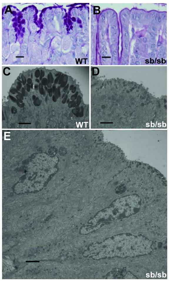
(A–B) High-magnification view of PAS-stained gastric foveolar cells from adult wild-type (A) and Foxq1sb/sb (B) mice. (C–D) Transmission electron microscopy of adult gastric foveolar cells, emphasizing the apical granular zone in wild-type (C) and Foxq1sb/sb (D) cells. (E) Full ultrastructural profile of Foxq1sa/sa gastric foveolar cells, indicating intact architecture except for apical granule deficiency. Scale bars: A–B, 60 μm; C–D, 1 μm; E, 2 μm.