Summary
Objective
To evaluate the scanning laser tomography characteristics of the optic nerve head in patients with primary open angle (POAG) glaucoma using the Heidelberg retina tomography (HRT II).
Design
A clinic-based retrospective study.
Participants
A total of 84 eyes of 42 POAG patients with good quality HRT II Images were studied at Charles Nicolle Hospital University department of Ophthalmology out-patient clinic, Tunis.
Methods
Characteristics of optic disc pattern of glaucoma patients were documented using the HRT II. Association of disc area with the other HRT parameters and inter-eye difference in the HRT parameters were assessed using simple and multiple regression analysis.
Main out-come measures
Disc area, cup area, rim area, cup-to-disc area ratio, cup volume, rim volume, mean cup depth, maximum cup depth and mean retinal nerve fibre layer thickness.
Results
Twenty-seven males and 15 females were studied. The mean age of glaucoma patients was 48.9±2.7 years. The mean disc area, cup area, cup-to-disc area ratio and rim area were 2.19±0.46 (range, 1.23 – 3.16mm2), 0.95±0.94 (range 0.08 – 2.15), 0.42±0.21(range 0.004–0.92), 1.25± 0.46 (range 0.18–2.64) respectively. Disc area was positively correlated to the cup area (p=0.001), rim area (p=0.001), cup to disc area ratio (p=0.03), and mean cup depth (p=0.02).The glaucoma diagnosis score was strongly correlated with the rim area (p< 0.001), cup area (p< 0.001), mean cup depth (p< 0.001) and cup disc area ratio (p< 0.001). Absolute inter eye parameter between the two eyes were positively correlated with disc area (p< 0.05).
Conclusions
There was a significant correlation of the parameters between the two eyes and between the disc area and some other HRT parameters.
Keywords: Diagnosis, disk area, disk cup, glaucoma, Scanning Laser Tomography
Introduction
Glaucoma is a major cause of irreversible blindness worldwide. Accurate and prompt detection of optic nerve damage is of tremendous importance in early diagnosis and prevention of blindness from glaucoma. Direct and indirect ophthalmoscopy and fundus photography have been widely used in the assessment of the glaucomatous optic nerve head damage. These techniques have been found to be very subjective and qualitative1. Over the years, facilities for a more objective and quantitative measurements of the optic nerve topography have been developed.
Confocal Scanning Laser Tomography is a robust, sophisticated diagnostic technique which produces 3-dimensional laser scan images of the optic nerve head and gives three dimensional cross-sectional images of the retina.
A Confocal scanning laser ophthalmoscope (CSLO) such as the Heidelberg retina tomogram, HRT II from Heidelberg Engineering, Heidelberg, Germany which is being studied gives a more precise measurement of various parameters of both the normal and diseased optic nerve head and retinal nerve fibre layer.2–4 This computerized morphometry of the optic nerve head is partially independent of the subjective evaluation by an examiner, it enables accurate and objective evaluation and follow up in eyes with pathology.
This technique has been used in determining the distributions and ranges of the optic nerve head parameters in large samples of normal subjects.5–9 The correct evaluation of the parameters studied by this technique and the knowledge of the instruments' limits are needed for an adequate interpretation of the results obtained. Among primary open angle glaucoma patients, little information has been provided regarding CSLO characteristics of the optic nerve head.
The aims of this present study were to evaluate the topographic characteristics of the optic nerve head by using the Heidelberg retina tomography (HRT II) in a clinic based population of glaucoma patients and to determine the relationships between the various optic nerve head parameters.
Materials and Methods
Topographic characteristics of eighty-four eyes of 42 patients who had diagnosis of primary open angle glaucoma (POAG) and had undergone Confocal Scanning laser tomography with good image quality were retrieved from the eye clinic of the Charles Nicolle University Department of Ophthalmology, Tunis where patients have been routinely screened and investigated. Patients demographic and background characteristics were also documented and analyzed. The research followed the tenets of the Declaration of Helsinki, and informed consent was obtained from the patients before being subjected to the diagnostic technique. Information retrieved was handled in accordance with permission obtained from the local research board.
The three topographic images which were obtained with the Heidelberg retina tomogram (Heidelberg Engineering HRT II) were retrieved from the computer. This consisted of the stereometric result of the Optic nerve head from 0°–360° at 2.5mm of 15°. Twelve HRT parameters obtained with routine analysis were documented: disc area (mm2), cup area (mm), rim area (mm2), cup-to-disc area ratio, cup volume(mm3), rim volume(mm3), mean cup depth, maximum cup depth (mm), height variation contour (mm), cup shape measure (mm), mean retinal nerve fiber layer (RNFL) thickness(mm), and RNFL cross-sectional area (mm2).
Good image quality was determined by brightness and clarity and appropriate centration of the image and a standard deviation of the mean topographic image being < 40microns. Only good quality topographic images were retrieved for analysis in this study. Topographic images from patients with media opacity, medical eye conditions such as diabetic or hypertensive retinopathy, or previous ocular trauma were excluded from the study analysis. Also, there were no significant ocular disease, history of previous laser or intra-ocular surgery and systemic diseases documented for any of the subjects whose eyes were studied.
Working diagnosis of primary open angle glaucoma was based on the presence of the following criteria: (1) An elevated IOP >21mmHg measured by applanation tonometry (2) an open anterior chamber angle on gonioscopy (3) a typical glaucomatous optic nerve head changes on ophthalmoscopy characterized by a glaucomatous shape of the neuro-retina rim, increase cup to disc ratio which is vertically higher than the horizontal, optic disc hemorrhages and localized or diffuse retinal nerve fiber layer defects (4) typical glaucomatous perimetric defects such as Bjerrum scotoma, nasal steps, and paracentral scotomata as documented by the Humphrey automated perimeter.
Two separate measurements collected over an average period of 9.5+/−2.8months were documented and evaluated. Univariate and multivariate regression analyses were performed to evaluate the associations between diagnosis (glaucoma or normal), optic disc area, cup disc area ratio, cup area, reference height variance, and rim area variability. All unreliable topographic images were excluded from the analysis.
Data were entered into SPSS version 11.1 for Windows software (SPSS, Inc., Chicago, IL.) Means ± SD were used for the description of quantitative data. One way analysis of variance was used to determine the relationship between disc area and other optic nerve head parameters. Pearson's correlation coefficients were calculated to determine correlations between two variables. A P value of less than 0.05 was considered to be statistically significant.
Results
There were 27males and 15 females in the study. The mean age of glaucoma patients was 48.9±2.7 years (range, 24 to 78years). The mean disc area, cup area, cup-to-disc area ratio and rim area were 2.19±0.46 (range, 1.23 – 3.16mm2), 0.95±0.94 (range 0.08 – 2.15), 0.42±0.21(range 0.004–0.92) and 1.25±0.46 (range 0.18–2.64) respectively.
When all the eyes included in this study are considered individually, linear regression analysis revealed that the disc area was positively correlated to the cup area (p=0.001), rim area (p=0.001), cup to disc area ratio (p=0.03), and mean cup depth (p=0.02) and the mean retina nerve fibre layer thickness. The disc height variation and the rim volume were negatively correlated to the disc area (Table 1).
Table 1.
Regression analysis of the relationship between disc area and other optic nerve head parameters in 84 eyes with POAG
| Parameter (Unit) | Coefficient | 95% CI | p value |
| Cup area (mm2) | 0.825 | 0.763, 0.887 | <0.001 |
| Cup disc area (mm2) | 0.219 | 0.027, 0.412 | 0.03 |
| Rim Area (mm2) | 1.085 | 1.036, 1.133 | <0.001 |
| Height Variation | −0.013 | −0.143, 0.118 | 0.85 |
| Rim Volume (mm3) | −0.192 | −0.430, 0.047 | 0.12 |
| Mean Cup depth (mm) | 0.182 | 0.031, 0.332 | 0.02 |
|
Mean Ret. Nr Fibre layer thickness (mm) |
−0.185 | −0.386, 0.016 | 0.07 |
| Glaucoma Score | −0.011 | −0.027, 0.006 | 0.21 |
Diagnosis of glaucoma based on CSLO assessment was positively correlated with rim volume and the rim area but negatively correlated to the cup area (Spearman correlation coefficient [rs]= −0.801, p< 0.001), cup disc area ratio (rs = −0.911, p<0.001), height variation (rs = −0.054, p = 0.49) and the mean cup depth (rs = −0.613, p<0.001). The negative correlation between the diagnosis of POAG and the disc area was not statistically significant (Table 2). The mean retinal nerve fibre layer thickness was not significantly correlated with the diagnosis of glaucoma.
Table 2.
Correlation between the optic nerve head parameters in 84 eyes with POAG
| Parameter | Correlation coefficient, p value | ||||||
| Disc area | Cup area | Cup disc area ratio |
Rim area | Height variation |
Rim Volume | Mean cup depth |
|
| Disc area | 1.000, <0.001 |
0.581, <0.001 |
0.342, <0.001 |
0.305, <0.001 |
0.001, 0.99 |
0.130, 0.094 |
0.398, <0.001 |
| Cup area | 0.581, <0.001 |
1.000, <0.001 |
0.932, <0.001, |
−0.584, <0.001 |
0.004, 0.96 |
−0.604, <0.001 |
0.714, <0.001 |
|
Cup disc area ratio |
0.342, <0.001 |
0.932, <0.001, |
1.000, <0.001 |
−0.770, <0.001 |
−0.007, 0.93 |
−0.757, <0.001 |
0.720, <0.001 |
| Rim area | 0.305, <0.001 |
−0.584, <0.001 |
−0.770, <0.001 |
1.000, <0.001 |
0.010, 0.90 |
0.869, <0.001 |
−0.459, <0.001 |
| Height variation | 0.001, 0.99 |
0.004, 0.96 |
−0.007, 0.93 |
0.010, 0.90 |
1.000, <0.001 |
0.310, <0.001 |
0.020, 0.79 |
| Rim Volume | 0.130, 0.094 |
−0.604, <0.001 |
−0.757, <0.001 |
0.869, <0.001 |
0.310, <0.001 |
1.000, <0.001 |
−0.383, <0.001 |
|
Mean cup depth |
0.398, <0.001 |
0.714, <0.001 |
0.720, <0.001 |
−0.459, <0.001 |
0.020, 0.79 |
−0.383, <0.001 |
1.000, <0.001 |
|
Mean retinal nerve fibre thickness |
−0.087, 0.26 |
−0.044, <0.001 |
−0.504, <0.001 |
0.448, <0.001 |
0.293, <0.001 |
0.634, <0.001 |
−0.041, 0.60 |
|
Glaucoma score |
−0.096, 0.21 |
−0.801, <0.001 |
−0.911, <0.001 |
0.866, <0.001 |
−0.054, 0.486 |
0.837, <0.001 |
−0.613, <0.001 |
The regression analysis when all the eyes of the 42 patients were clustered was as shown in Table 3. The disc area was significantly positively correlated to only the cup area p= 0.00, 95% confidence interval was 0.402 to 1.072 and the rim area p =0.00, 95% confidence interval was 0.966 to 1.449. Other parameters were not significantly correlated with the disc area p > 0.05.
Table 3.
Regression Analysis of the relationship between disc area and other optic nerve head parameters in clusters in 42 subjects.
| Parameters | Coefficient | 95%CI | P value |
| Cup area (mm2) | 0.734 | 0.402, 1.072 | <0.001 |
| Cup disc area (mm2) | 0.691 | −0.251, 1.633 | 1.46 |
| Rim Area (mm2) | 1.207 | 0.966, 1.448 | <0.001 |
| Height Variation | 0.067 | −0.036, 0.170 | 0.195 |
| Rim Volume (mm3) | −0.318 | −0.696, 0.060 | 0.097 |
| Mean Cup depth (mm) | 0.125 | −0.069, 0.320 | 0.230 |
| Mean Ret. Nr Fibre layer thickness (mm) | 0.009 | −0.175, 0.193 | 0.920 |
| Glaucoma Score | 0.008 | −0.025, 0.008 | 0.320 |
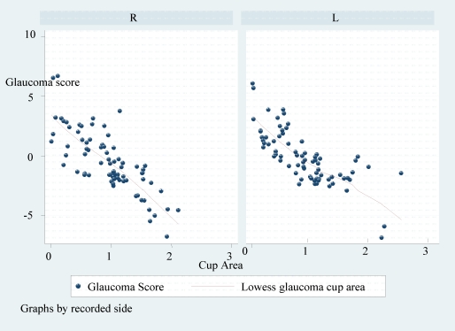
Figure 1 depicts the comparison in the correlation of the measurements of the optic nerve head parameters between the right and left eye.
Figure 1.
Lowess line plot showing the correlation of glaucoma score with the cup area between the right and left eye
There was a strong correlation of all the HRT II parameters between the right and the left eyes; the disc area mean value of right and left eyes was significantly positively correlated with inter eye absolute difference in the cup area, rim area, cup volume, and the rim volume. Absolute inter eye parameter between the two eyes were significantly positively correlated with disc area (p < 0.05).
The mean follow up period documented was 9.5±2.8 months (range of 6 to 28months). The comparison of the correlation of optic disc cup area with glaucoma probability score at presentation and at follow-up is as shown in Figure 2. A significant positive correlation was observed between the glaucoma probability score analysis and the cup disc area ratio, cup area and the rim area at presentation and follow-up. Spearman correlation coefficient [rs] =0. 967, p < 0.001
Figure 2.
Lowess line showing the comparisons of the correlation of glaucoma score with the cup area in clustered optic nerve head parameters at presentation and at follow up.
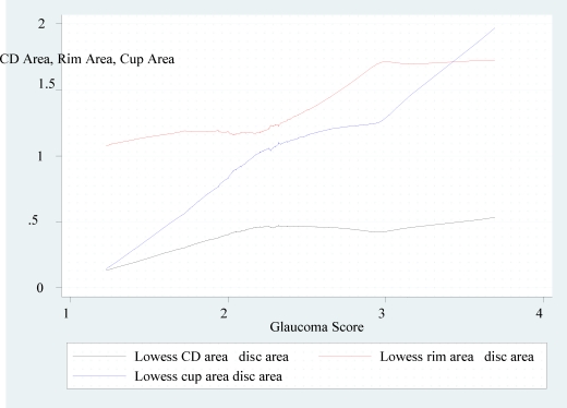
Graphical representation of the overall correlation between the glaucoma probability score analysis and other optic disc parameters were as shown in figure 3. The cup disc area ratio, cup area, and the rim area ratio are all positively correlated with the glaucoma probability score but the power of correlation is strongest with cup area followed by the rim area and weaker with the cup disc area ratio.
Figure 3.
Linear graph showing the correlations between the glaucoma score analysis and the cup disc area ratio, cup area and the rim area
Discussion
This study reported the descriptive statistics of the Heidelberg retina tomogram (Heidelberg Engineering HRT II) parameters in a clinic population of primary open angle glaucoma patients. There was no statistically significant difference between gender and within age-groups of the study population. Several researches had documented the optic nerve head characteristics in normal subjects and in disease with no significant gender nor age difference reported.10–14 The study population consisted mainly of an Arabian population dwelling in Tunisia, North African. A study conducted in the United states using HRT II on normal subjects documented that the disc area, cup area, cup to disc area ratio, cup volume, and maximum cup depth were significantly greater in African-Americans than in the whites with intermediate values for the Asians and Hispanics, though no significant sex predilection was documented.10 This study focused mainly on patients with a diagnosis of primary open angle glaucoma. Previous researchers have documented optic nerve head morphometry in eyes with POAG, ocular hypertension and normal eyes, while some have reported the correlation between these morphological findings in different topographical imaging technique.13–16
Considering the inter-correlation between the various disc parameters, a positive statistically significant correlation was found between the cup area, cup disc area ratio, rim area, mean cup depth and disc area. Other workers have documented similar findings in their studies.17–21 Several baseline topographic optic disc measurements were significantly associated with the development of POAG in both univariate and multivariate analyses, including larger cup-disc area ratio, mean cup depth, mean height contour, cup volume, reference plane height, and smaller rim area, rim area to disc area, and rim volume. This study revealed a strong positive correlation between the disc area and the rim area, cup area, cup disc area, and mean cup depth. Large discs had been known to be associated with larger cup, rim area and cup disc area ratio. Knowledge of the patient's disc area is of relevance when screening for primary open-angle glaucoma, as the vertical cup/disc ratio depends on the disc area.19–21 The result of this study showed variations in the power of positive correlation between the glaucoma probability score and other disc parameters. The cup area had the strongest correlation, while the rim are was also more strongly correlated with the glaucoma score than the cup disc area ratio. Britton, Jonas and other researchers also documented a similar report in their various studies.22–23
Moreover, it was found that the rim volume, height variation and the mean retinal nerve fibre layer were not significantly correlated with the disc area, though the disc area was negatively correlated with glaucoma diagnosis score, the correlation was not significant. Also the cup area was strongly positively correlated with cup disc area ratio but negatively correlated with the rim area, while it had no correlation with other disc parameters. The glaucoma probability score was significantly strongly positively correlated with the cup area, cup disc area ratio, rim area, mean cup depth and rim volume but it had no significant correlation with the disc area and height variation.
Many other studies have reported that the characteristics of optic nerve head are usually well correlated between right and left eyes in normal subjects.15,16 In the present report, absolute inter-eye differences in several HRT parameters were strongly or moderately correlated between the two eyes. Pertinent to diagnosis of primary open angle glaucoma are certain criteria such as a difference in the vertical cup-disc ratio of > 0.2 between the two eyes.
Optic nerve head parameter measurements of the glaucomatous eyes at presentation were similar to those of the follow-up visit. There was a significant strong positive correlation between the optic nerve head parameters at presentation and during follow-up. We can deduce from this that most of the POAG patients were well controlled on their anti-glaucoma treatment. The average follow up period documented was 2.5+/− 1.2years with a range of 6months to 5.2years.
This study is limited by its retrospective nature. Lack of detailed information on the subjects did not allow proper quantitative, statistical correlation between the clinical characteristics and CSLO features in glaucoma patients. In addition, it was not possible to relate our findings to that of HRT111 because of lack of data for the later in the study center by the time this study was conducted.
In conclusion, Confocal scanning laser tomography of the optic nerve head using HRT II is a very useful diagnostic tool in detecting and making appropriate diagnosis in primary open angle glaucoma. Most of the parameters measured are significantly strongly correlated with the diagnosis of glaucoma. The mean disc area and the height variation were not significantly related to making a diagnosis of POAG. Appropriate interpretations of the parameters studied are needed for making proper diagnosis of primary open glaucoma.
References
- 1.Varma R, Steinmann WC, Scott IU. Expert agreement in evaluating optic discs. Ophthalmology. 1992;99:215–221. doi: 10.1016/s0161-6420(92)31990-6. [DOI] [PubMed] [Google Scholar]
- 2.Burk ROW, Rohrschneider K, Takamoto T, Volcker HE, Schwartz B. Laser Scanning tomography and stereophotogrammetry in three-dimensional optic disc analysis. Graefes Arch Clin Exp Ophthalmol. 1993;231:193–198. doi: 10.1007/BF00918840. [DOI] [PubMed] [Google Scholar]
- 3.Dichtl A, Jonas JB, Mardin CY. Comparison between laser tomographic scanning evaluation and photographic measurement of the neuroretinal rim. Am J Ophthalmol. 1996;121:494–501. doi: 10.1016/s0002-9394(14)75423-6. [DOI] [PubMed] [Google Scholar]
- 4.Uchida H, Brigatti L, Caprioli J. Detection of structural damage from glaucoma with confocal scanning laser image analysis. Invest ophthalmol Vis Sci. 1996;37:2393–2401. [PubMed] [Google Scholar]
- 5.Vernon SA, Hawker MJ, Ainsworth G, et al. Laser scanning tomography of the optic nerve head in a normal elderly population: the Bridlington Eye Assessment Project. Invest ophthalmol Vis Sci. 2005;46:2823–2828. doi: 10.1167/iovs.05-0087. [DOI] [PubMed] [Google Scholar]
- 6.Durukan AH, Yucel I, Akar Y, Bayraktar MZ. Assessment of the optic nerve head topographic parameters with a confocal scanning laser ophthalmoscope. Clin Experiment Ophthalmol. 2004;32:259–264. doi: 10.1111/j.1442-9071.2004.00790.x. [DOI] [PubMed] [Google Scholar]
- 7.Uchida H, Yamamoto T, Araie M. Topographic characteristics of the optic nerve head measured with scanning laser tomography in normal Japanese subjects. Japan J Ophthalmol. 2005;49:469–476. doi: 10.1007/s10384-005-0248-2. [DOI] [PubMed] [Google Scholar]
- 8.Nakamura H, Maeda T, Suzuki Y, Inoue Y. Scanning laser tomography to evaluate optic discs of normal eyes. Japan J Ophthalmol. 1999;43:410–414. doi: 10.1016/s0021-5155(99)00089-1. [DOI] [PubMed] [Google Scholar]
- 9.Hermann MM, Theofylaktopoulos I, Bangard N, et al. Optic nerve morphometry in healthy adults using Confocal laser scanning tomography. Br J Ophthalmol. 2004;88:761–765. doi: 10.1136/bjo.2003.028068. [DOI] [PMC free article] [PubMed] [Google Scholar]
- 10.Zangwill LM, Weinreb RN, Berry CC. Confocal Scanning Laser Ophthalmoscopy Ancillary study to the Ocular Hypertension Treatment Study. Racial differences in optic disc topography: baseline result from the Confocal Scanning Laser Ophthalmoscopy Ancillary Study to the Ocular Hypertension Treatment Study. Arch Ophthalmol. 2004;122:22–28. doi: 10.1001/archopht.122.1.22. [DOI] [PubMed] [Google Scholar]
- 11.Tsai CS, Zangwill L, Gonzalez C, et al. Ethnic differences in optic nerve head topography. J Glaucoma. 1995;4:248–257. [PubMed] [Google Scholar]
- 12.Girkin CA, McGwin G, Jr, McNeal SF, DeLeon-Ortega J. Racial differences in the association between optic disc topography and early glaucoma. Invest Ophthalmol Vis Sci. 2003;44:3382–3387. doi: 10.1167/iovs.02-0792. [DOI] [PubMed] [Google Scholar]
- 13.Chi T, Ritch R, Stickler D. Racial differences in optic nerve head parameters. Arch Ophthalmol. 1989;107:836–839. doi: 10.1001/archopht.1989.01070010858029. [DOI] [PubMed] [Google Scholar]
- 14.Vernon SA, Hawker MJ, Ainsworth G, et al. Laser scanning tomography of the optic nerve head in a normal elderly population: the Bridlington Eye Assessment Project. Invest Ophthalmol Vis Sci. 2005;46:2823–2828. doi: 10.1167/iovs.05-0087. [DOI] [PubMed] [Google Scholar]
- 15.Iwase A, Suzuki Y, Araie M Tajimi Study group, author; Japan Glaucoma society, author. The prevalence of primary open-angle glaucoma in Japanese: the Tajimi Study. Ophthalmology. 2004;111:1641–1648. doi: 10.1016/j.ophtha.2004.03.029. [DOI] [PubMed] [Google Scholar]
- 16.Hermann MM, Theofylaktopoulos I, Bangard N. Optic nerve morphometry in healthy adults using Confocal laser scanning tomography. Br J Ophthalmol. 2004;88:761–765. doi: 10.1136/bjo.2003.028068. [DOI] [PMC free article] [PubMed] [Google Scholar]
- 17.Garway-Heath DF, Ruben ST, Viswanathan A, et al. Vertical cup/disc ratio in relation to optic disc size: its value in the assessment of the glaucoma suspect. Br J Ophthalmol. 1998;82:1118–1124. doi: 10.1136/bjo.82.10.1118. [DOI] [PMC free article] [PubMed] [Google Scholar]
- 18.Jonas JB, Gusek GC, Naumann GO. Optic disc, cup and neuroretinal rim size, configuration and correlations in normal eyes. Invest Ophthalmol Vis Sci. 1988;29:1151–1158. [PubMed] [Google Scholar]
- 19.Montgomery D. Clinical disc biometry in early glaucoma. Ophthalmology. 1993;100:52–56. doi: 10.1016/s0161-6420(13)31713-8. [DOI] [PubMed] [Google Scholar]
- 20.Wollstein G, Garway-Heath DF, Hitchings RA. Identification of early glaucoma cases with the scanning laser ophthalmoscope. Ophthalmology. 1998;105:1557–1563. doi: 10.1016/S0161-6420(98)98047-2. [DOI] [PubMed] [Google Scholar]
- 21.Caprioli J, Miller JM. Optic disc rim area is related to disc size in normal subjects. Arch Ophthalmol. 1987;105:1683–1685. doi: 10.1001/archopht.1987.01060120081030. [DOI] [PubMed] [Google Scholar]
- 22.Britton RJ, Drance SM, Schulzer M, et al. The area of the neuroretinal rim of the optic nerve in normal eyes. Am J Ophthalmol. 1987;103:497–504. doi: 10.1016/s0002-9394(14)74271-0. [DOI] [PubMed] [Google Scholar]
- 23.Jonas JB, Gusek GC, Naumann GO. Optic disc, cup and neuroretinal rim size, configuration and correlations in normal eyes. Invest Ophthalmol Vis Sci. 1988;29:1151–1158. [published errata appear in Invest Ophthalmol Vis Sci 1991; 32:1893 and 1992; 32:474–5] [PubMed] [Google Scholar]


