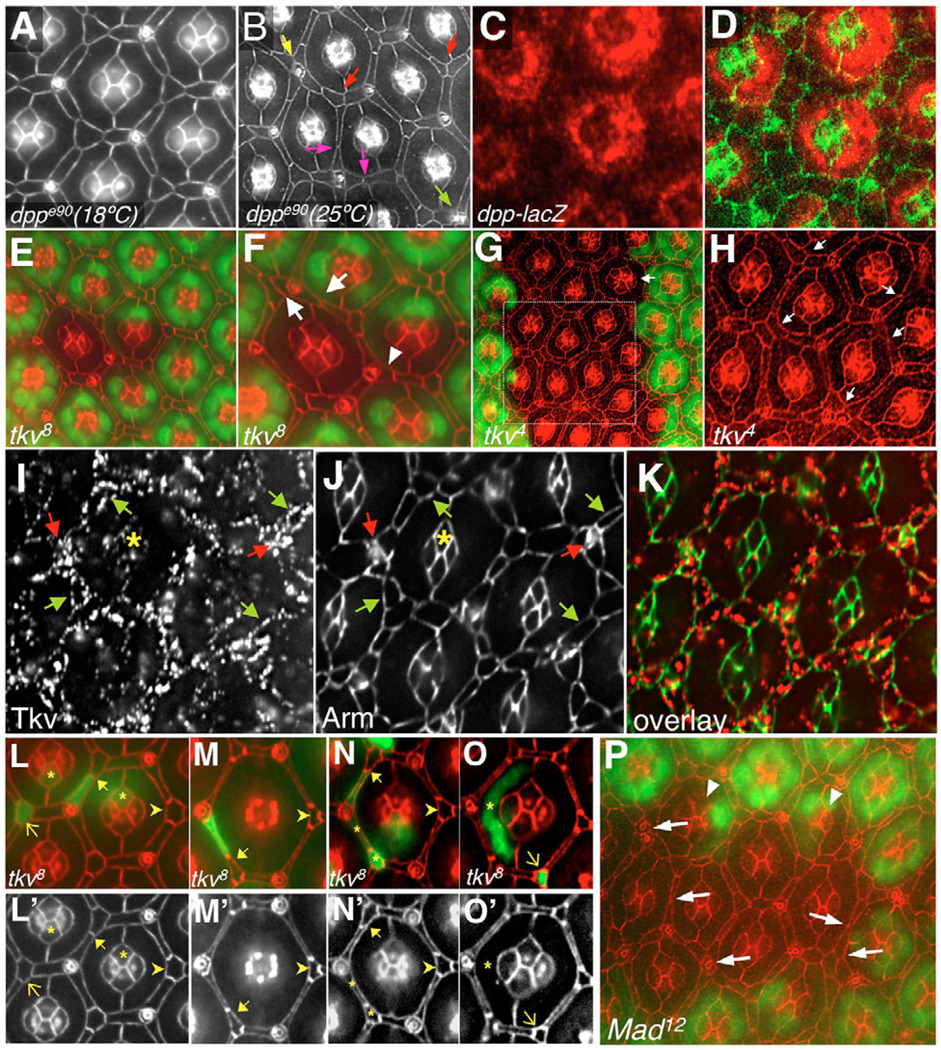
Fig. 2. Dpp signaling regulates IPC patterning in the Drosophila pupal retina.
All retinas are 42 hours APF and apical membrane profiles are highlighted with anti-Armadillo, except where noted. (A,B) Retinas from animals carrying the temperature-sensitive allele dppe90 raised at the permissive (A) or non-permissive (B) temperature. (B) Mutant retinas show: abnormal IPC:IPC contacts (pink arrows); 3°s, which failed to establish a correct position as a vertex of the hexagon (red arrows); 2°/3°s abnormally arranged around sensory bristles (yellow arrow); and aberrant bristle-bristle contacts (green arrow). (C,D) dpp was expressed in primary pigment cells at 26 hours APF (red). (E,F) tkv8 clone marked by the absence of nuclear GFP (green). (F) Magnification of the clonal tissue in E. Arrows in F point to typical 2°/3°s patterning defects; arrowhead points to ectopic 2°/3°. (G,H) tkv4 clone (cells marked as in E,F). H is a magnified view of the boxed region in G. Arrows in H indicate examples of typical 2°/3° defects. Arrow in G indicates a rare cone cell defect (five versus four). (I–K) Tkv localization at 26 hours APF. (I) Tkv protein was found primarily at the surface of IPCs (green arrows), sensory bristles (red arrows) and at lower levels in cone cells (asterisk). (L–O′) tkv8 single-cell clones were marked by the presence of GFP (green). Full arrows point to cases where tkv mutant 2°s failed to fully expand into their proper niche, as evidenced by their shortened apical profile, while wild-type neighboring 3°s elongated to compensate. Arrowheads point to examples of the apical profile characteristic of wild-type 3°s. Asterisks in N and N′ indicate how neighboring mutant cells typically show normal apical profiles. Thin arrows and asterisks in L,L′,O,O′ point to 3°s, cone cells and primary pigment cells, whose shape was not affected by the absence of Tkv activity. (P) A Mad12 clone marked by the absence of nuclear GFP (green). The arrows point to a subset of the 2°/3°s patterning defects and aberrant bristle-bristle contacts within the clone. Arrowheads indicate rare, abnormal cone cell clusters.

