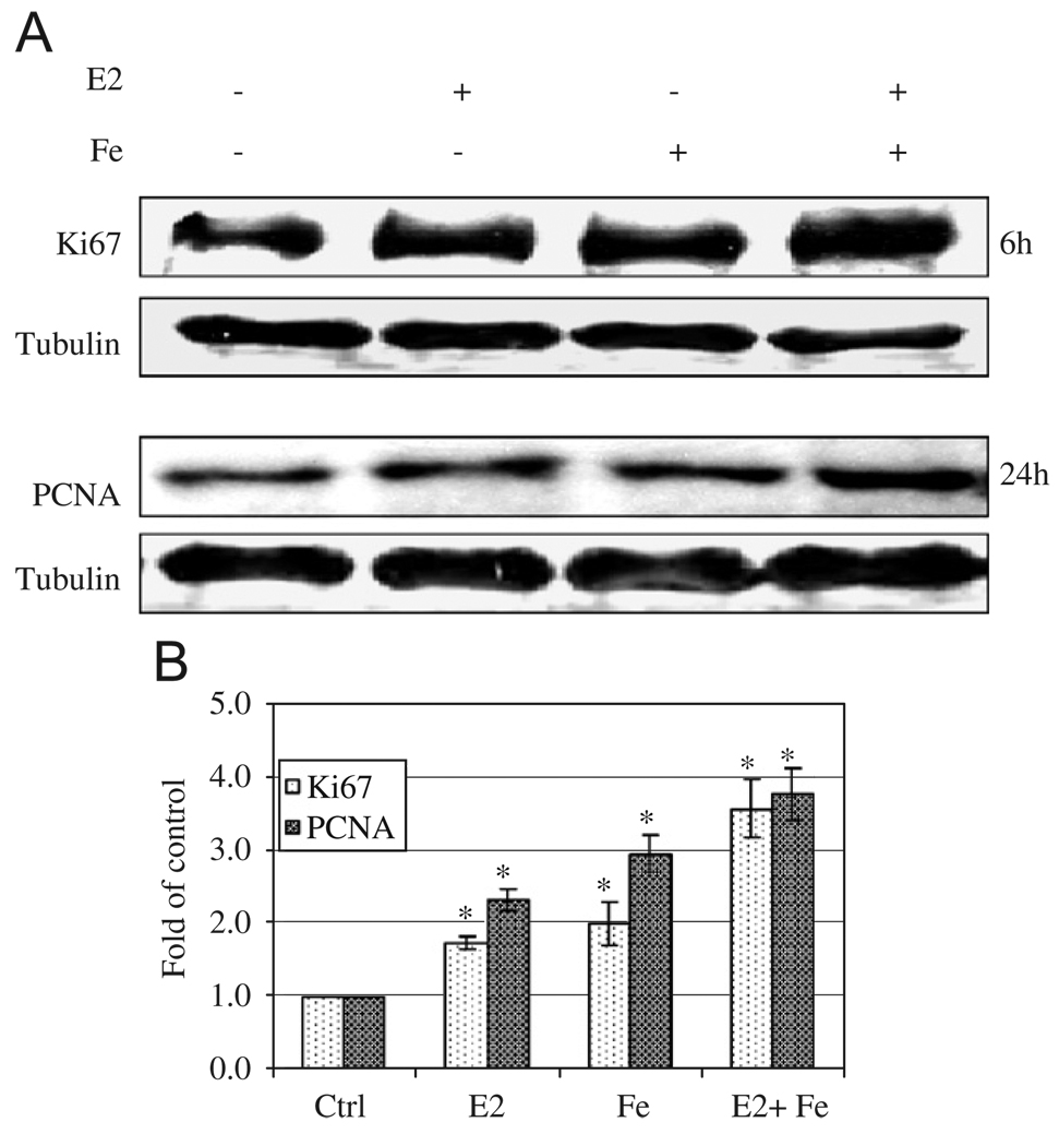
Fig. 5.
Enhancing effects of E2 and iron on Ki67 and PCNA, markers of cell proliferation. MCF-7 cells grown in 0.1% FBS α-MEM were treated with E2 (10−9 M), iron (10 µM) or E2 + iron cultured in 6-well dish for 6 and 24 h. Cells were collected and lysed using M-Per lysis buffer. Thirty µg of protein samples were loaded to each well for Ki67, PCNA, and β-tubulin Western blotting. A representative gel was shown from three replicates (A) and folds of increase over controls were shown in (B) after quantification by densitometry. *Significantly different from control for both Ki67 and PCNA, p < 0.05.

