Abstract
Given the reported common occurrence of Demodex dermatitis in the general population, Demodex dermatitis—considered as a separate condition from rosacea and seborrheic dermatitis—was evaluated in a retrospective case analysis.
Demodex folliculorum and Demodex brevis (collectively referred to as Demodex) are the most common permanent ectoparasites in man, occurring in 10 percent of skin biopsies and 12 percent of follicles.1,2 These commensal mites have four short pairs of legs on the anterior third of their bodies, are 0.3 to 0.4mm in length, and have a 14-day life cycle.1,3 Identification of these mites dates back to 1841 for D. folliculorum and 1963 for D. brevis.2,4 D. folliculorum is usually found in the upper canal of the pilosebaceous unit at a density of ≤5 organisms/cm2 and uses sebum as nourishment.5,6 Several mites, with heads directed toward the fundus, usually occupy a single follicle.2,7 D. brevis, on the other hand, burrows deeper into the sebaceous glands and ducts.1 Demodex is typically found on the face—cheeks, nose, chin, forehead, temples—and also on the balding scalp, neck, and ears.1,2 Although D. folliculorum is the more common of the two mites, D. brevis has a wider distribution on the body.2 Prevalence of both species increase with age, with men being more heavily colonized than women (23% vs 13%) and harboring more D. brevis than women (23% vs 9%).2 Presumably, Demodex passes to newborns through close physical contact after birth; however, due to low sebum production, infants and children lack significant Demodex colonization.1 Presence of mites in adolescents and young adults continues to be surprisingly low but increases from the second decade to the sixth decade of life and remains steady through the eighth decade.1,2 Demodex does not appear to be associated with acne vulgaris.8 Penetration of Demodex into the dermis or, more commonly, an increase in the number of mites in the pilosebaceous unit of >5/cm2, is thought to cause infestation, which triggers inflammation.5,9 Increased mite density has been correlated with increased readiness of lymphocytes to undergo apoptosis and with increased number of NK cells with Fc receptors.10 Significant decrease in absolute numbers of lymphocytes and T-cell subsets and significant increase in IgM levels have also been found in patients presenting with Demodex proliferation and facial skin lesions.11 These findings suggest that colonization of the skin with Demodex could be a reflection of the immune response of the host to the organism.10 Furthermore, Lacy et al12 recently reported that antigenic proteins related to a bacterium isolated from a D. folliculorum mite, Bacillus oleronius, have the potential to stimulate an inflammatory response in patients with papulopustular rosacea.12 Demodex is not easily detected in histological preparations5; therefore, skin surface biopsy (SSB) technique with cyanoacrylic adhesion is a commonly used method to measure the density of Demodex. It allows the collection of the superficial part of the horny layer and the contents of the pilosebaceous follicle13; however, it can fail to collect the complete biotope of D. folliculorum.9 Other sampling methods used in assessing the presence of Demodex by microscopy include adhesive bands, skin scrapings, skin impressions, expressed follicular contents, comedo extraction, hair epilation, and punch biopsies.7,14 The resulting number of mites measured varies greatly depending on the method selected.7 With modern and more sensitive assays, the prevalence of Demodex in skin samples approaches 100 percent; therefore, mere presence of Demodex does not indicate pathogenesis. Rather, of more importance in diagnosing Demodex pathology is the density of mites or their extrafollicular location.14
Numerous studies have reported elevated Demodex density in patients with rosacea and nonspecific signs and symptoms of facial dermatitis. Forton and Seys reported a mean mite density of 10.8/cm2 in 49 patients with rosacea and a density of 0.7/cm2 in 45 control patients.5 Similarly, Karincaoglu et al15 observed that, compared with control individuals, patients presenting with nonspecific facial symptoms such as facial pruritus with or without erythema, a seborrheic dermatitis-like eruption, perioral dermatitis-like lesions, and papulopustular and/or acneiform lesions without telangiectasia, flushing, or comedones had a significantly higher median mite density.15 Interestingly, examination of 388 follicles revealed the presence of Demodex in 42 percent of inflamed follicles and only 10 percent of noninflamed follicles (P=0.001).16 Increased number of Demodex mites has also been observed in perioral dermatitis, Grover's disease, eosinophilic folliculitis, blepharitis, papulovesicular facial eruptions, and papulopustular scalp eruptions.10,16
In 1961, Ayres and Ayres described a dry type of rosacea, which they termed rosacea-like demodicidosis.17 They proposed that this type of rosacea—characterized by flushing, dryness, numerous follicular scales, and a few usually small superficial vesicopustules—is caused by the proliferation of massive amounts of D. folliculorum, which responds favorably to treatment with a scabicide. Ayres later proposed that failure to wash the face and overuse of oily or creamy preparations supplies the Demodex mites with extra lipid nourishment. Secondarily, this promotes reproduction of mites in large numbers, which plugs the pilosebaceous ducts, leading to a rosacea-like facial eruption.18 Although elevated levels of Demodex occur in patients with rosacea and other facial skin conditions, no studies have proven a definitive relationship. It remains unknown if Demodex is the underlying cause of these conditions or if Demodex mite density increases due to inflammation of affected follicles.16
To complicate matters, clinical presentation of Demodex infestation is similar to that of rosacea and seborrheic dermatitis—facial flushing/blushing, erythema, telangiectasia, scaling and facial skin roughness on palpation, and centrofacial inflammatory lesions. Demodex dermatitis may in fact be distinct from rosacea and seborrheic dermatitis, as reported by one group.19
Given the high reported occurrence of Demodex dermatitis in the general population—2.4 cases/week in otherwise healthy individuals and the ninth most frequent diagnosis in 3,213 patients presenting with follicular scales and telangiectasia20—we further investigated the possibility that Demodex dermatitis is a separate condition from rosacea and seborrheic dermatitis. Advancing the knowledge of Demodex dermatitis, its occurrence, and treatment, will allow us to provide better and timelier care to patients who present with common symptoms of facial dermatitis.
This report represents a retrospective case review of patients treated with topical crotamiton 10% cream/lotion as monotherapy for a diagnosis of Demodex dermatitis involving the face. The diagnosis was made based on clinical grounds with or without supportive microscopy. Many of the subjects had been previously treated for rosacea with a variety of medical therapies and remained either unresponsive or were not satisfied with their previous response to treatment.
The methodology used in this report represents comprehensive clinical evaluation of the visible signs of the facial eruption as would be performed in “real world” clinical practice as opposed to more rigid criteria used only in formal clinical research. All cases were from the practice of a single investigator (JBB) allowing for greater consistency in clinical assessments at both baseline and follow up. Importantly, a controlled, prospective, randomized study evaluating Demodex dermatitis and treatment with topical crotamiton would be an important step in confirming the observations noted in this retrospective case analysis.
Patients and Methods
Medical records from 63 patients with unresolved facial redness/rash and visiting a private dermatology practice between June 2006 and August 2008 were examined retrospectively. The most common reasons for the initial dermatologic visit for these patients included follow-up visits for rosacea and seborrheic dermatitis; facial redness, rash, or itching; acne; and/or acneiform lesions. Nearly all patients presented with facial (i.e., cheeks, forehead, and/or chin) erythema (83%), followed by scaling (65%), dryness (56%), presence of papules or pustules (22%), greasiness (11%), roughness (10%), itchiness (6%), and thickening of the skin (5%). Initial observations identified a presence of rosacea, seborrheic dermatitis, or Demodex dermatitis.
When deemed appropriate, skin scrapings from the cheeks, forehead, chin, and/or neck were taken. Potassium hydroxide 10% solution (KOH) was applied to the glass side containing the scraping specimen, which was examined microscopically for the presence of Demodex mites. All patients included in this retrospective analysis were treated for Demodex dermatitis involving the face even though some tested negative for Demodex mites and others were not tested. The decision to treat these patients empirically was based on past medical history, recurring symptoms, and overall examination findings.
At both baseline and follow up, patients were evaluated using a global evaluation with comprehensive clinical assessment of visible signs of their facial eruption simulating “real world” clinical practice. The parameters included in the comprehensive clinical assessment were erythema, dryness, scaling, roughness, and/or papules/pustules. The grading scale at baseline and each follow-up visit was no improvement, some improvement (>25%), marked improvement (>50%), dramatic improvement (>75%), and complete improvement (100%).
Results
Patient demographics. Patients were between the ages of 15 and 85, with a mean age of 50.4 years and a median age of 51 years. The study group comprised 44.4 percent men and 55.6 percent women. All patients were Caucasian. Of 60 patients, 30 (50%) had a positive KOH test for Demodex mites, 17 patients (28.3%) had a negative test, and 13 patients (21.7%) were not tested. Records of the baseline visit of three patients were not available. All patients were seen for at least one follow-up visit after the baseline visit. The average time interval from the baseline visit to the first follow-up visit was 18.6 days (range 13–55 days). Half of the patients (32 of 63) were also seen for a second follow-up visit an average of 38.5 days (range 24–72 days) after the baseline visit or an average of 15.5 days (13–59 days) after the first follow-up visit.
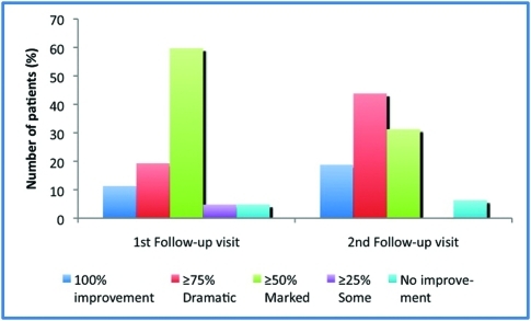
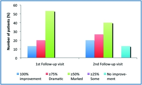
First follow-up visit. Use of crotamiton was beneficial in nearly all patients. At the first follow-up visit after baseline, 90.6 percent of patients (56/62) experienced at least a 50-percent reduction in erythema, dryness, scaling, roughness, and/or papules/pustules, and 4.8 percent of patients (3/62) experienced some improvement in symptoms compared with the baseline visit (Figure 1). Only three patients did not improve or remained the same. Of the 30 patients who tested negative for Demodex or were not tested with KOH, 87 percent (26/30) showed at least a 50-percent improvement in facial skin conditions (Figure 2).
Figure 1.
Facial skin improvement (all patients)
N=62 for first follow-up visit; N=32 for second follow-up visit
Figure 2.
Facial skin improvement in patients treated empirically (negative KOH test or not tested)
N=30 for first follow-up visit; N=15 for second follow-up visit
Nine of the 63 patients discontinued crotamiton after the first follow-up visit—two due to overwhelming improvements, three due to lack of improvement, and one due to burning. For 6 of the 63 patients, the dosage of crotamiton was lowered to once daily in the evening hours due to dramatic improvements.
Second follow-up visit. At the second follow-up visit, 93.8 percent of patients (30/32) experienced at least a 50-percent reduction in erythema, dryness, scaling, roughness, and/or papules/pustules compared with the baseline visit (Figure 1). The remaining two patients showed no improvement. Of the patients testing negative for Demodex or not tested at the baseline visit and seen at a second follow-up visit, nearly 9 out of 10 (86.7%) experienced at least a 50-percent improvement in erythema, dryness, scaling, roughness, and/or papules/pustules compared with observations at the baseline visit (Figure 2).
Due to the dramatic improvement in skin conditions, crotamiton was discontinued in 24 percent of patients and the dosage lowered from twice daily to once daily in 31 percent of patients at the second follow-up visit. Of the two patients who discontinued crotamiton after the first follow-up visit because of complete clearing, one maintained improved results and the other had no follow-up visit.
Improvement in erythema, dryness, scaling, roughness, and/or papules/pustules. Figures 3 to 7 illustrate the reduction in erythema, dryness, scaling, roughness, and/or papules/pustules from five representative patients treated with topical crotamiton twice daily. Selection of patients includes those who tested positive for the presence of Demodex and those treated empirically after a negative test or no test.
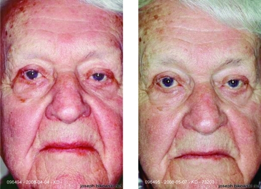
Figure 3.
Left photo: Baseline visit. Examination revealed red, dry, scaling and thickening of the skin on the forehead, nose, cheeks, and chin. Right photo: First follow-up visit one month later. Marked improvement on central third of face with topical crotamiton twice daily.
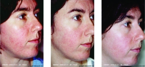
Figure 7.
Left photo: Baseline visit. Ten-year history of erythema and scaling on forehead, nose, cheeks, and chin. Middle photo: 16 days after baseline and topical crotamiton twice daily, at the first follow-up visit. At least a 75-percent improvement in erythema. Left photo: 30 days after baseline and topical crotamiton twice daily, at the second follow-up visit. Marked improvement, with a reduction in erythema on the forehead, nose, cheeks, and chin.
The patient (male, age 85 years) seen in Figure 3 presented with a facial “rash.” Palpation and examination revealed erythema, dryness, scaling, and thickening of the skin on the forehead, nose, cheeks, and chin. KOH test for Demodex mites was negative; however, clinical appearance and symptoms strongly suggested the possibility of Demodex infestation. The patient was treated with topical crotamiton twice daily, instructed to use a moisturizer, and returned four weeks later for a follow-up visit. The patient showed marked improvement in the central third of the face and was instructed to continue crotamiton application twice daily. A second follow-up visit four weeks later showed marked reduction in erythema, with no evidence of erythema detected on the forehead, nose, cheeks, and chin. Crotamiton application was decreased to once daily at bedtime.
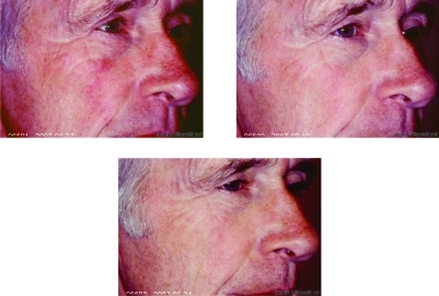
The patient (male, age 65 years) seen in Figure 4 presented with a four-month history of a nonpruritic eruption that started with erythema, scaling, and palpable dryness on the right and left infraorbital ridge and right malar eminence. The patient had a history of seborrheic dermatitis and tested positive for two Demodex mites on the right malar eminence. After two weeks of using topical crotamiton twice daily and a moisturizer, the patient experienced at least a 50-percent reduction in erythema. No objective evidence of erythema was detected elsewhere, including the forehead, left cheek, left malar eminence, and chin. Crotamiton application was continued twice daily for another two weeks. At the second follow-up visit, the patient showed at least a 75-percent reduction in erythema and dryness across the malar eminence (right side more so than left side). No objective evidence of erythema was detected elsewhere, including the forehead, cheeks, and chin. Application of crotamiton was reduced to once daily at bedtime.
Figure 4.
Left photo: Baseline visit. Erythema, dryness, and scaling on the left and right infraorbital ridge and right malar eminence. Patient had a history of seborrheic dermatitis and tested positive for two Demodex mites. Right photo: Two weeks after baseline and topical crotamiton twice daily, at the first follow-up visit. At least a 50-percent reduction in erythema. Bottom photo: Four weeks after baseline and crotamiton twice daily, at the second follow-up visit. Marked improvement, with 75-percent reduction in erythema and dryness across the malar eminences.
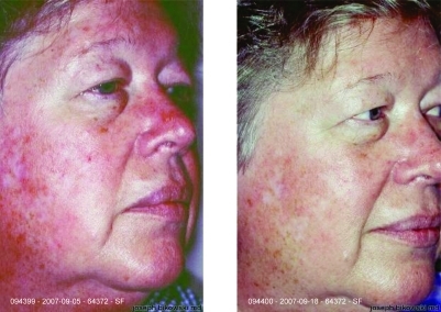
The baseline visit for the patient (female, age 61 years) in Figure 5 was a follow up for rosacea that remained unchanged. Erythema was present along with roughness on the forehead, nose, cheeks, and chin on palpation. The patient was treated empirically with topical crotamiton twice daily and a moisturizer. Two weeks later, the patient returned with a marked decrease in erythema, dryness, and scaling across the forehead, cheeks, and chin and was told to continue crotamiton twice daily. A second follow-up visit four weeks later revealed further improvement in erythema, dryness, and scaling across the forehead, cheeks, and chin. Crotamiton application was reduced to once daily. Interestingly, this patient had an irritant reaction to permethrin 5% previously, but tolerated crotamiton without difficulty.
Figure 5.
Left photo: Baseline visit. Examination revealed erythema and roughness on palpation of forehead, nose, cheeks, and chin. Right photo: First follow-up visit two weeks later with topical crotamiton application twice. Marked decrease in erythema, dryness, and scaling across forehead, cheeks, and chin.
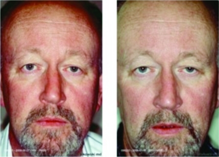
Figure 6 documents a 61-year-old male patient who presented with a history of long-standing erythematous, dry, scaling eruptions across the forehead, nose, cheeks, and chin. The patient was treated empirically for Demodex folliculitis and prescribed topical crotamiton twice daily on the entire face along with a moisturizing cleanser. Follow up two weeks later revealed no objective evidence of disease on the forehead and a decrease in erythema on the forehead, cheeks, chin, and dorsum of the nose. Crotamiton was continued twice daily.
Figure 6.
Left photo: Baseline visit. Examination revealed erythematous, dry, scaling eruption across the forehead, nose, cheeks, and chin. Right photo: First follow-up visit after 14 days of topical crotamiton twice daily. Decrease in erythema in the forehead, cheeks, chin, and dorsum of the nose-essentially no objective evidence of disease, with the exception of some roughness and dryness on the forehead.
The patient in Figure 7, a 34-year-old female, had a 10-year history of a red and itchy face with erythema and scaling on the forehead, nose, cheeks, and chin. She was prescribed topical crotamiton twice daily along with a moisturizer and gentle cleanser. KOH test was negative for the presence of Demodex. On follow up 16 days later, the patient demonstrated at least a 75-percent improvement in facial skin condition. Crotamiton was continued twice daily until the second follow-up visit 14 days later. Treatment resulted in a marked decrease in erythema on the forehead, nose, cheeks, and chin. Dosage of topical crotamiton was decreased to once daily.
Overall, patients with recurrent, unchanged erythema, dryness, scaling, roughness, and/or papules/pustules with or without evidence of Demodex mites markedly improved with the use of crotamiton twice daily. These findings strongly suggest that empirical treatment for Demodex dermatitis using crotamiton is beneficial for patients with these unresolved signs and symptoms. Since oftentimes rosacea, seborrheic dermatitis, and Demodex dermatitis cannot be distinguished from each other in patients with a red face, topical crotamiton should be considered as a component of the patient's initial treatment regimen.
Discussion
Use of topical crotamiton twice daily improved signs of facial dermatitis—i.e., erythema, dryness, scaling, roughness, and/or papules/pustules—in nearly all patients, regardless of the results of the KOH test. Nine out of 10 patients experienced at least a 50-percent improvement when examined at the first follow-up visit, about two weeks after the baseline visit. Similar results were observed at the second follow-up visit. Remarkably, of the 30 patients who tested negative for the presence of Demodex mites with KOH or who were treated empirically, 87 percent experienced at least a 50-percent improvement in facial skin eruptions at the first follow-up visit, and the same percentage experienced a similar improvement at the second follow-up visit.
Of the 63 patient charts examined, only two patients had a positive KOH test but did not respond to topical crotamiton treatment. One patient had problems with pruritic eruptions along with erythematous, dry, scaling eruptions; therefore, the underlying cause of erythema may be due to contact dermatitis rather than Demodex dermatitis. The other patient had been treated for rosacea for more than a year prior to initiating topical crotamiton. Rosacea had dramatically improved with azelaic acid 15%, but confluent erythema across the malar eminences and dorsum of the nose still persisted. It is likely that the continuing erythema is not a result of meaningful Demodex proliferation.
The fact that patients testing negative for Demodex mites and/or treated empirically with topical crotamiton improved, suggests that KOH testing may be unpredictable in identifying a meaningful presence of these mites. Our results, similar to those described by Ayres and Ayres, suggest that the presence of facial erythema, dryness, scaling, and roughness with or without papules/pustules is possibly due to proliferation of D. folliculorum,17 as evidenced by the improvement obtained with the use of topical crotamiton twice daily. Ayres separated this condition from seborrheic dermatitis and rosacea and coined the term rosacea-like demodicidosis.17,18 Based on our findings, we agree with these authors and with Pollotta et al19 that Demodex dermatitis is a separate condition from rosacea and seborrheic dermatitis.
In summary, our results demonstrate that presence of facial erythema, dryness, scaling, and roughness with or without papules/pustules may be a result of D. folliculorum proliferation and, in fact, is a separate condition from seborrheic dermatitis and rosacea. Patients with these skin conditions markedly improved with the use of topical crotamiton twice daily, regardless of results from a KOH test for the presence of Demodex mites. Crotamiton also possesses antipruritic properties, which may be helpful in cases associated with pruritis. Based on these findings, we recommend the use of topical crotamiton twice daily in patients with a chronic history of, or who present with, facial erythema, dryness, scaling, and roughness with or without papules/pustules.
References
- 1.Basta-Juzbasic A, Subic JS, Ljubojevic S. Demodex folliculorum in development of dermatitis rosaceiformis steroidica and rosacea-related diseases. Clin Dermatol. 2002;20(2):135–140. doi: 10.1016/s0738-081x(01)00244-9. [DOI] [PubMed] [Google Scholar]
- 2.Aylesworth R, Vance C. Demodex folliculorum and Demodex brevis in cutaneous biopsies. J Am Acad Dermatol. 1982;7:583–589. doi: 10.1016/s0190-9622(82)70137-9. [DOI] [PubMed] [Google Scholar]
- 3.Spickett SG. Studies on Demodex folliculorum Simon (1942). Life history. Parasitology. 1961;51:181–192. [Google Scholar]
- 4.Akbulatova LK. The pathogenic role of Demodex mite and the clinical form of demodicosis in man. Vest Derm Vener, Moscow. 1963;40:57–61. [PubMed] [Google Scholar]
- 5.Forton F, Seys B. Density of Demodex folliculorum in rosacea: a case-control study using standardized skin-surface biopsy. Br J Dermatol. 1993;128(6):650–659. doi: 10.1111/j.1365-2133.1993.tb00261.x. [DOI] [PubMed] [Google Scholar]
- 6.Baima B, Sticherling M. Demodicidosis revisited. Acta Derm Venereol. 2002;82(1):3–6. doi: 10.1080/000155502753600795. [DOI] [PubMed] [Google Scholar]
- 7.Bonnar E, Eustace P, Powell FC. The Demodex mite population in rosacea. J Am Acad Dermatol. 1993;28(3):443–448. doi: 10.1016/0190-9622(93)70065-2. [DOI] [PubMed] [Google Scholar]
- 8.Okyay P, Ertabaklar H, Savk E, Erfug S. Prevalence of Demodex folliculorum in young adults: relation with sociodemographic/hygienic factors and acne vulgaris. J Eur Acad Dermatol Venereol. 2006;20(4):474–476. doi: 10.1111/j.1468-3083.2006.01470.x. [DOI] [PubMed] [Google Scholar]
- 9.Forton F, Song M. Limitations of standardized skin surface biopsy in measurement of the density of Demodex folliculorum. A case report. Br J Dermatol. 1998;139(4):697–700. doi: 10.1046/j.1365-2133.1998.02471.x. [DOI] [PubMed] [Google Scholar]
- 10.Akilov OE, Mumcuoglu KY. Immune response in demodicosis. J Eur Acad Dermatol Venereol. 2004;18(4):440–444. doi: 10.1111/j.1468-3083.2004.00964.x. [DOI] [PubMed] [Google Scholar]
- 11.el-Bassiouni SO, Ahmed JA, Younis AI, et al. A study on Demodex folliculorum mite density and immune response in patients with facial dermatoses. J Egypt Soc Parasitol. 2005;35(3):899–910. [PubMed] [Google Scholar]
- 12.Lacey N, Delaney S, Kavanagh K, Powell FC. Mite-related bacterial antigens stimulate inflammatory cells in rosacea. Br J Dermatol. 2007;157(3):474–481. doi: 10.1111/j.1365-2133.2007.08028.x. [DOI] [PubMed] [Google Scholar]
- 13.Marks R, Dawber RP. Skin surface biopsy: an improved technique for the examination of the horny layer. Br J Dermatol. 1971;84(2):117–123. doi: 10.1111/j.1365-2133.1971.tb06853.x. [DOI] [PubMed] [Google Scholar]
- 14.Crawford GH, Pelle MT, James WD. Rosacea: I. Etiology, pathogenesis, and subtype classification. J Am Acad Dermatol. 2004;51(3):327–344. doi: 10.1016/j.jaad.2004.03.030. [DOI] [PubMed] [Google Scholar]
- 15.Karincaoglu Y, Bayram N, Aycan O, Esrefoglu M. The clinical importance of Demodex folliculorum presenting with nonspecific facial signs and symptoms. J Dermatol. 2004;31(8):618–626. doi: 10.1111/j.1346-8138.2004.tb00567.x. [DOI] [PubMed] [Google Scholar]
- 16.Vollmer RT. Demodex-associated folliculitis. Am J Dermatopathol. 1996;18(6):589–591. doi: 10.1097/00000372-199612000-00006. [DOI] [PubMed] [Google Scholar]
- 17.Ayres S Jr, Ayres S III. Demodectic eruptions (demodicidosis) in the human. 30 years' experience with two commonly unrecognized entities: Pityriasis folliculorum (Demodex) and acne rosacea (Demodex type) Arch Dermatol. 1961;83:816–827. doi: 10.1001/archderm.1961.01580110104016. [DOI] [PubMed] [Google Scholar]
- 18.Ayres S., Jr Demodex folliculorum as a pathogen. Cutis. 1986;37(6):441. [PubMed] [Google Scholar]
- 19.Pallotta S, Cianchini G, Martelloni E, et al. Unilateral demodicidosis. Eur J Dermatol. 1998;8(3):191–192. [PubMed] [Google Scholar]
- 20.Forton F, Germaux MA, Brasseur T, et al. Demodicosis and rosacea: epidemiology and significance in daily dermatologic practice. J Am Acad Dermatol. 2005;52:74–87. doi: 10.1016/j.jaad.2004.05.034. [DOI] [PubMed] [Google Scholar]


