With symptomatic complete heart block, the principal circulatory derangements are secondary to bradycardia.3, 4 Effective therapy, therefore, depends on increasing the ventricular rate. This has been achieved with pharmacologic agents or with electrical stimulation of the ventricles with internal or external pacemakers.
Recently, a different approach has been suggested by Ernst,1 based on the proposition that auriculo-ventricular conduction can be restored by transplantation of the sino-atrial (SA) node into the ventricular myocardium. Although the results of his studies with dogs supported the potential clinical use of this technique, Ernst took care to point out that he had not been able to carry out the crucial experiment necessary to establish its value. Such an experiment would necessitate the production of proved permanent complete heart block, and its subsequent effective treatment with SA nodal transplantation.
In the present study, SA nodal transplantation was carried out under circumstances in which success or failure could be ascertained with certainty. Complete heart block was established in 9 dogs, 2 to 9 weeks before nodal transplantation, to establish the fact that the heart block was permanent. The SA node was then transplanted, and the animals were observed for as long as 3 months for evidence of restored auriculo-ventricular conduction.
METHODS
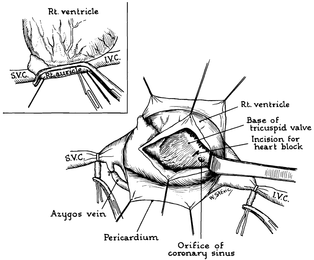
Thirty-seven mongrel dogs, weighing from 10 to 20 kilograms, were used. With the use of a previously described technique,4 complete heart block was produced (Fig. 1) with placement of the atriotomy so as to avoid the nutrient artery to the SA node.
Fig. 1.
First operation. Production of complete heart block by incision of the bundle of His during temporary inflow occlusion. Atrial incision is kept away from prospective pedicle.
Postoperatively, the animals were allowed to recover for an average of 28 days (range, 14 to 66 days) during which time daily pulse rates were counted and electrocardiographic confirmation of the complete heart block was obtained at intervals. In those animals with permanent idioventricular rhythm, SA nodal transplantation was carried out.
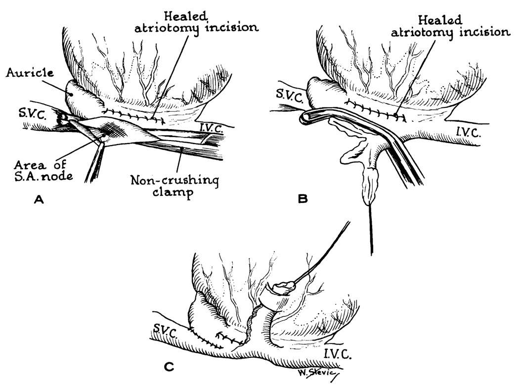
The original incision through the right fourth intercostal space was re-opened with the animal under Pentothal anesthesia. The technique of SA nodal transplantation was essentially the same as that described by Ernst.1 The SA node and its inferiorly based pedicle were drawn into a noncrushing clamp (Fig. 2, A). After dissecting the pedicle free, the resulting defect in the atrial wall was closed with continuous 5-0 arterial silk (Fig. 2, B). To insure completeness of excision of the SA node, a portion of the wall of the superior vena cava was included. This invariably produced narrowing of the inflow tract of the superior vena cava which was well tolerated.
Fig. 2.
Second operation. Technique used for S4 nodal transplant.
Vigorous bleeding from the tip of the pedicle was always observed. After noting the presence of auricular contractions throughout its length, the tip of the pedicle was implanted beneath the epicardium or within the myocardium of the right ventricular wall and fixed with a few 6-0 silk sutures (Fig. 2, C). The chest was closed with water-seal drainage. After SA nodal transplantation, pulse counts were obtained daily. Electrocardiograms were obtained every 7 to 10 days until the animals either died or were sacrificed. Complete autopsy was performed in every case. Particular attention was directed to the site of SA nodal transplantation, where multiple sections were obtained and stained with hematoxylin and eosin as well as trichrome stains.
RESULTS
Experimental Success Rate
Of the 37 dogs originally entered in the experiment, all but 9 were eventually eliminated for two reasons. (1) In 20 animals complete heart block either could not be produced or, more commonly, reversion to a sinus rhythm occurred subsequent to the original operation. (2) Eight additional animals died during or within a few days after the SA nodal transplantation.
Period of Observation After SA Nodal Transplantation
The 9 dogs meeting the full criteria of the experiment were followed (Table I) for an average of 65 days (range, 11 to 97 days). Six of the dogs were eventually sacrificed, and the other 3 died spontaneously.
TABLE I.
| NO | DAYS BETWEEN HEART BLOCK AND SA TRANSPLANT |
DAYS BETWEEN SA TRANS- PLANT AND DEATH |
HEART RATE PER MINUTE |
HEART FAILURE |
RETURN OF A-V CONDOC- TION |
|---|---|---|---|---|---|
| 1 | 28 | 38 | 24–30 | Yes | No |
| 2 | 30 | 11 | 50–55 | Yes | No |
| 3 | 19 | 77 | 32–44 | Yes | No |
| 4 | 14 | 78* | 35–55 | Yes | No |
| 5 | 14 | 97* | 48–54 | Yes | No |
| 6 | 66 | 76* | 35–55 | No | No |
| 7 | 28 | 59* | 48–50 | Yes | No |
| 8 | 27 | 66* | 45–55 | No | No |
| 9 | 24 | 84* | 50–65 | Yes | No |
Sacrificed.
Variations in Idioventricular Rhythm
Idioventricular rates ranged from 24 to 65 per minute, and in most dogs was 40 to 55 per minute. In a given animal, the rate varied little from day to day during the rest of its life. Similarly there was no electrocardiographic evidence in any dog of a change in the idioventricular rhythm after SA nodal transplant. The dissociation of auricular and ventricular beats was complete even after 97 days.
Autopsy Findings
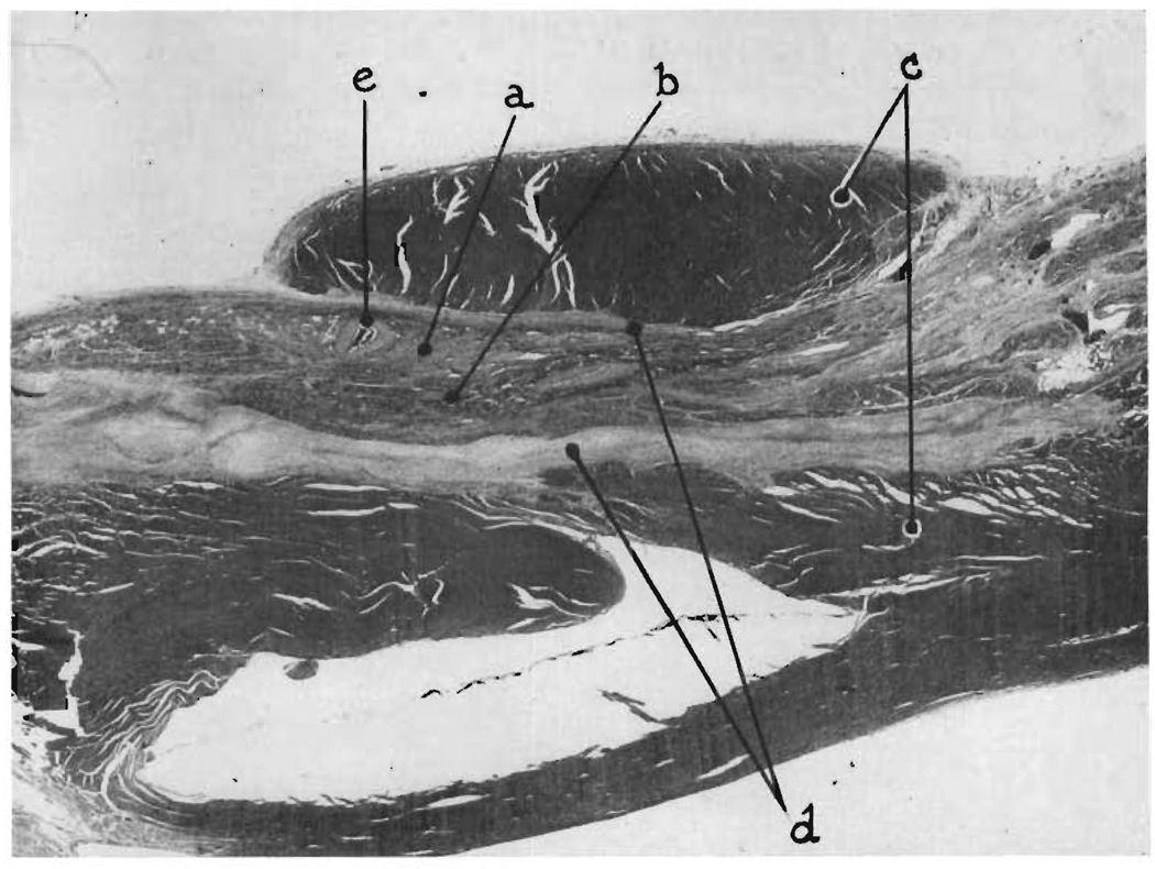
Seven of tile 9 animals had gross and/or microscopic evidence of various stages of left- and right-sided heart failure. The SA nodal transplant was always easily seen (Fig. 3), and attached to the surrounding tissues with a fibrous union. Auricular muscular or conduction tissue was identifiable, but sometimes atrophic. The subjacent myocardium often had some evidence of degeneration. Hyaline cartilage formation was occasionally observed.
Fig. 3.
Dog. No. 7, 8 weeks after SA nodal transplantation. Section through the transplantation site in the right ventricle. a, Purkinje fibers of SA. node. b, Viable auricular musole. c, Ventricular myocardium. d. Fibrous union. e. Nutrient artery.
Control Procedures
Five dogs not included in the definitive series were studied to determine (a) the effect of procainization and removal of the pedicle tip and (b) the possible role the contracting pedicle might have in mechanically stimulating the ventricle.
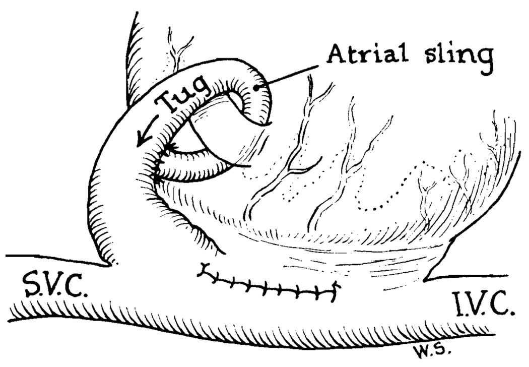
In 3 dogs, the SA nodal pedicle was prepared as described above. The tip was first injected with 1 per cent procaine and then excised. In one dog, the p waves disappeared for several hours, in another the amplitude was diminished, and, in the third, the auricular rate slowed. Similar alterations have been observed by Riberi and his associates2 with injection of procaine into the SA node. In 2 dogs, an auricular sling was devised (Fig. 4) immediately after the production of complete heart block. The ventricular contractions followed the auricular beat for 24 to 48 hours, at which time the idioventricular rate returned. At autopsy the auricular sling had become attenuated which accounted for the loss of mechanical stimulation.
Fig. 4.
Formation of auricular sling which at first mechanically stimulated ventricular beats. As the sling became attenuated, the idioventricular beat returned.
DISCUSSION
The recent ingenious experiments of Ernst,1 if confirmed, would have a far-reaching practical significance in the clinical therapy of complete heart block. However, two considerations have made it necessary to subject the method to further analysis. First, the concept that conduction can occur across the scar of the implantation site is contrary to well-accepted opinion on the requirements for cardiac excitation. Second, the experiments cited, while suggestive, did not conclusively prove the therapeutic value of SA nodal transplantation.
Aside from providing firm evidence that SA nodal transplants are not capable of ventricular stimulation, the present experiments indicate how easily systematic artefacts could be introduced into an evaluation of this sort. In order to perform a complete experiment in 9 animals, a total of 37 dogs were used. In 20 of these, permanent complete heart block could not be obtained. The usual sequence was the initial production of heart block with reversion to a sinus rhythm hours or days later. If the SA nodal transplant had been previously done, it might have been concluded that its presence accounted for the resumption of a sinus rhythm in two thirds of the animals studied.
An additional potential source of error could be mechanical stimulation of the ventricle by the mechanical tug of the transplanted pedicle. Although short-lived, this effect was demonstrated in the present study with the use of an auricular sling.
SUMMARY
SA nodal transplantation, for use in the treatment of complete heart block, has been evaluated. Complete heart block was surgically produced weeks or months before SA nodal transplantation. After transplantation, the animals were observed for as long as 97 days. Transplantation of the SA node had no influence on the pre-existent idioventricular rhythm. It is concluded that there is no justification for a clinical trial of this technique.
Acknowledgments
Aided by Grants A-6344 and A-6283 from the U. S. Public Health Service.
REFERENCES
- 1.Ernst RW. Pedicle Grafting of the Sino-Auricular Node to the Right Ventricle for the Treatment of Complete Atrioventricular Block. J. Thoracic Surg. 1962;44:681. [Google Scholar]
- 2.Riberi A, Shumacker HB, Jr, Kajikuri H, Grice PF, Boone RD. Ventricular Fibrillation in the Hypothermic State. Surgery. 1955;38:847. [PubMed] [Google Scholar]
- 3.Starzl TE, Gaertner RA, Baker RR. Acute Complete Heart Block in Dogs. Circulation. 1955;12:82. doi: 10.1161/01.cir.12.1.82. [DOI] [PMC free article] [PubMed] [Google Scholar]
- 4.Starzl TE, Gaertner RA. Chronic Heart Block in Dogs: An Experimental Study. Circulation. 1955;12:259. doi: 10.1161/01.cir.12.2.259. [DOI] [PMC free article] [PubMed] [Google Scholar]


