
Figure 4.
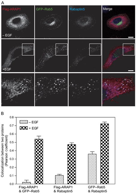
ARAP1 is recruited to a Rab5-positive compartment. A) Relative localization of ARAP1, Rab5 and Rabaptin 5. Serum-starved HeLa cells were treated with EGF for 5 min and stained for both ARAP1 and Rabaptin5, allowing visualization of ARAP1 (red), Rabaptin5 (blue) and Rab5 (green) in single cells. A higher magnification is shown in the inset. Scale bar, 10 μm. B) Colocalization of ARAP1 with Rab5 and Rabaptin5. Pearson coefficients were determined for the indicated combinations of proteins in 25 cells in which ARAP1, Rab5 and Rabaptin5 were simultaneously visualized. The errors bars represent the SEM.