Abstract
The genome of foot-and-mouth disease virus (FMDV) differs from that of other picornaviruses in that it encodes a larger 3A protein (>50% longer than poliovirus 3A), as well as three copies of protein 3B (also known as VPg). Previous studies have shown that a deletion of amino acids 93 to 102 of the 153-codon 3A protein is associated with an inability of a Taiwanese strain of FMDV (O/TAW/97) to cause disease in bovines. Recently, an Asian virus with a second 3A deletion (amino acids 133 to 143) has also been detected (N. J. Knowles et al., J. Virol. 75:1551-1556, 2001). Genetically engineered viruses harboring the amino acids 93 to 102 or 133 to 143 grew well in porcine cells but replicated poorly in bovine cells, whereas a genetically engineered derivative of the O/TAW/97 virus expressing a full-length 3A (strain A12) grew well in both cell types. Interestingly, a virus with a deletion spanning amino acid 93 to 144 also grew well in porcine cells and caused disease in swine. Further, genetically engineered viruses containing only a single copy of VPg were readily recovered with the full-length 3A, the deleted 3A (amino acids 93 to 102), or the “super” deleted forms of 3A (missing amino acids 93 to 144). All of the single-VPg viruses were attenuated in porcine cells and replicated poorly in bovine cells. The single-VPg viruses produced a mild disease in swine, indicating that the VPg copy number is an important determinant of host range and virulence. The association of VPg copy number with increased virulence in vivo may help to explain why all naturally occurring FMDVs have retained three copies of VPg.
Foot-and-mouth disease (FMD) is an extremely contagious viral disease of cattle, pigs, sheep, goats, and many wild animals. The disease is characterized by fever and vesicular lesions of the epithelium of the mouth, tongue, feet, and teats. The causal agent, FMD virus (FMDV), is a positive-stranded RNA virus that is the type species of the Aphthovirus genus of the Picornaviridae. The FMDV genome is over 8 kb in length and contains a protein cap (3B, also known as VPg) (27). During replication, the genome is expressed as a single open reading frame (ORF) that is processed into mature polypeptide products. Translation of the ORF begins with a proteinase (Lpro), which is followed in the ORF by the structural proteins (1A, 1B, 1C, and 1D), a short autoproteinase (2A), and the remaining nonstructural proteins (2B, 2C, 3A, 3B, 3Cpro, and 3Dpol). 3Cpro is responsible for proteolytic cleavage of the majority of the cleavage sites in the FMDV polyprotein (33), and 3Dpol is the core subunit of the picornavirus RNA-dependent RNA polymerase (7). Protein 3B, which is represented in three nonidentical copies in FMDV (8), is covalently bound to the 5′ end of the genome and antigenome, and functions in priming picornavirus RNA synthesis (see reference 34 for review). The functions of the nonstructural proteins 2B, 2C, and 3A are less well understood, although all three have hydrophobic domains (9, 26), and all have been found physically associated with intracellular membranes that proliferate in picornavirus-infected cells (2, 3, 29, 30, 32).
We have previously shown that a deletion in the 3A protein of FMDV is a characteristic of a virus (O/TAW/97) that devastated the Taiwanese pork industry in 1997 and that the deleted 3A is responsible for the virus' inability to cause disease in cattle (1). However, this deleted 3A does not interfere with production of an acute and highly and readily transmissible disease in pigs (5). The deletion in O/TAW/97 occurs at positions 93 to 102 of the 153-amino-acid 3A protein and is similar in size and position to deletions that were found in egg-adapted derivatives of FMDV that were developed for use as vaccines in South America (12). Interestingly, an investigation of the 3A coding regions of a number of Asian serotype O FMDVs revealed that viruses harboring this deletion have been circulating in Asia for more than 30 years and that viruses with 3As harboring a deletion spanning residues 133 to 143 have been circulating in Southeast Asia since the mid 1990s (18). Recently, Sobrino and coworkers have shown that adaptation of an FMDV isolate to cause disease in guinea pigs is dependent on a point mutation in 3A, adding further support to a critical role for 3A in determining FMDV host range and virulence (21).
The FMDV 3A protein differs markedly in size from the 3A protein of other picornaviruses (it is >50% larger than the 87-amino-acid 3A of poliovirus) (17). However, all 3A proteins contain a 15- to 20-amino-acid hydrophobic domain, located ca. 60 to 70 amino acids from the N terminus. Thus, the unique region of the FMDV 3A protein is located downstream of this hydrophobic domain, and it is this region that is altered in O/TAW/97, the egg-adapted FMDVs and in the Southeast Asian FMDVs. FMDV is also distinguished from other members of the Picornaviridae by the presence of three copies of 3B (see above). Thus, the 3ABBB coding region of prototype strains of FMDV has a length of 224 codons (9, 26), whereas the poliovirus 3AB coding region is only 109 codons in length (17). Finally, although not all three copies of FMDV 3B are needed to maintain infectivity (6), there are no reports of naturally occurring FMDV strains with fewer than three copies of 3B, a surprising finding, since FMDV is known to undergo homologous recombination (16), which should remove redundant genetic material.
Here we show that genetically engineered viruses with shortened 3As (deletions of amino acids 93 to 102 or 93 to 144) replicated poorly in bovine-derived cells but were able to replicate well in pig-derived cells in culture. Furthermore, these viruses caused a disease in pigs that was indistinguishable from the disease caused by a genetically engineered virus with a full-length 3A (shown here) and field-derived viruses (J. M. Pacheco and P. W. Mason, unpublished data). However, viruses lacking the first two copies of 3B were impaired in their ability to replicate in porcine cells in culture and caused attenuated disease in pigs. Thus, FMDV appears to have considerable flexibility in both regions of the genome, but both proteins appear to control the virus' pathogenic potential and host range.
MATERIALS AND METHODS
Cell lines, cDNAs, and viruses.
Baby hamster kidney (BHK) cells, strain 21, clone 13 (American Type Culture Collection), passages 62 to 66, were maintained as previously described (25). These cells were used to propagate viruses and perform plaque assays by standard techniques (25). Genome-length cDNA clones encoding FMDV genomes for a serotype A12 virus (pRMC35), as well as a derivative of this plasmid with the poly(C) to poly(A) region of O/TAW/97 substituted for the equivalent region of serotype A12 (designated pO/TAW/97Cn-An here), has been described elsewhere (1, 25). The serotype O virus isolated from a pig in Vietnam in 1997 (O/VIT/2/97) has been previously described (18). Accession numbers for the sequences of serotype A12, O/TAW/97, and the 3AB region of O/VIT/2/97 are as follows: M10975, AF308157, and AJ295002.
Construction of genome-length infectious cDNAs containing various 3A and 3B coding regions.
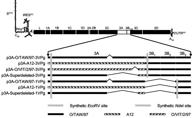
To facilitate exchange of 3A and 3B fragments, a derivative of plasmid pO/TAW/97Cn-An (1) was created by using PCR (15) to introduce silent changes that produced restriction endonuclease sites to facilitate exchange of 3A coding regions (Fig. 1). Specifically, an EcoRV site was introduced at the last codon of 2C and the first two codons of 3A (creating a CAG ATa TCA sequence encoding QIS, wherein the lowercasing indicates the base altered by mutagenesis) and an NdeI site was introduced at the second through fourth codons of VPg1 (3B, no. 1) (creating a CCa TAt GCT sequence encoding PYA, wherein lowercasing indicates the bases altered by mutagenesis). The resulting plasmid, designated p3A-O/TAW/97-3VPg, was then used to construct a derivative designated p3A-O/VIT/2/97-3VPg by PCR amplifying (with Herculase high-fidelity polymerase; Stratagene, La Jolla, Calif.) the 3A region of cDNA reverse-transcribed (SuperScript II RT; Life Technologies, Gaithersburg, Md.) from O/VIT/2/97 RNA (harvested from infected cell culture fluids by using TriZOL). The amplicon was created by using oligonucleotide primers containing the EcoRV and NdeI restriction endonuclease sites, allowing for substitution for the O/TAW/97 3A coding region in p3A-O/TAW/97-3VPg (Fig. 1). A similar strategy was utilized to substitute the serotype A12 3A coding region (amplified from cDNA plasmid pRMC35) for the O/TAW/97 3A in p3A-O/TAW/97-3VPg to create p3A-A12-3VPg (Fig. 1). Overlap PCR methodology (15) was then used to combine the 3A deletions in O/TAW/97 and O/VIT/2/97, producing a plasmid containing a 101-codon 3A designated p3A-Superdeleted-3VPg (Fig. 1). A similar PCR mutagenesis strategy (15) was utilized to delete the first two 3B coding regions in three of these plasmids (by introducing an NdeI site at the start of the third 3B coding region), producing three plasmids harboring one VPg only, designated in relation of the plasmid of origin as p3A-O/TAW/97-1VPg, p3A-A12-1VPg, and p3A-Superdeleted-1VPg (Fig. 1). After construction, all PCR-amplified regions were sequenced, confirming that only the desired mutations and/or deletions had been introduced.
FIG. 1.
Schematic diagram showing strategy for construction of chimeric cDNAs used to produce viruses with altered 3As and 3B deletions. The top portion of the figure indicates the position of 3AB on the FMDV genome. The bottom portion of the figure shows the structures of the 3A/B coding regions indicating the source of the viral cDNA used to construct the chimeras. The vertical hatched bars represent the restriction endonuclease sites introduced. The source of the coding regions in these chimeras is indicated by shading, and the source of the 5′ and 3′ untranslated regions is indicated by a single asterisk (O/TAW/97) or double asterisks (A12). The nomenclature of the plasmids is related to the 3A they harbor and number of 3Bs (VPgs) present. The first 80 N-terminal amino acids of 3A of the three viruses used in these studies were essentially identical (18). The sequences of VPg1, -2, and -3 of O/TAW/97 are as follows: GPYAGPLERQKPLKVKAELPQQE, GPYAGPMERQKPLKVKAKAPVVKE, and GPYEGPVKKPVALKVKAKNLIVTE.
In vitro RNA synthesis, transfection, and virus recovery.
Plasmids containing genome-length cDNAs were linearized at the unique NotI site found after the poly(A) tract and used as templates for RNA synthesis by using the MegaScript T7 RNA synthesis kit (Ambion, Austin, Tex.) according to the manufacturer's instructions. Transcripts were then transfected into BHK cells by using electroporation (20) or Lipofectin (Invitrogen, San Diego, Calif.). After transfection, virus was harvested from the transfected cells by freeze-thawing at 24 to 48 h posttransfection. Ten percent of the frozen-thawed lysates of these cells were then passaged on 35-mm-diameter BHK monolayers three subsequent times to recover high-titer viruses [to ensure elongation of the poly(C) tract (25]. Finally, these viruses were amplified a fourth time (again at a high multiplicity of infection [MOI]) on a 175-cm2 BHK monolayer, harvested when 90 to 95% of the cells displayed cytopathic effect (CPE) (reached between 16 and 24 h postinoculation), divided into aliquots, and stored at −70°C, and titers were determined by plaque assay (all of the viruses yielded titers of 1 × 10⁸ to 5 × 10⁸ PFU/ml). These BHK passage-4 stocks were used for all subsequent experiments, except for determination of intracellular RNA concentration, wherein a fifth passage followed by SDG virus purification was performed to obtain an innoculum with the required titer. Viruses were named according to the name of their plasmid of origin (e.g., v3A-O/TAW/97-3VPg, etc.).
Western blot analyses of 3A and 3B polypeptides in infected cells.
BHK cells were infected with viruses or transfected with plasmids encoding 3A, 3AB or 3ABBB of serotype A12, as previously described (22). The inoculation of the viruses was done at an MOI of 10, and when 60 to 70% of the cells displayed CPE, the cells were lysed with TNET buffer (10 mM Tris [pH 7.5], 150 mM NaCl, 1 mM EDTA, 1% [wt/vol] Triton X-100) for 20 min on ice; after clarification, the cell lysates were stored at −70°C. Samples containing 10 μg of infected cell lysate protein or 2 μg of protein from cells transfected with plasmids encoding 3A derivatives were resolved by sodium dodecyl sulfate-polyacrylamide gel electrophoresis gels (NuPAGE; Invitrogen) and electroblotted onto polyvinylidene difluoride membranes (Millipore) by standard methods. Immunoblocking buffer (1× phosphate-buffered saline-0.2% Tween 20-0.2% I-Block [Tropix, Bedford, Mass.]) was used for all of the blocking, washing (three times each), and incubation steps. All incubations were performed for 1 h at room temperature. After the blocking step, a polyclonal rabbit serum generated by using an Escherichia coli-expressed N-terminal fragment of 3A of O/TAW/97 (22) diluted 1/500 was used as the primary antibody. After being washed, the membranes were incubated with goat anti-rabbit phosphatase conjugate (KPL, Gaithersburg, Md.). After the washing steps described above and a rinsing with phosphate-buffered saline, the membranes were developed by using a chemiluminescent substrate (Duo-Lux; Vector Laboratories, Burlingame, Calif.) as recommended by the manufacturer.
Comparison of virus replication in fetal kidney cells.
Secondary cultures of fetal kidney cells were prepared from fetal bovine kidney (FBK) or fetal porcine kidney (FPK) (10, 11) and used at passage level 3. The ability of chimeric viruses to replicate in these cell types was determined by testing the ability of serial 10-fold dilutions of virus (starting with 107 PFU/ml [as measured previously on BHK cells]) to cause CPE on these cells. Analyses were performed side by side with BHK cells, and the resulting data were used to calculate the 50% tissue culture infectious dose(s) (TCID50) (14).
Determination of intracellular viral RNA concentration.
FPK or FBK cells were infected as described previously (22). Samples were collected at 0, 3, and 6 h postinoculation and processed for nonradioactive RNA hybridization. Integrated density values were determined from the resulting dot blots, and comparison of signals within the linear range of quantitation (as determined by the curve prepared from standard samples) were used to calculate the concentration of RNA in the cellular samples (22).
Swine inoculation and quantitative evaluation of porcine infectivity and pathogenicity.
The properties of chimeric viruses in vivo were evaluated by a method originally described by Burrows in 1966 (4) but infrequently used since that time. Briefly, for each virus, groups of four 20- to 40-kg outbred white pigs were intradermally inoculated in the heel bulb at two sites (inner and outer main digits) on each foot with a series of 10-fold dilutions of virus estimated to contain from 102 to 105 PFU/site. In the case of v3A-A12-1VPg and v3A-Superdeleted-1VPg, dilutions of 103 to 106 PFU/site were used since preliminary experiments indicated that these viruses were attenuated in their ability to form vesicles (results not shown). For the 7 days after inoculation, animals were carefully scored for the appearance of lesions at inoculation sites and at other sites, and the pig heel 50% infectious dose(s) (PHID50) was calculated from the results of 24-h observation (4) by standard methods (24). Vesicles were carefully tabulated each day, and the vesicle score was created by summing the following: one point for each affected digit, one point for vesicle(s) on the tongue, one point for vesicle(s) on the snout, one point for vesicle(s) on the lower lip, and one point for vesicle(s) on the carpal or tarsal area of one or more legs. A maximum lesion score of 12 was possible in these pigs because data collected from the eight injected toes were not included in these determinations. Once a vesicle appeared at a site, the site was scored “positive” on all subsequent days, even if the vesicle(s) at that site began to heal. Samples of blood and nasal secretions were collected daily and then used to determine the presence of virus in blood and in nasal secretions by determining the titers in multiwell plates by standard methods. An indirect enzyme-linked immunosorbent assay was used to detect blood immunoglobulin M (IgM). Briefly, blood samples were incubated in virus- and mock-coated wells on 96-well plates (coating was achieved by capturing virus with rabbit anti-O1 Manisa FMDV sera [IAH, Pirbright, England]), and bound IgM was detected with a goat anti-pig IgM conjugated with peroxidase (KPL) and ABTS [2,2′azinobis(3-ethylbenzthiazolinesulfonic acid)]. To confirm that the viruses that replicated in pigs had maintained the inoculated 3A/B genotype, viruses recovered from animals inoculated with v3A-Superdeleted-3VPg, v3A-O/TAW/97-1VPg, v3A-A12-1VPg, and v3A-Superdeleted-1VPg were sequenced throughout the entire 3A/B region after a single amplification in cell culture. The sequenced samples were obtained from virus isolated from blood (pigs 626, 627, 628, 630, 631, 632, 633, and 636) or nasal swabs (pigs 622, 623, 624, 625, 629, and 635). In the case of animal 634, no virus was recovered, so sequence data are not available. Sequencing was performed as previously described (20).
RESULTS
Viruses with large deletions in 3A or deletion of two copies of 3B grew nearly as well as wild-type virus in BHK cells.
Previous studies have revealed that FMDVs with deletions in two different regions of the 3A-coding region have been circulating in Asia over the last 33 years (18). These viruses include O/TAW/97, which caused a large outbreak in Taiwan in 1997, and a Vietnamese isolate from the same year (O/VIT/2/97; see Fig. 1). To facilitate the study of different 3A coding regions in the absence of other viral factors, we constructed a derivative of plasmid pO/TAW/97Cn-An containing unique restriction endonuclease sites bordering the 3A-coding region (see Materials and Methods). The resulting plasmid, which contains the S fragment of the serotype A12 virus and a poly(C) tract of 35 C residues, followed by the remainder of the genome of O/TAW/97, contained an EcoRV and an NdeI endonuclease site bordering the 3A coding region (Fig. 1). Virus obtained from in vitro-generated RNAs derived from this plasmid (designated p3A-O/TAW/97-3VPg) had properties indistinguishable from those of the virus recovered from the plasmid containing the naturally occurring codons bordering the 3A coding region (results not shown).
Using p3A-O/TAW/97-3VPg as a genetic background, we developed a panel of cDNA-containing plasmids with the structures shown in Fig. 1. Synthetic RNAs produced from these cDNAs were all able to produce infectious viruses in BHK cell cultures, which were used to grow high-titer stocks of all of these viruses. During the recovery of working stocks of these viruses, several high-MOI passages were utilized (see Materials and Methods), opening the possibility that revertants or pseudorevertants could have been obtained during passage. However, we did not detect any differences in recovery of viruses from these transfections that were indicative of the selection of these types of revertants. Despite the presence of deletions in the 3AB region of the genomes of several of these viruses, they all grew well in BHK cells (titers of 1 × 10⁸ to 5 × 10⁸ PFU/ml). As expected from the fact that all of these viruses encode exactly the same capsid, the physical particle/PFU ratios were indistinguishable for all of these viruses (data not shown).
Deletions within 3A and/or deletion of two copies of 3B does not alter processing of 3A/B.
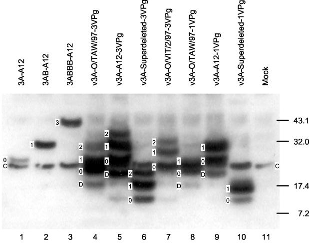
To determine whether deletions in the 3A or 3B coding regions altered the processing of 3A or 3B, we analyzed the proteolytic cleavage products of our seven chimeric viruses in vitro. Changes in processing were not expected since the cleavage sites for 3C between 2C/3A, 3A/3B1, 3B1/3B2, 3B2/3B3, and 3B3/3C were conserved among all three viruses used as the sources of the cDNAs for our chimeras. However, like Beck and coworkers (6), we felt that altered processing was an important possibility to address in the recombinant viruses we generated. As previously described, 3B is too small to be readily resolved by standard polyacrylamide gel electrophoresis (6), so detection of 3B was limited to its presence in partially processed intermediates. Figure 2 shows a Western blot developed with a rabbit polyclonal serum that recognizes the conserved N-terminal portion of 3A (see Materials and Methods). To facilitate identification of 3A/B products, “0” is used to designate 3A, “1” is used to designate 3AB, “2” is used to designate 3ABB, “3” is used to designate 3ABBB, and “D” is used to identify what appears to be a degradation product of 3A. In all lysates, including the one from uninfected cells, this antibody reacted with a cellular protein designated “C.” Cells infected with v3A-A12-3VPg contained 3A (“0”), 3AB (“1”) (both in correspondence with transfected proteins marker) and a weaker 3ABB (“2”). A faster-migrating band, which probably represents degraded-3A (“D”; see above), is also visible in this sample. As expected, cells infected with v3A-A12-1VPg contain only three of the four polypeptides present in cells infected with v3A-A12-3VPg, including degraded 3A (i.e., D), 3A (i.e., 0), and 3AB (i.e., 1). Cells infected with v3A-O/TAW/97-3VPg contained four 3A products—i.e., D, 0, 1, and 2—corresponding to degraded-3A, 3A, 3AB, and 3ABB, respectively, but the migration of the nonspecific cellular band (i.e., C) prevents clear resolution of 3A (i.e., 0) and 3AB (i.e., 1) in this lysate. Cells infected with v3A-O/TAW/97-1VPg contained only three of the four bands present in cells infected with v3A-O/TAW/97-3VPg, including degraded 3A (i.e., D), 3A (i.e., 0), and 3AB (i.e., 1). Cells infected with v3A-O/VIT/2/97-3VPg contained three polypeptides marked 0, 1, and 2 corresponding to 3A, 3AB, and 3ABB, respectively (degraded 3A cannot be seen in this lane). Cells infected with v3A-Superdeleted-3VPg contained three polypeptides marked 0, 1, and 2 corresponding to 3A, 3AB, and 3ABB, respectively (degraded 3A cannot be seen in this sample). Cells infected with v3A-Superdeleted-1VPg contained two polypeptides, marked 0 and 1, corresponding to 3A and 3AB. The possibility that the particularly strong C band in this cell lysate corresponded to another form of 3A was excluded by staining of this blot with a pool of monoclonal antibodies against 3A (results not shown). Additional analyses with a pool of monoclonal antibodies against 3B and a polyclonal rabbit sera against the C-term of 3A of O/TAW/97 confirmed the assignment of products shown in Fig. 2 (results not shown). Taken together, these results show that deletions in 3A and/or absence of VPg1 and VPg2 do not result in aberrant processing of 3A or 3AB in BHK cells.
FIG. 2.
Western blot showing 3A and 3B proteins from lysates of BHK cells that had been infected with chimeric virus (lanes 4 to 10), mock infected (lane 11), or transfected with plasmids encoding 3A, 3AB, and 3ABBB (lanes 1 to 3, respectively). The numbers on the right represent molecular mass standards in kilodaltons. Bands were identified with a specific rabbit polyclonal serum against N-terminal portion of 3A of O/TAW/97. Band designations: 0, 3A; 1, 3AB; 2, 3ABB; 3, 3ABBB; D, a degraded fragment of 3A; C, a cellular protein recognized by the polyclonal antibody in all of the lanes.
Viruses with a single VPg replicate in pig- but not in bovine-derived cells.
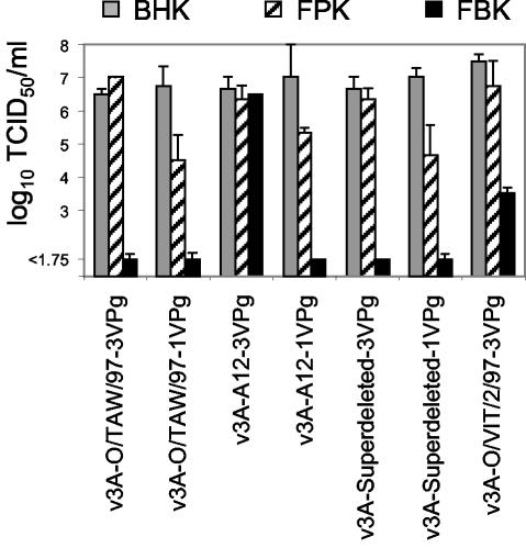
All seven viruses showed similar ability to grow in BHK cells, yielding similar titers of virus at 16 to 24 h postinoculation (see above). The calculated number of infectious viral particles produced per BHK cell is ≥50, a result similar to results obtained with wild-type and chimeric viruses in our laboratory. To determine species specificity in vitro, an assay was developed to measure the minimum infectious dose of virus able to propagate an infection on various cell types (Fig. 3). For these experiments, multiwell plates with BHK, FPK, and FBK cells were infected with 10-fold dilutions of virus and examined to determine the lowest dose of virus able to cause complete CPE at 48 h, and these results were expressed as TCID50/milliliter. These experiments revealed that all of the viruses tested showed equal ability to cause CPE in BHK cells at 48 h postinoculation (values close to 107 TCID50/ml). The viruses harboring three VPgs caused CPE as well in FPK as in BHK cells, whereas the TCID50/milliliter of the single-VPg virus was ∼100 times lower in FPK cells. In FBK cells, only the virus with three VPgs and the full-length 3A (v3A-A12-3VPg) caused CPE as well as it did in BHK and FPK cells, whereas the virus with three VPgs and 3A missing amino acids 133 to 143 (v3A-O/VIT/2/97-3VPg) required a 1,000-fold-greater inoculum to cause CPE; all of the remaining viruses were unable to cause CPE at any of the dilutions tested.These results indicate that a full-length 3A and three VPgs are needed for an effective replication in bovine-derived cells. However, in porcine cells, virus growth was independent of 3A length and slightly reduced by deletion of the first two VPgs.
FIG. 3.
Ability of the chimeric viruses to cause CPE in cells of different origin in vitro. Representative data showing replication of the indicated viruses in secondary bovine and porcine kidney cells side-by-side with BHK cells in two separate experiments. TCID50 values were determined, starting with viruses at dilutions containing 107 PFU/ml determined previously in BHK cells (see Materials and Methods). Bars and extended bars represent values obtained in two independent experiments.
The level of intracellular viral RNA correlates with 3A length and 3B copy number.
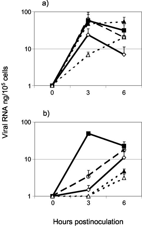
It has been previously shown that viruses with deletions in 3A display reduced RNA synthesis in cells of bovine origin (13, 22, 28). To study the amount of RNA present at early times postinfection, five of our chimeric viruses were used to infect FPK and FBK cells, and samples were processed to determine intracellular viral RNA concentrations. In FPK cells, all four viruses with three VPgs achieved similar levels of intracellular RNA at 3 h postinfection (Fig. 4a). At 6 h postinfection, intracellular RNA values were either equal to or lower than this value, with the latter phenomenon possibly due to CPE removing cells from the monolayer prior to harvest and/or RNA degradation (Fig. 4a). Cells infected with v3A-Superdeleted-1VPg produced less RNA at 3 h postinfection than cells infected with the three-VPg viruses, although at 6 h postinfection the level of RNA achieved was similar to that achieved by v3A-A12-3VPg. In FBK cells, only the virus with the full-length 3A (v3A-A12-3VPg) achieved RNA levels similar to those obtained for FPK cells at 3 and 6 h postinoculation (Fig. 4b). In the case of cells infected with v3A-O/VIT/2/97-3VPg there was a reduction in RNA concentration at 3 h postinoculation, although at 6 h postinoculation the level of RNA was similar to that obtained with v3A-A12-3VPg at 3 h postinoculation, suggesting a delay in RNA synthesis for this virus (Fig. 4b). In bovine cells infected with v3A-O/TAW/97-3VPg, the concentration of RNA was very low at 3 h postinoculation and did not reach the 3-h value of v3A-A12-3VPg at 6 h postinoculation (Fig. 4b). With the two viruses harboring the big deletion, with three or one VPg, no RNA was detected at 3 h postinoculation in FBK cells, and lower values than those for the other viruses were detected at 6 h postinoculation in these cells (Fig. 4b). These results suggest that synthesis of FMDV RNA in bovine cells is dependent on a full-length 3A and three VPgs, whereas the deletion of two copies of VPg results in a delay in RNA synthesis in porcine cells.
FIG. 4.
Levels of viral RNA detected after infection of secondary porcine and bovine fetal kidney cells with selected chimeric viruses. Amounts of RNA recovered from monolayers of porcine (a) and bovine (b) kidney cells, infected with v3A-A12-3VPg (▪), v3A-O/TAW/97-3VPg (⋄), v3A-O/VIT/2/97-3VPg (○), v3A-Superdeleted-3VPg (▴), or v3A-Superdeleted-1VPg (▵) at an MOI of 10 are shown. At each time, total RNA was extracted from the cells, and dot hybridization was used to determine the amount of RNA present, which is expressed in terms of nanograms per 100,000 cells. The results and error bars represent two independent values obtained in different experiments.
Viruses with a single VPg are attenuated in pigs.
To evaluate the influence of 3A length and 3B copy number on porcine infectivity and pathogenicity, six chimeric viruses were diluted and inoculated into the heel bulbs of pigs. After 24 h, careful inspection of the inoculation site was performed, and these observations were used to calculate the dose of virus (in PFU) that is able of cause a lesion to form at the inoculation site. These PHID50 values are shown in Table 1. As can be seen from these data, three-VPg viruses with complete (A12) or nearly complete (O/TAW/97) 3As exhibited very similar specific infectivities in pigs, with the number of PFU needed to cause a vesicle to form being similar to that observed with field-derived viruses (Pacheco and Mason, unpublished). However, for the virus with the superdeleted form of 3A, ∼30-fold more PFU were needed to form vesicles than for either the full-length 3A virus or the 3A virus with amino acids 93 to 102 deleted (Table 1). Table 1 also shows that all of the viruses with a single VPg displayed much lower infectivities, with >100,000 PFU needed to produce a vesicle. Vesicles formed at the sites inoculated with v3A-O/TAW/97-3VPg and v3A-A12-3VPg were similar in size to each other and to those formed by the natural isolate (O/TAW/97), whereas vesicles formed by v3A-Superdeleted-3VPg, v3A-O/TAW/97-1VPg, and v3A-A12-1VPg were smaller, a finding indicative of a slower replication in dermal cells. No vesicles were detected at the sites inoculated with up to 1,000,000 PFU of v3A-Superdeleted-1VPg.
TABLE 1.
Influence of 3A length and 3B copy number on the ability of viruses to establish infection in pigs
| Virus | Specific infectivity (PFU/PHID50)a | Total inoculated dose (PHID50/pig) | Total inoculated dose (PFU/pig) |
|---|---|---|---|
| v3A-O/TAW/97-3VPg | 1,300 | 170 | 2.2 × 10⁵ |
| v3A-A12-3VPg | 1,000 | 720 | 7.2 × 10⁵ |
| v3A-Superdeleted-3VPg | ≥32,000 | ≤13.75 | 4.4 × 10⁵ |
| v3A-O/TAW/97-1VPg | ≥130,000 | ≤3 | 3.9 × 10⁵ |
| v3A-A12-1VPg | ≥630,000 | ≤2 | 1.3 × 10⁶b |
| v3A-Superdeleted-1VPg | >1,500,000 | <0.8 | 1.2 × 10⁶b |
Using lesion data from all four inoculated animals obtained at 24 h postinoculation, PHID50 values were determined by the method of Reed and Muench (24). Lower PFU/PHID50 values indicate that a virus is more infectious in pigs.
For these viruses, 10 times more PFU were used for the inoculation because we expected them to have a lower infectivity (based on preliminary experiments [data not shown]).
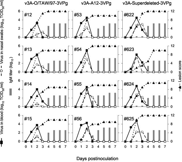
Quantitative comparison of disease produced in these animals over the 7-day period after inoculation is confounded by differences in infectious dose inoculated into these animals. Specifically, animals receiving the viruses with three VPgs and the long versions of 3A received a higher number of pig infectious doses (Table 1), which could have made their disease more severe, although it could also be argued that, once an animal is infected, the severity of the disease is independent of dose, as reported for FMDV over a century ago (19). This supposition is supported by our finding that disease progressed in a very similar pattern in all 12 pigs inoculated with three-VPg viruses, as shown in Fig. 5, although the 8 pigs inoculated with v3A-O/TAW/97-3VPg and v3A-A12-3VPg were given a dose ∼30 times higher, in terms of PHID50, and had twice as many lesions at the inoculation sites on day one postinoculation relative to the other four pigs (Table 1).
FIG. 5.
Comparison of disease induced in pigs by selected 3VPg-containing chimeric viruses (v3A-O/TAW/97-3VPg, v3A-A12-3VPg, and v3A-Superdeleted-3VPg). For these experiments, animals were inoculated intradermally in the heel of the bulb with 105 PFU of virus (as described in Table 1). Viremia, virus in nasal secretions, and IgM titers are expressed in the left axes; the lesion scores based on the presence of vesicle(s) in toes, tongue, lips, snout, carpus, or tarsus are expressed in the right axes.
Figure 5 shows that all 12 animals inoculated with three-VPg viruses in these three experiments displayed an acute and synchronous disease. Viremia was detected at 24 h postinoculation and lasted 2 or 3 days, with a peak of >103 TCID50/ml on day 2 after inoculation. The amount of virus recovered in nasal secretions was more variable than the viremia data, but virus was isolated on days 2 and/or 3 after inoculation in all 12 animals. Clinical signs of FMD were evident at 1 or 2 days postinfection (dpi) and reached the maximum score at 3 to 5 dpi, with values of ≥9. Specific IgM was detected at 3 to 4 dpi, at or near the time that virus was no longer detectable in blood. The simultaneous development of disease in these animals indicated that the infection and/or the disease was produced by inoculation of these 12 pigs and not by contagion from cohoused pigs. The pattern of disease produced by these three viruses is similar to the disease produced by direct inoculation with the natural isolate from which they were derived (O/TAW/97; Pacheco and Mason, unpublished).
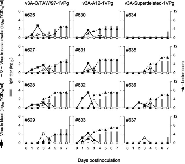
Figure 6 shows that the pattern of disease in all 12 pigs inoculated with one-VPg viruses was milder than that observed in animals inoculated with viruses containing three copies of VPg (Fig. 5). Among the four animals inoculated with v3A-O/TAW/97-1VPg, viremia was detected at 24 h postinoculation in three animals (pigs 626, 627, and 628) and lasted for 3 days, albeit at levels lower than those observed in pigs inoculated with any of the three-VPg-containing viruses. In the fourth pig (animal 629), viremia was never detected. Interestingly, the recovery of the virus from nasal secretions was similar to that observed with v3A-O/TAW/97-3VPg. Clinical signs (away from the inoculation site) were not detected until 3 dpi, and a maximum score of 7 was reached on days 5 and 6 postinoculation. Specific IgM were detected at 4 dpi, the day after the last when virus was detected in blood. Because animal 629 did not show replication in the site of the inoculation and no virus was detected in blood, it is not possible to determine whether the infection detected on day 3 was due to inoculation or contagion. Of the four animals inoculated with v3A-A12-1VPg, two (pigs 632 and 633) showed viremia at 24 h postinoculation that lasted for 2 and 3 days. In the other two animals (pigs 630 and 631) viremia was detected only on day 2 postinoculation. Only pig 632 displayed a viremia of >103 TCID50/ml. Isolation of virus in nasal secretions was similar to the single-VPg virus described above (v3A-O/TAW/97-1VPg). Clinical signs were detected on days 1, 2, and 3 after inoculation and never reached values of >7. Specific IgM was detected at 4 dpi, the last day viremia was detected, or on the day after the last day viremia was detected. Among the four animals inoculated with v3A-Superdeleted-1VPg, viremia was detected only in one animal (pig 636) on days 2 and 3 postinoculation with values that were ≤101.7 TCID50/ml. Virus in nasal secretions was detected in three animals (pigs 635, 636, and 637) for only a single day, with values of 101.7 TCID50/ml. Clinical signs were detected only in two animals (pigs 635 and 636) starting on day 3 postinoculation and reached a peak on day 6 postinoculation, with a maximum score of 7. Specific IgM was detected in animals 635 and 636 at 4 dpi, but in the other two animals IgM was not detectable until 7 dpi. Since none of these animals showed any detectable vesicle(s) at the sites inoculated with more than 106 PFU of v3A-Superdeleted-1VPg, we were not able to obtain a precise PFU/PHID50 value (Table 1). Furthermore, of the two pigs that failed to show viremia or any vesicles, only one showed detectable viral replication (animal 637 [virus recovered in nasal swabs]). Thus, it is possible that one animal (pig 634) did not become infected and that its seroconversion was only due to the antigen present in the initial inoculum. Based on these data, it is impossible to determine whether more than two pigs (animals 635 and 636) in this group became infected by inoculation or contagion and whether one animal (pig 634) ever became infected. Taken together, these animal studies demonstrate that although the removal of the first two copies of VPg significantly attenuates FMDV, the resulting single-VPg viruses are able to cause disease in pigs.
FIG. 6.
Comparison of disease induced in pigs by 1VPg-containing chimeric viruses (v3A-O/TAW/97-1VPg, v3A-A12-1VPg, and v3A-Superdeleted-1VPg). For these experiments, animals were inoculated intradermally in the heel of the bulb with 105 or 106 PFU of virus (as described in Table 1). Viremia, virus in nasal secretions, and IgM titers are expressed in the left axes; lesion scores based on presence of vesicle(s) in toes, tongue, lips, snout, carpus, or tarsus are expressed in the right axes.
Due to the lower specific infectivity (PFU/PHID50) of v3A-Superdeleted-3VPg and the one-VPg viruses, it was possible that the observed disease was produced by a contaminating virus or a virus that was selected in the animals due to the presence of a specific, adaptive mutation. To confirm that the disease was produced by the same genotype of virus inoculated into these animals, viruses isolated from samples of blood or nasal swabs that had been amplified by a single passage in BHK cells were sequenced through the entire 3AB region (including the bordering regions of 2C and 3C). Sequence data were obtained from viruses recovered from all pigs inoculated with v3A-Superdeleted-3VPg, v3A-O/TAW/97-1VPg, v3A-A12-1VPg, and v3A-Superdeleted-1VPg (except for pig 634, from which no virus was isolated). These analyses revealed no changes in encoded amino acid residues; however, two silent mutations were detected in the 3A coding regions of viruses isolated from pig 629 (A to G at base 378 of 3A) and pig 630 (A to G at base 27 of the single VPg). Taken together, these data demonstrate that the VPg copy number is an important determinant in host range and virulence of FMDV in pigs.
DISCUSSION
Among the picornaviruses, FMDV is noted for its broad host range. The virus also has unique genomic features in 3A/B (see the introduction), including the presence of redundant copies of 3B. VPg number has been reported to influence RNA synthesis and production of infectious FMDV particles in culture (6). Previous studies have shown that the length of 3A is related to species specificity. Deletions in 3A have been related to host range by studies showing that passage of FMDV in embryonated eggs produces viruses with 3A deletions that were attenuated for bovines (13, 23, 28). However, Sagedahl et al. (28) noted that these egg-adapted viruses remained virulent for pigs, and a similar deletion in 3A in a pig-virulent virus, O/TAW/97, was shown to be associated with the inability of this virus to cause disease in bovines (1). These data led us to propose that O/TAW/97 and O/VIT/2/97 could have been derived from live attenuated vaccine strains of FMDV that reverted to virulence (18).
To address the importance of 3A/B in host range and particularly in pig virulence, we created genetically engineered viruses with different 3A lengths and 3B copy numbers, in the background of an isolate of FMDV that is highly virulent in pigs (O/TAW/97). The 3As tested included a full-length 3A from a bovine-virulent virus, a 3A with a 10-amino-acid deletion (from O/TAW/97), a 3A with an 11-amino-acid deletion (from O/VIT/2/97) (18), and a superdeleted 3A, lacking 52 amino acids, created by joining the deletions of the former two viruses. In addition, we generated viruses from several of these recombinant viruses lacking two of the three copies of 3B. All recombinants had identical 5′ and 3′ untranslated regions, P1 (capsid), and P2, as well as nonstructural proteins 3C and 3D. Since we did not sequence the entire genome of the viruses used to conduct these studies, we cannot exclude that the phenotypes observed could have been altered by second-site mutations. However, the ready recovery of viruses from all of the cDNAs shown here and the high-MOI passaging used to produce viral stocks argue against that possibility.
All of the viruses generated (including those with one VPg) showed similar abilities to grow in BHK cell monolayers, reaching titers of between 1 × 10⁸ and 5 × 10⁸ PFU/ml; these results were similar to those for the field isolate O/TAW/97 (results not shown), in contrast to what was reported by Beck and coworkers (6), who detected 25- to 50-fold fewer infectious viral particles produced in viruses with a single functional VPg than in three-VPg viruses. In addition, all of our viruses, including O/TAW/97, produced plaques of indistinguishable shape and size on BHK monolayers (results not shown). Furthermore, changes in 3A size and 3B copy number did not appear to alter the processing of 3A in infected BHK cells. Thus, in our hands, large changes in 3A and 3B do not significantly affect the ability of FMDV to grow in highly susceptible BHK cells. Although it is unclear why our data differ from those of Beck and coworkers (6), it is worth noting that those investigators used VPg knockouts (replacing the functional Y residue with an F), whereas we performed complete deletions of the first two VPgs.
Evaluation of our panel of viruses in cells of bovine or porcine origin revealed that 3A length could affect replication in bovine cells. Consistent with previously cited findings for 3A deletions (see the introduction), all viruses with 3A deletions were significantly attenuated in FBK cells, with reduced levels of RNA early in infection and an inability to spread in bovine cells. In FPK cells, on the other hand, 3A length had no detectable effect in virulence in vitro. The evaluation of VPg copy number demonstrated that VPg copy number moderately (porcine) or severely (bovine) attenuated virus replication and spread in kidney cell cultures. Since we did not produce more than one type of single-VPg virus, it is possible that the reason for the observed phenotype of our single-VPg virus is an inherent “suboptimal” nature of the third copy of VPg. However, this seems unlikely, since Beck and coworkers were unable to detect any phenotypic difference among VPg knockouts (see above) that retained a single functional copy of VPg in position 1, 2, or 3 (6). Furthermore, during the passages of our viruses through animals, which should have favored the selection of better-performing 3B genotypes, we did not detect any mutations in this region of the genome.
For three-VPg viruses, however, large changes in 3A reduced infectivity in pigs in vivo. Despite this reduced initial infectivity, suggesting a reduced ability of the virus to replicate in dermal cells, the virus with this large deletion produced a disease of similar severity and levels of virus replication and shedding as viruses with full-length or nearly full-length 3As. Studies with animals with single-VPg viruses revealed that they were >1,000-fold less infectious than three-VPg-containing viruses in terms of their ability to establish a vesicular lesion at the inoculation site. However, all three single-VPg viruses were able to cause a systemic infection and were shed by the inoculated animals, but the disease was milder than that observed with viruses containing all three VPgs.
Thus, FMDV appears to have considerable flexibility in both regions of the genome, but both 3A and 3B appear to influence the virus's pathogenic potential and host range. We may conclude that, as has been demonstrated previously (1, 5), the length of 3A plays a role for species specificity, since only the virus with a full-length 3A (v3A-A12-3VPg) replicated well in bovine-derived cells. However, this virus, although able to cause small lesions in the site of inoculation and viremia in an inoculated cow, did not cause the severe disease observed in animals inoculated with bovine-derived viruses (unpublished data). For swine, drastic 3A length reduction reduced infectivity slightly without altering pathogenicity but deletion of the first two VPgs reduced both infectivity and pathogenicity. The demonstration that one-VPg viruses are attenuated for both of these species help to explain why all naturally occurring FMDV strains harbor three copies of 3B, since three copies of VPg would provide better virus transmission and dissemination of the disease.
The facts that changes in 3A length have little or no effect on virus growth in hamster-derived cells known for a defective antiviral response and that viruses with deletions in 3A can be selected in embryonated eggs (13, 23, 28) and mice (Q. Zhao, unpublished data) have interesting implications for interaction with host cells. They suggest that portions of 3A and redundant copies of 3B that are not needed to propagate viral nucleic acids provide an additional function in viral transmission and pathogenesis. It is still unclear why populations of viruses with deleted 3A overgrow parental populations in embryonated eggs or mice.
Our findings that 3A length and 3B copy number also affect viral growth in porcine and bovine kidney cells support the role of the “nonessential” portions of 3A or “redundant” copies of 3B in an intimate interaction with unknown host factors or a role in optimization of infection in cells of the natural host animal. Since 3A has been proposed to anchor the RNA replication complex to cellular membranes (3, 31), the truncated 3As may interact in a different way in different membranes of different species, giving altering viral RNA replication. Since it seemed likely that the truncation sites selected in vivo identified viable limits of 3A interaction with membranes (O/TAW/97) or ability to be cleaved from 3B by 3C (O/VIT/2/97), we utilized this pair of “natural” deletions to produce a viable “superdeleted” virus.
Until our report of the truncated 3A in O/TAW/97, all reported sequences of field isolates of FMDV had complete 3As. This may reflect the observed need for complete 3As for bovine virulence and the fact that most isolates sequenced were of bovine origin. Thus, the previously observed stability of 3A and 3B copies across all seven serotypes of FMDV suggest that both genetic elements are required for efficient transmission among cattle. However, viruses that may have originated under selection in alternative species (see above) that have deletions in 3A are still infectious for pigs, but the spread of these has been limited to areas of Asia. Therefore, the recent appearance of a pig-specific virus may be a peculiar adaptation of the viruses with 3A deletions to new intensive pig raising and an oral transmission route made possible by feeding of animal waste products and the introduction of live-attenuated vaccines (see above). Taken together, these data are consistent with the possibility that, until recently, FMDV has evolved as a ruminant virus.
The finding that viruses with shortened 3As are useful as vaccines for bovines was responsible, in part, for our desire to produce and evaluate viruses with larger 3A deletions, as well as fewer copies of 3B. Specifically, we hoped that these viruses could be useful as live-attenuated vaccines for FMD, overcoming the pig virulence of egg-adapted viruses with short deletions in 3A (see the introduction). The results of our studies indicate that we were not able to generate a pig-attenuated vaccine by means of deletion of 3A and/or 3B. Furthermore, our studies imply that 3A deletions achieved by laboratory passage are unlikely to produce completely safe vaccine candidates, confirming the decision made over two decades ago to discontinue the development of egg-adapted FMDV vaccines in favor of chemically inactivated vaccines.
Acknowledgments
We thank Sabrina Boettcher for technical assistance, the Plum Island Animal Disease Center (PIADC) Animal Caretakers for assistance with animal experiments,and Emiliana Brocchi (Istituto Zooprofilattico Sperimentale della Lombardia e dell Emilia-Romagna) for providing the monoclonal antibodies used in identifying different forms of 3AB. We also thank Qizu Zhao (Lanzhou Veterinary Research Institute, PIADC, and University of Texas Medical Branch) for producing the virus V3A-A12-3VPg and for helpful discussions.
This study was partially supported by the Agricultural Research Service of the U.S. Department of Agriculture (USDA; CRIS project 1940-32000-035-00D) and by grant 99-35204-7949 from the National Research Initiative Competitive Grants Program of USDA/CSREES.
REFERENCES
- 1.Beard, C. W., and P. W. Mason. 2000. Genetic determinants of altered virulence of Taiwanese foot-and-mouth disease virus. J. Virol. 74:987-991. [DOI] [PMC free article] [PubMed] [Google Scholar]
- 2.Bienz, K., D. Egger, and L. Pasamontes. 1987. Association of polioviral proteins of the P2 genomic region with the viral replication complex and virus-induced membrane synthesis as visualized by electron microscopic immunocytochemistry and autoradiography. Virology 160:220-226. [DOI] [PubMed] [Google Scholar]
- 3.Bienz, K., D. Egger, Y. Rasser, and W. Bossart. 1983. Intracellular distribution of poliovirus proteins and the induction of virus-specific cytoplasmic structures. Virology 131:39-48. [DOI] [PubMed] [Google Scholar]
- 4.Burrows, R. 1966. The infectivity assay of foot-and-mouth disease virus in pigs. J. Hyg. 64:419-429. [DOI] [PMC free article] [PubMed] [Google Scholar]
- 5.Dunn, C. S., and A. I. Donaldson. 1997. Natural adaption to pigs of a Taiwanese isolate of foot-and-mouth disease virus. Vet. Rec. 141:174-175. [DOI] [PubMed] [Google Scholar]
- 6.Falk, M. M., F. Sobrino, and E. Beck. 1992. VPg gene amplification correlates with infective particle formation in foot-and-mouth disease virus. J. Virol. 66:2251-2260. [DOI] [PMC free article] [PubMed] [Google Scholar]
- 7.Flanegan, J. B., and D. Baltimore. 1977. Poliovirus-specific primer-dependent RNA polymerase able to copy poly(A). Proc. Natl. Acad. Sci. USA 74:3677-3680. [DOI] [PMC free article] [PubMed] [Google Scholar]
- 8.Forss, S., and H. Schaller. 1982. A tandem repeat gene in a picornavirus. Nucleic Acids Res. 10:6441-6450. [DOI] [PMC free article] [PubMed] [Google Scholar]
- 9.Forss, S., K. Strebel, E. Beck, and H. Schaller. 1984. Nucleotide sequence and genome organization of foot-and-mouth disease virus. Nucleic Acids Res. 12:6587-6601. [DOI] [PMC free article] [PubMed] [Google Scholar]
- 10.Freshney, R. I. 1987. Culture of animal cells: a manual of basic technique. ARL, Inc., New York, N.Y.
- 11.George, V. G., J. C. Hierholzer, and E. W. Ades. 1996. Cell culture, p. 3-23. In H. O. Kangro (ed.), Virology methods manual. Academic Press, Inc., San Diego, Calif.
- 12.Giraudo, A. T., E. Beck, K. Strebel, P. A. de Mello, J. L. La Torre, E. A. Scodeller, and I. E. Bergmann. 1990. Identification of a nucleotide deletion in parts of polypeptide 3A in two independent attenuated aphthovirus strains. Virology 177:780-783. [DOI] [PubMed] [Google Scholar]
- 13.Giraudo, A. T., A. Sagedahl, I. E. Bergmann, J. L. La Torre, and E. A. Scodeller. 1987. Isolation and characterization of recombinants between attenuated and virulent aphthovirus strains. J. Virol. 61:419-425. [DOI] [PMC free article] [PubMed] [Google Scholar]
- 14.Hierholzer, J. C., and R. A. Killington. 1996. Virus isolation and quantitation, p. 25-46. In H. O. Kangro (ed.), Virology methods manual. Academic Press, Inc., San Diego, Calif.
- 15.Higuchi, R., B. Krummel, and R. K. Saiki. 1988. A general method of in vitro preparation and specific mutagenesis of DNA fragments: study of protein and DNA interactions. Nucleic Acids Res. 16:7351-7367. [DOI] [PMC free article] [PubMed] [Google Scholar]
- 16.King, A. M. Q., D. McCahon, W. R. Slade, and J. W. I. Newman. 1982. Recombination in RNA. Cell 29:921-928. [DOI] [PMC free article] [PubMed] [Google Scholar]
- 17.Kitamura, N., B. L. Semler, P. G. Rothberg, G. R. Larsen, C. J. Adler, A. J. Dorner, E. A. Emini, R. Hanecak, J. J. Lee, S. van der Werf, C. W. Anderson, and E. Wimmer. 1981. Primary structure, gene organization and polypeptide expression of poliovirus RNA. Nature 291:547-553. [DOI] [PubMed] [Google Scholar]
- 18.Knowles, N. J., P. R. Davies, T. Henry, V. O'Donnell, J. M. Pacheco, and P. W. Mason. 2001. Emergence in Asia of foot-and-mouth disease viruses with altered host range: characterization of alterations in the 3A protein. J. Virol. 75:1551-1556. [DOI] [PMC free article] [PubMed] [Google Scholar]
- 19.Loeffler, F., and P. Frosch. 1964. Report of the Commission for Research on Foot-and-Mouth Disease, p. 64-68. In N. Hahon (ed.), Selected papers on virology. Prentice-Hall, Englewood Cliffs, N.J.
- 20.Mason, P. W., S. V. Bezborodova, and T. M. Henry. 2002. Identification and characterization of a cis-acting replication element (cre) adjacent to the internal ribosome entry site of foot-and-mouth disease virus. J. Virol. 76:9686-9694. [DOI] [PMC free article] [PubMed] [Google Scholar]
- 21.Nunez, J. I., E. Baranowski, N. Molina, C. M. Ruiz-Jarabo, C. Sanchez, E. Domingo, and F. Sobrino. 2001. A single amino acid substitution in nonstructural protein 3A can mediate adaptation of foot-and-mouth disease virus to the guinea pig. J. Virol. 75:3977-3983. [DOI] [PMC free article] [PubMed] [Google Scholar]
- 22.O'Donnell, V. K., J. M. Pacheco, T. M. Henry, and P. W. Mason. 2001. Subcellular distribution of the foot-and-mouth disease virus 3A protein in cells infected with viruses encoding wild-type and bovine-attenuated forms of 3A. Virology 287:151-162. [DOI] [PubMed] [Google Scholar]
- 23.Parisi, J. M., P. Costa Giomi, P. Grigera, P. Auge de Mello, I. E. Bergmann, J. L. La Torre, and E. A. Scodeller. 1985. Biochemical characterization of an aphthovirus type 0(1) strain campos attenuated for cattle by serial passages in chicken embryos. Virology 147:61-71. [DOI] [PubMed] [Google Scholar]
- 24.Reed, L., and H. Muench. 1938. A simple method of estimating fifty percent endpoints. Am. J. Hyg. 27:493-497. [Google Scholar]
- 25.Rieder, E., T. Bunch, F. Brown, and P. W. Mason. 1993. Genetically engineered foot-and-mouth disease viruses with poly(C) tracts of two nucleotides are virulent in mice. J. Virol. 67:5139-5145. [DOI] [PMC free article] [PubMed] [Google Scholar]
- 26.Robertson, B. H., M. J. Grubman, G. N. Weddell, D. M. Moore, J. D. Welsh, T. Fischer, D. J. Dowbenko, D. G. Yansura, B. Small, and D. G. Kleid. 1985. Nucleotide and amino acid sequence coding for polypeptides of foot-and-mouth disease virus type A12. J. Virol. 54:651-660. [DOI] [PMC free article] [PubMed] [Google Scholar]
- 27.Rueckert, R. R. 1996. Picornaviridae: the viruses and their replication, p. 609-654. In B. N. Fields, D. M. Knipe, and P. M. Howley (ed.), Fields virology, 3rd ed. Lippincott-Raven Publishers, Philadelphia, Pa.
- 28.Sagedahl, A., A. T. Giraudo, P. A. De Mello, I. E. Bergmann, J. L. La Torre, and E. A. Scodeller. 1987. Biochemical characterization of an aphthovirus type C3 strain Resende attenuated for cattle by serial passages in chicken embryos. Virology 157:366-374. [DOI] [PubMed] [Google Scholar]
- 29.Schlegel, A., T. H. Giddings, Jr., M. S. Ladinsky, and K. Kirkegaard. 1996. Cellular origin and ultrastructure of membranes induced during poliovirus infection. J. Virol. 70:6576-6588. [DOI] [PMC free article] [PubMed] [Google Scholar]
- 30.Semler, B. L., C. W. Anderson, R. Hanecak, L. F. Dorner, and E. Wimmer. 1982. A membrane-associated precursor to poliovirus VPg identified by immunoprecipitation with antibodies directed against a synthetic heptapeptide. Cell 28:405-412. [DOI] [PubMed] [Google Scholar]
- 31.Takegami, T., B. L. Semler, C. W. Anderson, and E. Wimmer. 1983. Membrane fractions active in poliovirus RNA replication contain VPg precursor polypeptides. Virology 128:33-47. [DOI] [PubMed] [Google Scholar]
- 32.Towner, J. S., T. V. Ho, and B. L. Semler. 1996. Determinants of membrane association for poliovirus protein 3AB. J. Biol. Chem. 271:26810-26818. [DOI] [PubMed] [Google Scholar]
- 33.Vakharia, V. N., M. A. Devaney, D. M. Moore, J. J. Dunn, and M. J. Grubman. 1987. Proteolytic processing of foot-and-mouth disease virus polyproteins expressed in a cell-free system from clone-derived transcripts. J. Virol. 61:3199-3207. [DOI] [PMC free article] [PubMed] [Google Scholar]
- 34.Wimmer, E. 1982. Genome-linked proteins of viruses. Cell 28:199-201. [DOI] [PubMed] [Google Scholar]


