Abstract
We have designed a glycodendritic structure, BH30sucMan, that blocks the interaction between dendritic cell-specific intercellular adhesion molecule 3-grabbing nonintegrin (DC-SIGN) and Ebola virus (EBOV) envelope. BH30sucMan inhibits DC-SIGN-mediated EBOV infection at nanomolar concentrations. BH30sucMan may counteract important steps of the infective process of EBOV and, potentially, of microorganisms shown to exploit DC-SIGN for cell entry and infection.
Ebola virus (EBOV) causes hemorrhagic fever and is considered to be one of the most lethal pathogens known. The risk of a widespread epidemic episode, or even its potential use in bioterrorism, has stimulated research on the agent and the development of vaccine programs (18). Due to its extraordinary virulence, EBOV is classified as a biosafety level 4 pathogen; however, the generation of pseudotyped recombinant retroviral particles has recently allowed the exploration of important aspects of EBOV biology using biosafety level 2 facilities (21). Despite these efforts, there is no specific cure for EBOV infection.
Our group and others (1, 17) have previously demonstrated that EBOV binds to C-type lectins dendritic cell-specific intercellular adhesion molecule 3-grabbing nonintegrin (DC-SIGN) and liver/lymph node SIGN (L-SIGN) and utilizes these molecules as mediators to gain access to certain cell types. Furthermore, DC-SIGN and L-SIGN can act as trans-receptors to efficiently capture infective particles and transmit the infection to susceptible cells (1). DC-SIGN, originally cloned as a human immunodeficiency virus (HIV) gp120-binding protein (7), is expressed exclusively on immature dendritic cells and confers upon these cells the ability to facilitate, in trans, HIV infection of activated CD4+ T cells (8). L-SIGN is a closely related molecule expressed on the surface of endothelial cells in the liver, lymph node sinuses, and placental villi (3). DC-SIGN and L-SIGN bind ligands containing oligomannose glycans through the C-terminal carbohydrate recognition domain. It is highly likely that EBOV, in a manner similar to that of HIV, subverts the physiological role of these C-type lectins to achieve important steps in its infectious process, these molecules being critical for the initial steps of viral infection at the mucosal level and subsequent dissemination throughout the body. Therefore, DC- and L-SIGN can be considered potential new targets for the design of compounds acting as viral entry inhibitors. The low affinity of carbohydrate interactions, compensated for in nature by the multivalent presentation of carbohydrate units, has encouraged the design of carbohydrate multivalent structures to interfere with biological processes in which carbohydrates are involved. Among these multivalent systems, glycodendrimers are one of the most popular systems (4), and they have been used previously in several processes, including blockage of viral receptors (13, 15). Previous works with glycodendritic structures, based on the hyperbranched polymer BoltornH30 (Perstorp Specialty Chemicals), have shown a number of advantages, including low cytotoxicity, high solubility in physiological media, and relatively low cost (2).
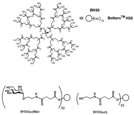
In order to inhibit EBOV DC-SIGN interaction, we have designed a glycodendritic structure based on BoltornH30 (Fig. 1), presenting 32 mannose units linked through a succinyl spacer, BH30sucMan. We tested this compound in an experimental model using an EBOV-GP-pseudotyped lentivirus (1). Pseudotyped viral vectors have been successfully used in a wide variety of experimental applications (16), including characterization of the receptors used by EBOV (1, 5, 6, 17, 19, 21).
FIG. 1.
Structure of BoltornH30 (BH30) (top) and schematic representation of BH30sucMan and BH30sucL (bottom).
The lentiviral vector pNL4-3.Luc.R-E-, obtained from Nathaniel Landau (11) (through the AIDS Research and Reference Reagent Program, Division of AIDS, National Institute of Allergy and Infectious Diseases, National Institutes of Health), was used for production of vesicular stomatitis virus G and EBOV Zaire GP pseudotypes (1) in a transient transfection protocol using 293T cells as previously described (20). The expression plasmid for the GP of the Zaire strain of EBOV (i.e., EBOV-GP) was kindly provided by A. Sanchez (Centers for Disease Control and Prevention) (21). BH30sucMan was prepared as described elsewhere (2). Jurkat cells stably transduced with DC-SIGN were infected with EBOV-GP-pseudotyped lentiviral vectors expressing luciferase using a multiplicity of infection of 0.1. A range of concentrations of BH30sucMan was added simultaneously along with the control compound BH30sucL (without mannoses) (Fig. 1). After 48 h, cells were lysed and assayed for luciferase expression. The 50% inhibitory concentrations (IC50) were calculated with Graphpad Prism software version 3.0. The inhibitory activity of BH30sucMan for the in trans function of DC-SIGN was studied on K562 erythroleukemia cells stably expressing DC-SIGN (14). Cells were incubated by rotation for 60 min at room temperature with supernatants containing Ebola Zaire-pseudotyped retroviruses in the presence or absence of BH30sucMan (500 nM) and controls. After this step, cells were extensively washed with phosphate-buffered saline-1 mM CaCl2-0.5% bovine serum albumin, resuspended in fresh medium, and plated onto HeLa cell monolayers. After 48 h, K562 cells were removed, the monolayer of HeLa cells was washed twice, and luciferase activity was measured.
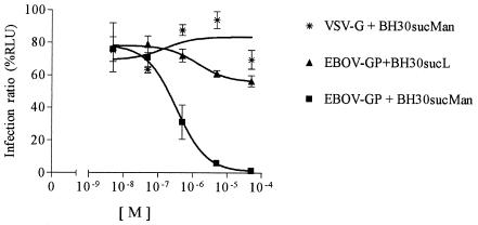
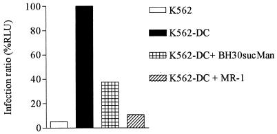
Results of our experiments showed that BH30sucMan was able to selectively inhibit DC-SIGN-mediated EBOV infection in an efficient dose-dependent manner (IC50, 337 nM), whereas it did not affect infection mediated by a DC-SIGN-independent viral envelope such as vesicular stomatitis virus G (1). Infection was also inhibited by the monosaccharide α-methyl-d-mannopyranoside in a dose-dependent manner with an IC50 of 1.27 mM (data not shown). The control structure, BH30sucL, which does not present sugar units, only showed a limited effect at concentrations beyond 10−5 M, which is most likely explained by nonspecific interactions with receptors at the cell surface (Fig. 2). Similar results were obtained in parallel experiments by using L-SIGN-expressing cells (data not shown). As a further proof of the specific action of the glycodendritic structure, BH30sucMan did not show any inhibitory effect in infection experiments using DC-SIGN-negative cell lines, such as HeLa, which are known to be susceptible to EBOV infection (data not shown). In the experiments in trans, in which a more complex series of events such as internalization and presentation of the viral particle to susceptible cells can take place (12), BH30sucMan also showed a significant reduction of DC-SIGN-mediated infection in trans at levels comparable to the inhibition shown in cis (Fig. 3).
FIG. 2.
Inhibition of DC-SIGN-mediated EBOV-GP pseudotyped viruses in cis, using Jurkat cells expressing DC-SIGN and BH30sucMan or BH30sucL. Results are shown as the proportion of inhibition in comparison to control (no compound), and the values correspond to the means of three experiments ± standard error of the mean.
FIG. 3.
Inhibition of infection of EBOV-GP pseudotyped viruses mediated in trans by DC-SIGN. K562 cells (negative control) and K562-expressing DC-SIGN (K562-DC) were used to bind and transfer viral particles to HeLa cells in the presence or absence of BH30sucMan and the anti-DC-SIGN monoclonal antibody MR-1 (14). One representative experiment out of three is shown.
We have shown that BH30sucMan is a potent inhibitor of EBOV infection mediated by DC-SIGN both in cis (Fig. 2) and in trans (Fig. 3), presumably due to the same mechanisms of inhibiting the interaction between the lectin and the viral envelope. A carbohydrate-dependent inhibitory effect has been demonstrated in these experiments; also, a multivalent effect of two orders of magnitude is shown, since monovalent mannose was able to inhibit this interaction but at millimolar concentrations.
Blocking EBOV interaction with lectin receptors in the mucosal and endothelial territories is a reasonable goal and also a model for other microorganisms such as HIV (8), cytomegalovirus (10), or Mycobacterium tuberculosis (9), any of which could potentially exploit a similar mechanism. Additionally, the use of these glycodendritic structures will allow us to better understand the molecular basis of this interaction as well as the design of more potent and selective inhibitors.
Acknowledgments
We thank Perstorp Specialty Chemicals for the generous gift of Boltorn polymers. K562-expressing DC-SIGN cells and the MR-1 monoclonal antibody were generously provided by A. L. Corbí (Centro de Investigaciones Biológicas, CSIC, Madrid, Spain).
This research was supported by DGI grant no. BQU2002-03734 to J.R. and grants FIS 01/1430 and FIPSE 3026/99 to R.D.
REFERENCES
- 1.Alvarez, C. P., F. Lasala, J. Carrillo, O. Muniz, A. L. Corbi, and R. Delgado. 2002. C-Type lectins DC-SIGN and L-SIGN mediate cellular entry by Ebola virus in cis and in trans. J. Virol. 76:6841-6844. [DOI] [PMC free article] [PubMed] [Google Scholar]
- 2.Arce, E., P. M. Nieto, V. Díaz, R. García-Castro, A. Bernad, and J. Rojo. 2003. Glycodendritic structures based on Boltorn hyperbranched polymers and their interactions with Lens culinaris lectin. Bioconjug. Chem. 14:817-823. [DOI] [PubMed] [Google Scholar]
- 3.Bashirova, A. A., T. B. H. Geijtenbeek, G. C. F. van Duijnhoven, S. J. van Vliet, J. B. G. Eilering, M. P. Martin, L. Wu, T. D. Martin, N. Viebig, P. A. Knolle, V. N. KewalRamani, Y. van Kooyk, and M. Carrington. 2001. A dendritic cell-specific intercellular adhesion molecule 3-grabbing nonintegrin (DC-SIGN)-related protein is highly expressed on human liver sinusoidal endothelial cells and promotes HIV-1 infection. J. Exp. Med. 193:671-678. [DOI] [PMC free article] [PubMed] [Google Scholar]
- 4.Bezouška, K. 2002. Design, functional evaluation and biomedical applications of carbohydrate dendrimers (glycodendrimers). J. Biotechnol. 90:269-290. [DOI] [PubMed] [Google Scholar]
- 5.Chan, S. Y., C. J. Empig, F. J. Welte, R. F. Speck, A. Schmaljohn, J. F. Kreisberg, and M. A. Goldsmith. 2001. Folate receptor-α is a cofactor for cellular entry by Marburg and Ebola viruses. Cell 106:117-126. [DOI] [PubMed] [Google Scholar]
- 6.Chan, S. Y., R. F. Speck, M. C. Ma, and M. A. Goldsmith. 2000. Distinct mechanisms of entry by envelope glycoproteins of Marburg and Ebola (Zaire) viruses. J. Virol. 74:4933-4937. [DOI] [PMC free article] [PubMed] [Google Scholar]
- 7.Curtis, B. M., S. Scharnowske, and A. J. Watson. 1992. Sequence and expression of a membrane-associated C-type lectin that exhibits CD4-independent binding of human immunodeficiency virus envelope glycoprotein gp120. Proc. Natl. Acad. Sci. USA 89:8356-8360. [DOI] [PMC free article] [PubMed] [Google Scholar]
- 8.Geijtenbeek, T. B., D. S. Kwon, R. Torensma, S. J. van Vliet, G. C. van Duijnhoven, J. Middel, I. L. Cornelissen, H. S. Nottet, V. N. KewalRamani, D. R. Littman, C. G. Figdor, and Y. van Kooyk. 2000. DC-SIGN, a dendritic cell-specific HIV-1-binding protein that enhances trans-infection of T cells. Cell 100:587-597. [DOI] [PubMed] [Google Scholar]
- 9.Geijtenbeek, T. B., S. J. van Vliet, E. A. Koppel, M. Sanchez-Hernandez, C. M. Vandenbroucke-Grauls, B. Appelmelk, and Y. van Kooyk. 2003. Mycobacteria target DC-SIGN to suppress dendritic cell function. J. Exp. Med. 197:7-17. [DOI] [PMC free article] [PubMed] [Google Scholar]
- 10.Halary, F., A. Amara, H. Lortat-Jacob, M. Messerle, T. Delaunay, C. Houles, F. Fieschi, F. Arenzana-Seisdedos, J. F. Moreau, and J. Dechanet-Merville. 2002. Human cytomegalovirus binding to DC-SIGN is required for dendritic cell infection and target cell trans-infection. Immunity 17:653-664. [DOI] [PubMed] [Google Scholar]
- 11.He, J., S. Choe, R. Walker, P. Di Marzio, D. O. Morgan, and N. R. Landau. 1995. Human immunodeficiency virus type 1 viral protein R (Vpr) arrests cells in the G2 phase of the cell cycle by inhibiting p34cdc2 activity. J. Virol. 69:6705-6711. [DOI] [PMC free article] [PubMed] [Google Scholar]
- 12.Kwon, D. S., G. Gregorio, N. Bitton, W. A. Hendrickson, and D. R. Littman. 2002. DC-SIGN-mediated internalization of HIV is required for trans-enhancement of T-cell infection. Immunity 16:135-144. [DOI] [PubMed] [Google Scholar]
- 13.Landers, J. J., Z. Cao, I. Lee, L. T. Piehler, P. P. Myc, A. Myc, T. Hamouda, A. T. Galecki, and J. R. Baker, Jr. 2002. Prevention of influenza pneumonitis by sialic acid-conjugated dendritic polymers. J. Infect. Dis. 186:1222-1230. [DOI] [PubMed] [Google Scholar]
- 14.Relloso, M., A. Puig-Kröger, O. Muñiz, J. L. Rodríguez-Fernández, G. de la Rosa, N. Longo, J. Navarro, M. A. Muñoz-Fernández, P. Sánchez-Mateos, and A. Corbí. 2002. DC-SIGN (CD209) expression is IL-4 dependent and is negatively regulated by IFN, TGF-β, and anti-inflammatory agents. J. Immunol. 168:2634-2643. [DOI] [PubMed] [Google Scholar]
- 15.Reuter, J. D., A. Myc, M. M. Hayes, Z. Gan, R. Roy, D. Qin, R. Yin, L. T. Piehler, R. Esfand, D. A. Tomalia, and J. R. Baker, Jr. 1999. Inhibition of viral adhesion and infection by sialic acid-conjugated dendritic polymers. Bioconjug. Chem. 10:271-278. [DOI] [PubMed] [Google Scholar]
- 16.Sanders, D. A. 2002. No false start for novel pseudotyped vectors. Curr. Opin. Biotechnol. 13:437-442. [DOI] [PubMed] [Google Scholar]
- 17.Simmons, G., J. D. Reeves, C. C. Grogan, L. H. Vandenberghe, F. Baribaud, J. C. Whitbeck, E. Burke, M. J. Buchmeier, E. J. Soilleux, J. L. Riley, R. W. Doms, P. Bates, and S. Pohlmann. 2003. DC-SIGN and DC-SIGNR bind Ebola glycoproteins and enhance infection of macrophages and endothelial cells. Virology 305:115-123. [DOI] [PubMed] [Google Scholar]
- 18.Sullivan, N. J., A. Sanchez, P. E. Rollin, Z. Y. Yang, and G. J. Nabel. 2000. Development of a preventive vaccine for Ebola virus infection in primates. Nature 408:605-609. [DOI] [PubMed] [Google Scholar]
- 19.Wool-Lewis, R. J., and P. Bates. 1998. Characterization of Ebola virus entry by using pseudotyped viruses: identification of receptor-deficient cell lines. J. Virol. 72:3155-3160. [DOI] [PMC free article] [PubMed] [Google Scholar]
- 20.Yang, S., R. Delgado, S. R. King, C. Woffendin, C. S. Barker, Z. Y. Yang, L. Xu, G. P. Nolan, and G. J. Nabel. 1999. Generation of retroviral vector for clinical studies using transient transfection. Hum. Gene Ther. 10:123-132. [DOI] [PubMed] [Google Scholar]
- 21.Yang, Z., R. Delgado, L. Xu, R. F. Todd, E. G. Nabel, A. Sanchez, and G. J. Nabel. 1998. Distinct cellular interactions of secreted and transmembrane Ebola virus glycoproteins. Science 279:1034-1037. [DOI] [PubMed] [Google Scholar]


