It has been known that the liver graft is protective of or tolerogenic for concomitantly transplanted organs.1 Whether allograft antigen delivery into the portal vein, instead of the systemic venous system, is an important factor is controversial.2 We have previously shown that small intestine had histologically less severe rejection when transplanted as part of a multivisceral graft than when transplanted alone.3 It could not be determined if this difference was due to the ameliorating effect of the other organs in the multivisceral complex, or if it was the consequence of the disparate intestinal venous drainage in the two experimental models, or if both factors contributed. This study was carried out to directly compare the small bowel as part of a multivisceral graft with the isolated intestinal graft with ponal drainage vs systemic drainage.
MATERIALS AND METHODS
Animals
Inbred male Lewis rats (RT11, LEW) and Brown Norway rats (RTln, BN) weighing 200 to 300 g were obtained from Harlan Sprague Dawley, Inc (Indianapolis, Ind), and were used as recipients and donors, respectively.
Operation
Multivisceral Transplantation (MVTx)
The operative procedure has been described in detail.4 An en bloc graft including liver, pancreas, stomach, omentum, small intestine, and colon was transplanted orthotopically by anastomosis between the donor and recipient suprahepatic vena cava and infrahepatic vena cava. Aortic continuity was reconstructed with a side-to-end anastomosis between the donor and recipient aorta using an interposition aortic graft. Gastrointestinal continuity was restored by anastomoses at the stomach above and between the rectum of the graft and recipient below.
Orthotopic Small Intestine Transplantation With Portal Drainage (SITX-PP)
The entire donor small intestine from the ligament of Treitz to the ileocecal valve was transplanted by end-to-side vascular anastomosis between graft aorta and recipient infrarenal aorta, and graft and recipient portal veins at the distal site of the confluence of the splenic vein. The entire recipient small intestine was resected and intestinal continuity was restored by proximal and distal intestinal anastomoses.
Orthotopic Small Intestine Transplantation With Caval Drainage (SITX-PC)
The surgical procedure was identical to that above except the end-to-side venous anastomosis was performed between graft portal vein and recipient infrarenal vena cava.
Immunosuppression
Intramuscular FK 506 (Fujisawa Pharmaceutical Company, Ltd, Osaka, Japan) was diluted in normal saline and injected at a dose of 0.64 mg/kg/d for 14 days starting the day of surgery.
Histologic Examination
Two or three animals from each transplant group were killed between 100 days and 140 days after grafting. The small bowel graft including Peyer’s patches and mesenteric lymph node was examined grossly and histopathologically.
RESULTS
Animal Survival
Without treatment, median survival for each group was 10.0 days (MVTx n = 5), 10.5 days (SITX-PP n = 9), and 12.0 days (SITX-PC n = 10). These were not significantly different. When FK 506 was used at the dose of 0.64 mg/kg/d for 14 days after transplantation, two animals in the MVTX group died early with ileus and chylothorax and one animal in the SITX-PC group died 42 days after transplantation with rejection. All other animals survived more than 100 days. These differences in raw survival were not significant in any of the three models.
Body Weight Gain and Histologic Examination
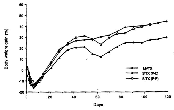
After the initial operative weight loss, the best gain was after MVTX. After isolated small bowel transplantation, weight gain was better with portal drainage than with caval drainage (Fig 1). Histopathologic examination of the grafts from long surviving animals showed differences in the intestinal component of the multivisceral grafts and the isolated small bowel grafts. The Peyer’s patches and mesenteric lymph nodes in isolated small bowel grafts of both portal and caval drainage groups were completely depleted and replaced by scar. The epithelial intestinal components were relatively intact, but villous blunting, cryptitis, and lymphoid depletion of the lamina propria were common in these isolated small bowel grafts of both groups. In contrast, the small intestine in multivisceral grafts was completely protected.
Fig 1.
Body weight gain (mean) after transplantation of multivisceral grafts (MVTx), isolated small bowel graft with portal drainage (SITX-PP), and caval drainage (SITX-PC).
DISCUSSION
In one of our publications, we summarized the evidence that there is a diminution of immunologic response by draining transplanted organs into the portal system. The hypothesis was that the liver modified either allograft antigens or immunoreactive cells in the host. We were unable to show this effect in dogs or pigs using kidney grafts as the test organ.2 The results were similar in the present study. Distortion of lymphoid architecture associated with chronic rejection was observed with equal severity and frequency in long surviving animals bearing isolated intestinal grafts drained into either the portal or caval systems. These findings were absent when the intestine was part of a multi visceral graft.
However, the behavior of the isolated intestine recipient was different, depending on the route of the intestinal graft venous outflow. Rats with portal drainage of their isolated intestinal grafts had better weight gain than after caval drainage and were healthier.
This was not surprising. It has been shown in nontransplant models that optimum liver structure and function depend on hormones (especially insulin), nutrients, and other substances found in high concentration in splanchnic venous blood.5 Depriving the liver of this kind of blood causes hepatic atrophy, organelle deterioration, and subtle loss of function. When all of this portal blood is taken from the liver, the full consequences of an Eck fistula can be expected, which are less extreme in humans than in animals.5 Diversion in dogs of intestinal blood only, or of pancreaticoduodenosplenic blood only causes similar but less pronounced liver abnormalities than with total diversion.6
This is the essence of the so-called hepatotrophic concept of liver physiology. The concept has relevance to intestinal transplantation as has been demonstrated by Schraut et al7 and not clearly by Schaffer et al.8 In Schraut’s experiments, rats with caval (but not portal) drainage exhibited hyperammonemia, a minor reduction of the liver weight, characteristic change in plasma amino acid concentrations, significant retardation of postoperative weight gain, and histologic appearance of the liver in the direction of those but less extreme than those caused by Eck fistula.
These results as well as prior knowledge about liver physiology mean that there is no major immunologic advantage to portal drainage of isolated intestinal transplants, but that this is an important provision for maintenance of normal metabolic homeostasis. The results also show that the nonintestinal component of a multivisceral graft (presumably the liver) is tolerogenic for the intestine.
Acknowledgments
Supported by research grants from the Veterans Administration and project grant No. DK 29961 from the National Institutes of Health, Bethesda, Maryland.
References
- 1.Kamada N. Experimental Liver Transplantation. Boca Raton, FL: CRC Press, Inc; 1988. [Google Scholar]
- 2.Mazzoni G, Benichou J, Porter KA, et al. Transplantation. 1977;24:268. doi: 10.1097/00007890-197710000-00006. [DOI] [PMC free article] [PubMed] [Google Scholar]
- 3.Murase N, Demetris J, Matsuzaki T, et al. Surgery. 1991;110:87. [PMC free article] [PubMed] [Google Scholar]
- 4.Murase N, Demetris J, Kim DG, et al. Surgery. 1990;108:880. [PMC free article] [PubMed] [Google Scholar]
- 5.Starzl TE, Porter KA, Francavilla A. Curr Probl Surg. 1983;20:687. doi: 10.1016/s0011-3840(83)80010-0. [DOI] [PMC free article] [PubMed] [Google Scholar]
- 6.Starzl TE, Lee IY, Porter KA, et al. Surg Gynecol Obstet. 1975;140:381. [PMC free article] [PubMed] [Google Scholar]
- 7.Schraut WH, Abraham RL, Lee KW. Surgery. 1986;99:193. [PubMed] [Google Scholar]
- 8.Schaffer, et al. Surgery. 1988;104:518. [PubMed] [Google Scholar]


