Abstract
OBJECTIVE
To verify whether the leptin gene epigenetic (DNA methylation) profile is altered in the offspring of mothers with gestational impaired glucose tolerance (IGT).
RESEARCH DESIGN AND METHODS
Placental tissues and maternal and cord blood samples were obtained from 48 women at term including 23 subjects with gestational IGT. Leptin DNA methylation, gene expression levels, and circulating concentration were measured using the Sequenom EpiTYPER system, quantitative real-time RT-PCR, and enzyme-linked immunosorbent assay, respectively. IGT was assessed after a 75-g oral glucose tolerance test (OGTT) at 24–28 weeks of gestation.
RESULTS
We have shown that placental leptin gene DNA methylation levels were correlated with glucose levels (2-h post-OGTT) in women with IGT (fetal side: ρ = −0.44, P ≤ 0.05; maternal side: ρ = 0.53, P ≤ 0.01) and with decreased leptin gene expression (n = 48; ρ ≥ −0.30, P ≤ 0.05) in the whole cohort. Placental leptin mRNA levels accounted for 16% of the variance in maternal circulating leptin concentration (P < 0.05).
CONCLUSIONS
IGT during pregnancy was associated with leptin gene DNA methylation adaptations with potential functional impacts. These epigenetic changes provide novel mechanisms that could contribute to explaining the detrimental health effects associated with fetal programming, such as long-term increased risk of developing obesity and type 2 diabetes.
The fetal programming or Barker's hypothesis states that increased risk of developing obesity and diabetes in adulthood originates from fetal exposure to detrimental environments such as impaired glucose tolerance (IGT) and gestational diabetes mellitus (GDM) (1). However, the molecular mechanisms connecting pre- and perinatal exposure to high glucose and insulin levels with an increased long-term risk for health have not yet been identified. Our hypothesis is that the reprogramming of the newborn's energy metabolism through epigenetic changes or adaptation in response to a detrimental fetal environment contributes to explaining the long-term interindividual variability in obesity and diabetes risk. Therefore, comparing epigenetic differences between placental tissues exposed or not to IGT could help better define the role of epigenetics in fetal programming.
Epigenetics denotes molecular mechanisms independent of the DNA sequence that refer to the heritable, but also reversible, regulation of gene transcription (2). Epigenetic marks are subjected to reprogramming in response to both stochastic and environmental stimuli, such as changes in diet and in utero environment (3). Indeed, epigenetic marks can be mitotically stable and enduring, producing long-term changes to gene expression (4) and phenotypic variability. The DNA methylation that takes place at position 5′ of the cytosine (Cmet) pyrimidine ring is the most stable and best understood epigenetic mark (5).
Leptin is a well-recognized obesity and diabetes candidate gene for which proximal promoter demethylation has been shown to induce its gene transcription in mature adipocytes (6). This adipocytokine is involved in energy metabolism and insulin sensitivity control and is expressed and secreted by the placenta during pregnancy (7). It follows that leptin expression and plasma levels are increased in obesity and diabetes, as well as during pregnancy (8,9). Accordingly, leptin is a strong candidate gene for DNA methylation studies to verify whether epigenetic mechanisms are affected by glucose metabolism dysregulation during pregnancy. The goals of this study were, thus, to assess whether leptin promoter DNA methylation shows adaptation (or variability) to IGT during pregnancy and to what extent these changes have functional consequences on leptin gene expression and circulating levels.
RESEARCH DESIGN AND METHODS
Forty-eight women with a singleton pregnancy were recruited in the Saguenay area from a founder population of French-Canadian origin (self-reported and confirmed by the last name and first language). Women aged >40 years, with pregestational diabetes or other disorders known to affect glucose metabolism and those with a positive history of alcohol and/or drug abuse during the current pregnancy were excluded. The Chicoutimi Hospital Ethics Committee approved the project. All women provided written informed consent before their inclusion in the study in accordance with the Declaration of Helsinki.
Anthropometric variables (BMI and waist and hip girth) and blood pressure were measured using standardized procedures (10). Glucose was evaluated using a Beckman analyzer (model CX7; Beckman Coulter, Fullerton, CA), and insulin measurements were performed using a radioimmunoassay method (ADVIA Centaur, Siemens). Serum leptin levels were measured using an ELISA as recommended by the manufacturer (B-Bridge International). IGT was defined as a 2-h glucose ≥7.8 mmol/l after a 75-g oral glucose tolerance test (OGTT) performed at 24–28 weeks of gestation. IGT in women was controlled with diet only (n = 14) or with diet and insulin treatment (n = 7). Two mothers with IGT did not receive any treatment.
Placental tissue sampling
Placental tissues were sampled in the minutes after delivery (on average, <15 min) and kept in RNALater (Qiagen, Valencia, CA) at −80°C until nucleic acid extraction. Tissue biopsy samples were taken from the fetal and the maternal sides. The former consisted of the intervillous tissues and chorionic villi, and the latter consisted mainly of fetal villous tissue but may also contained tissue of maternal origin in the deciduas basalis (basal plate). Analyses were performed on both sides independently.
Leptin DNA methylation and mRNA level measurements
DNA and RNA were purified using the All Prep DNA/RNA/Protein Mini Kit (Qiagen). RNA quality was assessed with Agilent 2100 bioanalyzer RNA Nano Chips (Agilent Technologies). On average, the RNA showed good integrity (mean RNA integrity number = 7.63).
The gold standard Sequenom EpiTYPER system was used to determine base-specific cytosine methylation levels (11). This assay combines sodium bisulfite DNA conversion chemistry (EpiTect Bisulfite Kits; Qiagen), PCR amplification (Pyromark PCR kit; Qiagen) of the target sequence, and base-specific cleavage. Sodium bisulfite preferentially deaminates unmethylated cytosine residues to thymines, whereas methylcytosines remain unmodified. Consequently, the base-specific cleavage pattern will be affected by the presence of methylated cytosine within the original DNA sequence. The cleavage products were analyzed by matrix-assisted laser desorption ionization/time of flight mass spectrometry in which the size ratio of the cleaved products provides quantitative methylation estimates for each CpG site within the target sequence (11). The PCR primers for leptin gene CpG island locus amplification were: 5′-ACCACCCCCAAATTTT-3′ and 5′-AGATTAGTAGAGAAGGAGGAAGGA-3′. One of the 96 DNA samples (from the fetal side, IGT group) did not amplify and was therefore not analyzed.
cDNA was generated from total RNA using a random primer hexamer provided with the High Capacity cDNA Archive kit (Applied Biosystems, Foster City, CA). Equal amounts of cDNA were run in duplicate and amplified in a 20-μl reaction containing 10 μl of 2× Universal PCR Master Mix (Applied Biosystems). Primers and TaqMan probes were obtained from Applied Biosystems (Leptin: Hs00174877_m1; Applied Biosystems). Each sample was calibrated to the YWHAZ housekeeping gene (endogenous control; YWHAZ: Hs00237047_m1) (12,13). Relative quantification estimations were performed using a 7500 Real-Time PCR System as recommended by the manufacturer (Applied Biosystems).
Statistical analyses
Women with normal glucose tolerance (NGT) and IGT were matched for maternal age and first trimester BMI. Student t tests were applied to determine whether mean group differences were statistically significant (P < 0.05).
Fetal and maternal side mean locus DNA methylation for each sample was calculated among the cytosines with mean DNA methylation levels >10% across samples. Leptin/YWHAZ Ct ratio (1/x) values were used for correlation analyses.
First, the correlation between the dependent variables (leptin DNA methylation, mRNA, and circulating levels) and possible confounders (age, BMI, weight changes, and insulin levels) was tested. Leptin DNA methylation, mRNA, and cord blood leptin levels were not correlated with any of these possible confounders. However, third trimester serum leptin levels were correlated with first trimester BMI (ρ = 0.39, P = 0.01). On the other hand, leptin cord blood levels were found to be correlated with birth weight (ρ = 0.47, P = 0.01). Accordingly, first trimester BMI and birth weight were included in statistical models testing association with third trimester serum and cord blood leptin levels, respectively. Second, statistical interaction between 2-h post-OGTT glucose levels and IGT status has been tested using the general linear model. Significant interactions were found for leptin DNA methylation (fetal side placenta: pinter = 0.035; maternal side placenta: pinter = 0.029) but not for leptin mRNA and circulating levels. Accordingly, correlation analyses for leptin DNA methylation were conducted in both groups separately. Correlations were tested using (partial) Spearman rank correlation coefficients (ρ). P values were two-sided. Statistical analyses were performed using SAS software (version 9.1.3).
RESULTS
Table 1 shows women's and newborn's physical and metabolic characteristics. On average, women with IGT gained less weight during pregnancy and had decreased third trimester serum leptin levels compared with the NGT group. Cord blood leptin levels were decreased by almost 35% in the IGT group. No other significant difference between the two groups was found. Nevertheless, third trimester serum and cord blood leptin levels were negatively correlated with 2-h post-OGTT glucose levels (ρ = −0.57, P = 0.0002 and ρ = −0.33, P = 0.043, respectively). Therefore, placental leptin gene DNA methylation and expression levels have been assessed in both the fetal and the maternal sides of the placenta to determine whether they were also dysregulated according to 2-h post-OGTT glucose levels and IGT status.
Table 1.
Characteristics of women and newborns
| NGT | IGT | |
|---|---|---|
| n | 25 | 23 |
| Mother's age (years) | 28.1 ± 0.7 | 29.0 ± 0.7 |
| 1st T BMI (kg/m2) | 24.3 ± 0.7 | 25.2 ± 0.7 |
| 1st T fasting glycemia (mmol/l) | 4.36 ± 0.08 | 4.31 ± 0.08 |
| 1st T fasting insulin (mU/l) | 5.56 ± 0.72 | 6.48 ± 0.74 |
| 1st T fasting serum leptin levels (ng/ml)† | 28.9 ± 3.3 | 25.8 ± 3.6 |
| Weight gain between 1st and 3rd T (kg) | 13.5 ± 2.68 | 8.7 ± 3.63* |
| 3rd T fasting serum leptin levels (ng/ml)§ | 44.1 ± 4.72 | 25.7 ± 5.1* |
| Maternal side placenta mean leptin DNA methylation levels (%) | 28.1 ± 1.4 | 29.4 ± 1.4 |
| Maternal side placenta leptin mRNA levels (AU) | 1.00 ± 0.02 | 1.01 ± 0.02 |
| Birth weight (kg) | 3.35 ± 0.10 | 3.27 ± 0.10 |
| Cord blood leptin levels (ng/ml)‡ | 11.1 ± 1.2 | 7.2 ± 1.3* |
| Fetal side placenta mean leptin DNA methylation levels (%) | 28.5 ± 1.1 | 28.8 ± 1.2 |
| Fetal side placenta leptin mRNA levels (AU) | 1.11 ± 0.02 | 1.10 ± 0.02 |
Data are mean ± SD. IGT was defined as a 2-h glucose ≥7.8 mmol/l after a 75-g OGTT performed at 24–28 weeks of gestation. AU, arbitrary units; 1st T, first trimester of gestation (between weeks 11 and 14); 2nd T, second trimester of gestation (between weeks 24 and 28); 3rd T, third trimester of gestation (between weeks 36 and 37).
*P < 0.0001 (unpaired t test).
†n = 35 (NGT = 19 and IGT = 16).
§n = 43 (NGT = 23 and IGT = 20).
‡n = 39 (NGT = 21 and IGT = 18).
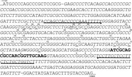
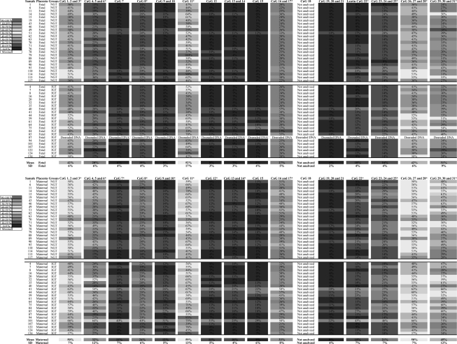
DNA methylation analyses targeted the region of the leptin gene promoter that has already been shown to be subjected to demethylation associated with leptin gene transcription activation during preadipocyte maturation into adipocyte (6). In fact, our analyses covered 31 of 42 cytosines located within the leptin gene proximal promoter CpG island (Fig. 1, supplementary Table 1, available in an online appendix at http://care.diabetesjournals.org/cgi/content/full/dc10-1024/DC1). Mean DNA methylation levels >10% were observed for 20 and 26 CpG sites in the fetal and the maternal placental sides, respectively (Fig. 2). Within the same placental side, CpG site pairwise DNA methylation correlation was moderate to high (ρ ≥0.50, P ≤ 0.0001). However, most of the correlation between fetal side and maternal side CpG methylation did not reach the level of statistical significance (ρ ≤0.30, P > 0.05) (data not shown).
Figure 1.
Leptin gene CpG island proximal promoter region. The forward CpG island sequence is shown in italics and delimited by hyphens. The leptin exon 1 sequence is in bold type. Arrows indicate both PCR primer sequences. Underlined cytosines have already been associated with demethylation and leptin gene transcription activation (6). The sequence has been numbered relative to the first leptin gene codon (exon 2; Ensembl release 56 [September 2009]). The CpG sites have been numbered relative to the 3′ of the amplicon. The CpG no. 31 corresponds to −204 position in Melzner et al. (6).
Figure 2.
DNA methylation levels for each CpG site and sample analyzed. *Cytosines that contributed to the summary statistic.
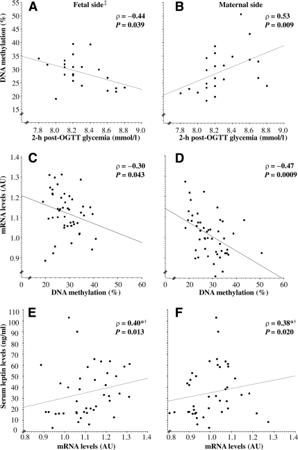
No significant difference was found between groups for mean leptin gene DNA methylation levels (Table 1). However, mean DNA methylation was found to be significantly correlated with glucose levels (2-h post-OGTT) in the IGT group (Fig. 3A and B). Interestingly, the correlation coefficient showed opposite trends between fetal and maternal placental tissues (Fig. 3A and B). Of note, most CpG sites provided very similar results when analyzed separately (data not shown). No correlation between leptin DNA methylation and glucose levels 2-h post-OGTT was observed in the NGT group (n = 25) (data not shown).
Figure 3.
Correlations between leptin DNA methylation and 2-h post-OGTT glucose levels, within the IGT group (A and B), leptin DNA methylation and mRNA levels (C and D); and leptin mRNA levels and circulating concentrations measured at the end of the third trimester (between weeks 36 and 37) of pregnancy (E and F). IGT was defined as a 2-h glucose ≥7.8 mmol/l after a 75-g OGTT performed between 24 and 28 weeks of gestation. ‡Leptin DNA methylation n = 47 (NGT = 25 and IGT = 22). *Adjusted for first trimester BMI. †n = 43 (IGT [−] = 23 and IGT [+] = 20). AU, arbitrary units.
The functional impacts of the observed leptin gene DNA methylation variability on leptin gene transcription and circulating levels were then assessed. On the one hand, we found that the leptin gene DNA methylation was associated with decreased leptin gene mRNA levels in both types of placental tissues (Fig. 3C and D). On the other hand, although increased leptin gene expression levels in both the fetal and the maternal sides were correlated with third trimester circulating leptin levels (corrected for first trimester BMI) (Fig. 3E and F), the correlations between DNA methylation and circulating (third trimester serum and cord blood) concentrations were not found to be significant.
CONCLUSIONS
To the best of our knowledge, this is the first study in humans focusing on the effect of gestational IGT (GDM) on the newborn epigenetic profile. The most important finding is that placental DNA methylation is correlated with mother's glycemia during pregnancy. Interestingly, the leptin DNA methylation showed adaptation only within the group above the IGT diagnostic criteria threshold (2-h post-OGTT glycemia >7.8 mmol/l), suggesting that the leptin gene epigenetic profile will be affected by plasma glucose levels only above a critical threshold.
Although the fetal programming hypothesis is now well recognized, only a few studies have provided direct evidence that may support this concept. Of these, Tobi et al. (14) have shown that DNA methylation at several genome loci harboring genes implicated in growth, development, and energy metabolism (including the leptin gene) was dysregulated in human peripheral blood mononuclear cells obtained from adult subjects who had experienced prenatal exposure to famine decades before. We have observed that the correlations between 2-h post-OGTT glucose and placental DNA methylation performed at the end of second trimester suggest that IGT-related DNA methylation adaptations are long lasting. Although, DNA methylation adaptations may have arisen during critical windows of fetal development (first trimester) (15), we cannot rule out the possibility that the placental DNA methylation adaptation may also be related to the mother's hyperglycemia at the end of gestation. Nevertheless, these results suggest that dysregulation of the DNA methylation profile may potentially have long-term consequences and suggest that the IGT-related DNA methylation changes we have observed have the potential to account for a long-term and possibly transgenerational (16,17) risk to develop obesity and type 2 diabetes associated with fetal programming. Accordingly, if GDM could be detected earlier and treated rapidly, IGT would probably be prevented along with epigenetic changes. This finding reinforces the need to identify early GDM markers.
Interestingly, the correlations between 2-h post-OGTT glucose levels and leptin DNA methylation showed opposite trends in fetal and maternal sides suggesting that both placental sides are differently affected by the mother's IGT. Although the biological reasons will not be simple to explain, we can hypothesize that the maternofetal glucose transport dysregulation associated with GDM (18) contributes to expose both sides of the placenta to different glucose and insulin concentrations. In accordance and contrary to what we have observed in maternal circulation, cord blood glucose and insulin levels failed to be correlated with each other, suggesting that glucose metabolism is fairly different on both sides of the placenta and may thus have a distinctive influence on the epigenetic profile. Furthermore, although the 2-h post-OGTT glycemia explains between 19 and 28% of the DNA methylation variability at the leptin gene locus, a significant portion of the variability remains to be explained by other factors. Identifying those factors will be the focus of further studies and could possibly explain the DNA methylation differences between both sides of the placenta.
We have also shown that epigenetic variability at the leptin gene locus has functional consequences. However, although placental leptin gene expression levels have been associated with its circulating concentration, leptin gene DNA methylation itself has not been associated with its plasma levels. This raises the question about the influence of the placenta on leptin circulating levels. Although this influence is not obvious to assess, our results suggest that placental leptin gene expression could account for up to 16% of the variance of maternal leptin circulating levels. Nevertheless, it is likely that the impact of placental leptin DNA methylation variability on leptin circulating levels could have been missed because of the masking effect of other factors such as the adipose tissue leptin production. In all, the placental contribution to leptin circulating levels is small but significant, and the relationship between leptin DNA methylation and circulating levels does not seem to be as straightforward as first thought. Nevertheless, the placental leptin levels could have been affected by its DNA methylation variability, but the leptin concentration could not be measured at tissue level in this study. Additional studies are therefore needed to explore this possibility.
It is well accepted that pregnancy is associated with an increased circulating leptin concentration (8), although it is still unclear whether plasma leptin concentration shows differences according to GDM status. A number of studies have been published reporting either increased, decreased, or unchanged concentrations in GDM (19–22). Discrepancies between studies can be explained by GDM treatment (diet and exercise versus insulin) as well as mother's and newborn's adiposity and weight gain differences among studies. Nevertheless, our results are in accordance with three studies reporting hypoleptinemia in similar cohorts of women with mild GDM (23,24) and newborns of mothers with GDM without insulin treatment (25), suggesting that leptin has beneficial effects on the mother's glucose disposal and insulin sensitivity. This could also have short- and long-term consequences on mothers' and newborns' health.
One limitation of the current study is the lack of significant differences in placental leptin DNA methylation and mRNA levels between NGT and IGT groups. Although 48 samples (total 96) can be considered a large sample size for DNA methylation and transcriptomic analyses with this very high phenotyping quality, it is possible that the study was short of statistical power to find significant group differences. Also, we studied women with intermediate glucose intolerance instead of women with GDM. Therefore, we hypothesize that our results should at least remain unchanged with the more stringent GDM diagnosis criteria and may be even more significant. Unfortunately, we cannot verify this last hypothesis with our data because most of the women with glucose perturbations in our study sample had only a moderate glucose increase after the second trimester 75-g OGTT. None of them fulfilled the American Diabetes Association criteria for GDM. Nevertheless, the most important finding was clearly to have shown significant correlations between leptin DNA methylation and 2-h post-OGTT glucose concentration, a recognized indicator of glucose metabolism status during pregnancy.
In summary, our results give a better understanding of the molecular mechanisms involved in fetal programming associated with pregnancy-related IGT. Because DNA methylation can be mitotically stable, leptin gene DNA methylation adaptations to IGT and the parallel transcriptional response that we have observed could have profound short- and long-term phenotypic effects. These effects could therefore contribute to explaining why newborns exposed to a detrimental fetal environment (such as GDM) have an increased risk of developing obesity and type 2 diabetes later in life. Although it is largely speculative, one hypothesis is that increasing maternal glycemia leads to fetal leptin gene DNA demethylation, which leads to higher mRNA levels and subsequently higher leptin levels, possibly promoting leptin resistance and obesity development. Overall, these data provide supportive evidence for potential unfavorable molecular adaptations to the fetal environment. They also concur to support the idea that other genes could contribute to clarifying the concept of molecular fetal programming.
Supplementary Material
Acknowledgments
This project was supported by ECOGENE-21 (principal investigator: Dr. Daniel Gaudet, MD, PhD, Montreal University), the Canadian Institutes of Health Research (CIHR team in community genetics grant CTP-82941), Fonds de la Recherche en Santé du Québec (FRSQ), and Diabète Québec. S.-P.G. is supported by a CIHR/Frederick Banting and Charles Best Canada Graduate Scholarship-Master's Awards and by the FRSQ.
No potential conflicts of interest relevant to this article were reported.
L.B. conceived the study design, performed data analysis/interpretation, and wrote the manuscript. S.T., S.-P.G., and A.M. performed data collection and revised/edited the manuscript, M.S. participated in study design conception. J.St.-P. and P.P. participated in study design conception and revised/edited the manuscript. D.B. participated in study design conception, performed data analysis/interpretation, and revised/edited the manuscript.
We acknowledge the contribution of Sébastien Claveau, MSc; Nadia Mior; and Chantale Aubut, Research Nurse, for their dedicated work in this study. We also express our gratitude to Robert Sladek, MD, PhD, McGill University, and Céline Bélanger, Chicoutimi Hospital, for their thoughtful revision of the manuscript.
Footnotes
The costs of publication of this article were defrayed in part by the payment of page charges. This article must therefore be hereby marked “advertisement” in accordance with 18 U.S.C. Section 1734 solely to indicate this fact.
References
- 1.Barker DJ. Fetal origins of cardiovascular disease. Ann Med 1999;31(Suppl. 1):3–6 [PubMed] [Google Scholar]
- 2.Henikoff S, Matzke MA. Exploring and explaining epigenetic effects. Trends Genet 1997;13:293–295 [DOI] [PubMed] [Google Scholar]
- 3.Jaenisch R, Bird A. Epigenetic regulation of gene expression: how the genome integrates intrinsic and environmental signals. Nat Genet 2003;33(Suppl.):245–254 [DOI] [PubMed] [Google Scholar]
- 4.Kangaspeska S, Stride B, Métivier R, Polycarpou-Schwarz M, Ibberson D, Carmouche RP, Benes V, Gannon F, Reid G. Transient cyclical methylation of promoter DNA. Nature 2008;452:112–115 [DOI] [PubMed] [Google Scholar]
- 5.Bird A. DNA methylation patterns and epigenetic memory. Genes Dev 2002;16:6–21 [DOI] [PubMed] [Google Scholar]
- 6.Melzner I, Scott V, Dorsch K, Fischer P, Wabitsch M, Brüderlein S, Hasel C, Möller P. Leptin gene expression in human preadipocytes is switched on by maturation-induced demethylation of distinct CpGs in its proximal promoter. J Biol Chem 2002;277:45420–45427 [DOI] [PubMed] [Google Scholar]
- 7.Zavalza-Gómez AB, Anaya-Prado R, Rincón-Sánchez AR, Mora-Martínez JM. Adipokines and insulin resistance during pregnancy. Diabetes Res Clin Pract 2008;80:8–15 [DOI] [PubMed] [Google Scholar]
- 8.Frühbeck G. A heliocentric view of leptin. Proc Nutr Soc 2001;60:301–318 [DOI] [PubMed] [Google Scholar]
- 9.Phillips DI, Fall CH, Cooper C, Norman RJ, Robinson JS, Owens PC. Size at birth and plasma leptin concentrations in adult life. Int J Obes Relat Metab Disord 1999;23:1025–1029 [DOI] [PubMed] [Google Scholar]
- 10.Lohman TG, Roche AF, Martorell R. Anthropometric Standardization Reference Manual. Champaign, IL, Human Kinetics, 1988, p. 55–80 [Google Scholar]
- 11.Ehrich M, Nelson MR, Stanssens P, Zabeau M, Liloglou T, Xinarianos G, Cantor CR, Field JK, van den Boom D. Quantitative high-throughput analysis of DNA methylation patterns by base-specific cleavage and mass spectrometry. Proc Natl Acad Sci USA 2005;102:15785–15790 [DOI] [PMC free article] [PubMed] [Google Scholar]
- 12.Meller M, Vadachkoria S, Luthy DA, Williams MA. Evaluation of housekeeping genes in placental comparative expression studies. Placenta 2005;26:601–607 [DOI] [PubMed] [Google Scholar]
- 13.Murthi P, Fitzpatrick E, Borg AJ, Donath S, Brennecke SP, Kalionis B. GAPDH, 18S rRNA and YWHAZ are suitable endogenous reference genes for relative gene expression studies in placental tissues from human idiopathic fetal growth restriction. Placenta 2008;29:798–801 [DOI] [PubMed] [Google Scholar]
- 14.Tobi EW, Lumey LH, Talens RP, Kremer D, Putter H, Stein AD, Slagboom PE, Heijmans BT. DNA methylation differences after exposure to prenatal famine are common and timing- and sex-specific. Hum Mol Genet 2009;18:4046–4053 [DOI] [PMC free article] [PubMed] [Google Scholar]
- 15.Weaver JR, Susiarjo M, Bartolomei MS. Imprinting and epigenetic changes in the early embryo. Mamm Genome 2009;20:532–543 [DOI] [PubMed] [Google Scholar]
- 16.Margetts BM, Mohd Yusof S, Al Dallal Z, Jackson AA. Persistence of lower birth weight in second generation South Asian babies born in the United Kingdom. J Epidemiol Community Health 2002;56:684–687 [DOI] [PMC free article] [PubMed] [Google Scholar]
- 17.Drake AJ, Walker BR. The intergenerational effects of fetal programming: non-genomic mechanisms for the inheritance of low birth weight and cardiovascular risk. J Endocrinol 2004;180:1–16 [DOI] [PubMed] [Google Scholar]
- 18.Osmond DT, King RG, Brennecke SP, Gude NM. Placental glucose transport and utilisation is altered at term in insulin-treated, gestational-diabetic patients. Diabetologia 2001;44:1133–1139 [DOI] [PubMed] [Google Scholar]
- 19.Kleiblova P, Dostalova I, Bartlova M, Lacinova Z, Ticha I, Krejci V, Springer D, Kleibl Z, Haluzik M. Expression of adipokines and estrogen receptors in adipose tissue and placenta of patients with gestational diabetes mellitus. Mol Cell Endocrinol 2010;314:150–156 [DOI] [PubMed] [Google Scholar]
- 20.Sagawa N, Yura S, Itoh H, Kakui K, Takemura M, Nuamah MA, Ogawa Y, Masuzaki H, Nakao K, Fujii S. Possible role of placental leptin in pregnancy: a review. Endocrine 2002;19:65–71 [DOI] [PubMed] [Google Scholar]
- 21.Simmons D, Breier BH. Fetal overnutrition in polynesian pregnancies and in gestational diabetes may lead to dysregulation of the adipoinsular axis in offspring. Diabetes Care 2002;25:1539–1544 [DOI] [PubMed] [Google Scholar]
- 22.Yilmaz O, Kucuk M, Ilgin A, Dagdelen M. Assessment of insulin sensitivity/resistance and their relations with leptin concentrations and anthropometric measures in a pregnant population with and without gestational diabetes mellitus. J Diabetes Complications 2010;24:109–114 [DOI] [PubMed] [Google Scholar]
- 23.Festa A, Shnawa N, Krugluger W, Hopmeier P, Schernthaner G, Haffner SM. Relative hypoleptinaemia in women with mild gestational diabetes mellitus. Diabet Med 1999;16:656–662 [DOI] [PubMed] [Google Scholar]
- 24.McLachlan KA, O'Neal D, Jenkins A, Alford FP. Do adiponectin, TNFα, leptin and CRP relate to insulin resistance in pregnancy? Studies in women with and without gestational diabetes, during and after pregnancy. Diabetes Metab Res Rev 2006;22:131–138 [DOI] [PubMed] [Google Scholar]
- 25.Ng PC, Lam CW, Lee CH, Wong GW, Fok TF, Wong E, Ma KC, Chan IH. Leptin and metabolic hormones in infants of diabetic mothers. Arch Dis Child Fetal Neonatal Ed 2000;83:F193–F197 [DOI] [PMC free article] [PubMed] [Google Scholar]
Associated Data
This section collects any data citations, data availability statements, or supplementary materials included in this article.


