Abstract
Purpose
The aim of this study was to evaluate possible predictive variables for the outcome of shock wave lithotripsy (SWL) of renal stones in a single center.
Materials and Methods
Between March 2008 and March 2010, a retrospective review was performed of 115 patients who underwent SWL for solitary renal stones. The patients' characteristics and stone size, location, skin-to-stone distance (SSD), and Hounsfield units (HU) of stone were reviewed. The impact of the possible predictors on the disintegration of the stones was evaluated by logistic regression analysis. Receiver operator characteristic (ROC) curves were generated to compare the predictive powers of the variables.
Results
Seventy-nine patients (68.7%) had successful outcomes, whereas 36 patients (31.3%) had residual stones. Significant differences were found in the mean size and mean HU of the stones (size: 8.34±3.58 mm vs. 13.57±5.41 mm, p<0.001; HU: 675.29±254.34 vs. 1,075.00±290.41, p<0.001). In the unadjusted model, age, stone size, and stone density were significant predictors. In the reduced model, stone density and size were significant predictors for the outcome of SWL. The area under the ROC curve (AUC) was significantly higher for stone density and size than for the other parameters, but the AUC between stone density and size did not differ significantly (stone density: 0.874, stone size: 0.827, p=0.388).
Conclusions
Stone density and size were significant predictors of the outcome of SWL for renal stones less than 2.0 cm in diameter. We should consider HU and stone size when making decisions on the treatment of renal stones.
Keywords: Kidney calculi, Lithotripsy, X-ray computed tomography
INTRODUCTION
Extracorporeal shock wave lithotripsy (SWL) has been the most common treatment of choice, especially for small renal stones (<2 cm), since its introduction by Chaussy et al [1]. The effectiveness of SWL on kidney stones varies from 69.5% up to 99% [2-4]. Failure to fragment by SWL could result in unnecessary exposure of the renal parenchyme to shock waves and the requirement for an alternative procedure, which increases medical cost [5]. Therefore, it is important to identify patients who would benefit most from SWL before treatment. There have been many reports on the factors predicting stone disintegration by SWL. In particular, radiographic findings have been studied, such as the skin-to-stone distance (SSD) and the Hounsfield unit (HU) for measuring the density of the stone on noncontrast computed tomography (NCCT) [6,7]. Patient characteristics such as body mass index (BMI) [6] have also been reported as significant predictors of the results of SWL.
In this study, we evaluated possible predictive variables for the outcome of SWL of renal stones to help to better define the indications for SWL treatment and to determine the efficiency with which we can check HU and stone size.
MATERIALS AND METHODS
Between March 2008 and March 2010, 115 patients (71 men and 44 women) with solitary renal stones were evaluated. Patients were included if they had a stone of 0.5-2.0 cm in the longest dimension in a plain film. Patients who had multiple stones on the same side, patients with a stone size >20 mm in maximal diameter, patients with radiolucent stones, cases followed up elsewhere, and cases that required a stent or developed steinstrasse and active urosepsis during the therapy were excluded.
NCCT was performed for all patients with a multislice helical computed tomography (CT) scanner (64-channel, multi-detector computed tomography, Aquilion®, Toshiba, Tokyo, Japan). The images were obtained by use of the high-quality mode at 300 mA, 120 kVp, and 5 mm collimation.
The patients underwent SWL with the electromagnetic lithotripter (Compact Delta®, Dornier, Wessling, Germany) under fluoroscopy. In each treatment, the maximum number of shock waves was limited to 3,000 (mean shock waves number: 2,900) at a maximum energy of 3.0 kVs increasing gradually from 0.1 kV. Repeated treatment was carried out if inadequate fragmentation was observed. The result of treatment was evaluated by KUB at 1 month after the last lithotripsy. If there were residual fragments larger than 3 mm after three sessions per week, the case was considered as a failure.
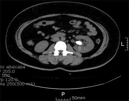
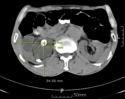
Multiple variables including patient characteristics such as sex and age and calculus characteristics such as location, size, density, and SSD were collected. Stone location was categorized as lower calyx and non lower calyx. For the measurement of stone density, three regions of interest with a diameter of 2 mm were drawn on the stone at an axial plane of the NCCT where the stone length was the longest. The mean number of HU calculated from the 3 regions represented the density of the stone on NCCT (Fig. 1). [6]. The SSD was calculated by measuring three distances from the stone to the skin at 0°, 45°, and 90° by using radiographic calipers, and the average of these three values was calculated to represent the SSD for each stone as described in the literature (Fig. 2) [6].
FIG. 1.
Three regions of interest with a diameter of 2 mm were drawn on the stone in the axial plane of NCCT where the stone length was the longest. The mean number of HU calculated from the 3 regions represents the density of the stone. NCCT: non-contrast computed tomography, HU: Hounsfield unit.
FIG. 2.
Measurement of the skin-to-stone distance at 0°, 45°, and 90° on an axial scan of NCCT. NCCT: non-contrast computed tomography.
Statistical analysis was performed by using Student's t-test for continuous data and Fisher's exact test or chi-square test for categorical data. We used logistic regression analysis to determine the factors influencing the outcome of treatment. The impacts of variables were assessed by logistic regression analysis and those variables with a significant association with SWL outcome were further evaluated by multivariate (logistic regression) analysis. A 5% level of significance was used for all statistical testing, and all statistical tests were two-sided. Receiver operator characteristic (ROC) curves were generated to compare the predictive power of the variables. Medcalc software ver. 9.6.40 (Medcalc, Mariakerke, Belgium) was used for the data analysis.
RESULTS
The mean age of the patients was 50.5±14.0 years. The mean size and density of the stones were 10.1+5.0 mm and 799.5±321.5 HU, respectively.
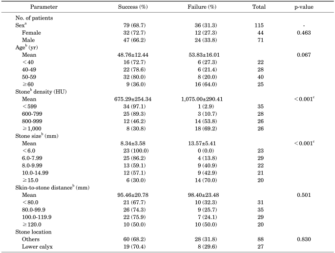
Of the 115 patients studied, 79 (68.7%) patients had successful outcomes, whereas 36 (31.3%) patients had residual stones. Between the two groups, significant differences were found in the size and HU of the stones. The mean HU of the success and failure groups were 675.29±254.34 HU and 1,075.00±290.41 HU (p<0.001), and the mean sizes of the stone were 8.34±3.58 mm in the success group and 13.57±5.41 mm in the failure group (p<0.001), respectively. However, patient age, SSD, and location of lower calyceal stones did not differ significantly between the two groups (Table 1).
TABLE 1.
Comparisons of characteristics between the shock wave lithotripsy success and failure groups
HU: Hounsfield units, a: chi-square test, b: Student's t-test, c: p<0.05
In the univariate analysis, stone density and stone size were significant predictors of the outcome of SWL (Table 2). There were no significant differences in the prediction of outcome of SWL according to SSD or lower calyceal stone location.
TABLE 2.
Influence of patient and stone characteristics on failure of disintegration by shock wave lithotripsy
OR: odds ratio, Adjusted model: adjusted for sex, age, stone density, stone size, skin-to-stone distance, and stone location, Reduced model: adjusted for all confounders after analysis of unadjusted model, a: p<0.05 in unadjusted model, b: p<0.05 in adjusted model, c: p<0.05 in reduced model
In the multivariable analysis taking all factors into account, stone density and size were strongly associated with the outcome of SWL (Table 2). No other factors were significant. When sex and lower calyceal stone location were eliminated in the reduced model, stone density and size were still significant predictors of the outcome of SWL (Table 2).
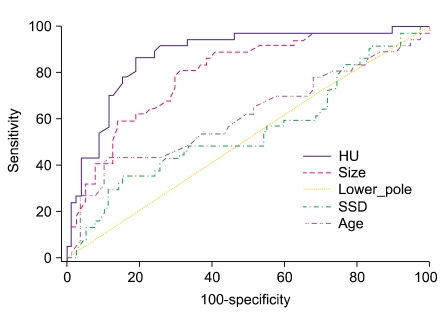
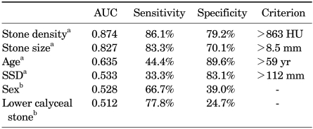
The ROC curves of all parameters were obtained for the prediction of an unsuccessful outcome of SWL (Fig. 3). Stone density showed that 863 HU was the ideal cutoff value for the prediction of failure of SWL with sensitivity of 86.1% and specificity of 79.2% (95% confidence interval) (Table 3). Stone size showed that 8.5 mm is the ideal cutoff value for the prediction of failure of SWL with sensitivity of 83.3% and specificity of 70.1% (95% confidence interval) (Table 3). In the comparison of ROC curves to test the statistical significance of the difference among the areas under different ROC curves, the area under the ROC curve (AUC) was significantly higher for stone density and size than for the other parameters, but the AUC between stone density and size did not differ significantly (stone density: 0.874; stone size: 0.827, p=0.388) (Fig. 3).
FIG. 3.
Comparison of ROC curves to test the statistical significance of the difference between the area under different ROC curves. In pairwise comparison of all predictors for the outcome of shock wave lithotripsy, stone density and stone size was not different (AUC difference: 0.0465, p=0.388). ROC: receiver operator characteristic, AUC: area under the ROC curve.
TABLE 3.
Comparison of receiver operating characteristic curve analysis for the prediction of outcome of shock wave lithotripsy
AUC: area under the receiver operating characteristic curve, SSD: skin-to-stone distance, a: continous variables, b: categorical variables
DISCUSSION
Although NCCT has become the most sensitive and accurate imaging modality for the diagnosis of urinary calculi [8-12], the intravenous pyelogram (IVP) is still widely used in Korea. Until 2 years ago, the Korean health insurance review agency prohibited the use of computed tomography as the initial imaging modality in the diagnosis of stone disease. However, IVP has many limitations in detecting urinary calculi owing to the interference with bowel gas or bony structures, and it also exposes patients to a risk of renal insufficiency and allergy by administration of contrast material [13]. Also, IVP has a limitation in diagnosis of radiolucent and small stones.
By contrast, NCCT is noninvasive and it can detect not only radiolucent and small stones but also other diseases of the urinary tract and other organs (e.g., renal mass, duplicated ureter, bladder mass, gall bladder stone, etc.) [14]. NCCT can precisely localize the site of the stone without the use of contrast. Besides, recent studies have used high-resolution CT protocols to predict the outcome of SWL. For example, Gupta et al concluded that the worst outcome was in patients with a calculus density >750 HU and a stone diameter of >1.1 cm, because 77% of those patients needed more than three sessions of SWL and the clearance rate was 60% [15].
The factors associated with the outcome of SWL have been discussed in many studies over the past decade [16-21]. Stone characteristics, such as size and location, have been reported as significant predictors of the results of SWL by other authors [22,23]. However, lower calyceal stone location was not a significant predictive variable in this study. This is because of the different patient populations in the studies. Also, the definition of success of SWL in our study was to fragment the stones into pieces of less than 3 mm and not to excrete them all. This may be another explanation for the difference in results.
There have been reports that the composition of the stones is related to the fragmentation of the stone by SWL [24]. However, knowing the stone composition before treatment is difficult and may not be sufficient to allow for prediction of the response to SWL. Therefore, pre-SWL radiographic examination should focus on those radiographic characteristics that can influence SWL outcome rather than stone composition.
We also found that the density of the stone was a significant predictor of SWL outcome. To date, a few studies have reported that stone density is a significant predictive factor for SWL outcome. For example, Pareek et al found that the mean HU values of stones were significantly higher in patients with residual stones [25,26], and Joseph et al found a positive correlation between the number of shock waves required to treat a stone and its HU value [27]. Wang et al suggested cutoff values (the stone density >900 HU and volume >700 mm3) for predicting SWL failure [28]. In the present study, the cutoff value was >863 HU, which is lower than the cutoffs reported in other studies. El-Nahas et al suggested that the differences in the cutoff values that predicted extracorporeal SWL failure may be due to different inclusion criteria, the use of different CT protocols, or the measurement of different endpoints (e.g., failure of disintegration, the need for multiple sessions, or rate of residual stones) in these studies [7].
Several patient characteristics have been suggested to influence SWL results. Abe et al reported that older patients are likely to have difficulty in successful SWL [29]. In the univariate and multivariable analysis in the present study, however, age was not a significant factor. Abdel-Khalek et al also showed that age is not a significant predictor of SWL outcome [19]. Other studies have reported that higher BMI and SSD are significant predictors for SWL failure [6,30]. In the present study, SSD did not reach statistical significance even in the univariate analysis. Our patients' relatively lower SSD than those of Western patients could be the reason for that. Also, we could not get exact information on the patients' BMI for many patients in real practice. Therefore, we did not consider BMI as a patient characteristic. However, because this study was performed retrospectively and the number of patients studied was not large enough, further studies with large numbers of patients and a standardized CT protocol are needed to clarify this important point.
CONCLUSIONS
NCCT is noninvasive and useful for obtaining a lot of information about the patient and urinary calculi. In this study, stone density and size were significant predictors of the outcome of SWL for renal stones less than 2.0 cm in diameter. We should consider HU and stone size when making decisions on the treatment of renal stones.
Footnotes
This work was supported by grant from Inje University, 2008.
The authors have nothing to disclose.
References
- 1.Chaussy C, Brendel W, Schmiedt E. Extracorporeally induced destruction of kidney stones by shock waves. Lancet. 1980;2:1265–1268. doi: 10.1016/s0140-6736(80)92335-1. [DOI] [PubMed] [Google Scholar]
- 2.Lingeman JE, Newman D, Mertz JH, Mosbaugh PG, Steele RE, Kahnoski RJ, et al. Extracorporeal shock wave lithotripsy: the Methodist Hospital of Indiana experience. J Urol. 1986;135:1134–1137. doi: 10.1016/s0022-5347(17)46016-2. [DOI] [PubMed] [Google Scholar]
- 3.Ehreth JT, Drach GW, Arnett ML, Barnett RB, Govan D, Lingeman J, et al. Extracorporeal shock wave lithotripsy: multicenter study of kidney and upper ureter versus middle and lower ureter treatments. J Urol. 1994;152:1379–1385. doi: 10.1016/s0022-5347(17)32425-4. [DOI] [PubMed] [Google Scholar]
- 4.Cass AS. Comparison of first generation (Dornier HM3) and second generation (Medstone STS) lithotriptors: treatment results with 13,864 renal and ureteral calculi. J Urol. 1995;153:588–592. doi: 10.1097/00005392-199503000-00006. [DOI] [PubMed] [Google Scholar]
- 5.Lingeman JE, Woods JR, Toth PD. Blood pressure changes following extracorporeal shock wave lithotripsy and other forms of treatment for nephrolithiasis. JAMA. 1990;263:1789–1794. [PubMed] [Google Scholar]
- 6.Kim JH, Moon YT. Predicting the therapeutic effect of extracorporeal shockwave lithotripsy by non-enhanced computed tomography in renal stones. Korean J Urol. 2008;49:252–256. [Google Scholar]
- 7.El-Nahas AR, El-Assmy AM, Mansour O, Sheir KZ. A prospective multivariate analysis of factors predicting stone disintegration by extracorporeal shock wave lithotripsy: the value of high-resolution noncontrast computed tomography. Eur Urol. 2007;51:1688–1693. doi: 10.1016/j.eururo.2006.11.048. [DOI] [PubMed] [Google Scholar]
- 8.Dalla Palma L, Pozzi-Mucelli R, Stacul F. Present-day imaging of patients with renal colic. Eur Radiol. 2001;11:4–17. doi: 10.1007/s003300000589. [DOI] [PubMed] [Google Scholar]
- 9.Katz DS, Lane MJ, Sommer FG. Non-contrast spiral CT for patients with suspected renal colic. Eur Radiol. 1997;7:680–685. doi: 10.1007/BF02742925. [DOI] [PubMed] [Google Scholar]
- 10.Wang LJ, Ng CJ, Chen JC, Chiu TF, Wong YC. Diagnosis of acute flank pain caused by ureteral stones: value of combined direct and indirect signs on IVU and unenhanced helical CT. Eur Radiol. 2004;14:1634–1640. doi: 10.1007/s00330-004-2309-1. [DOI] [PubMed] [Google Scholar]
- 11.Yilmaz S, Sindel T, Arslan G, Ozkaynak C, Karaali K, Kabaalioglu A, et al. Renal colic: comparison of spiral CT, US and IVU in the detection of ureteral calculi. Eur Radiol. 1998;8:212–217. doi: 10.1007/s003300050364. [DOI] [PubMed] [Google Scholar]
- 12.Arac M, Celik H, Oner AY, Gultekin S, Gumus T, Kosar S. Distinguishing pelvic phleboliths from distal ureteral calculi: thin-slice CT findings. Eur Radiol. 2005;15:65–70. doi: 10.1007/s00330-004-2511-1. [DOI] [PubMed] [Google Scholar]
- 13.Preminger GM, Vieweg J, Leder RA, Nelson RC. Urolithiasis: detection and management with unenhanced spiral CT--a urologic perspective. Radiology. 1998;207:308–309. doi: 10.1148/radiology.207.2.9577473. [DOI] [PubMed] [Google Scholar]
- 14.Ahn SS, Lee SH, Kang I. The value of non-enhanced spiral CT in the diagnosis of suspected urolithiasis. Korean J Urol. 2002;43:1008–1013. [Google Scholar]
- 15.Gupta NP, Ansari MS, Kesarvani P, Kapoor A, Mukhopadhyay S. Role of computed tomography with no contrast medium enhancement in predicting the outcome of extracorporeal shock wave lithotripsy for urinary calculi. BJU Int. 2005;95:1285–1288. doi: 10.1111/j.1464-410X.2005.05520.x. [DOI] [PubMed] [Google Scholar]
- 16.Kim HS, Jang SW, Jeong YB, Kim YG, Kim JS. The usefulness of unenhanced helical computerized tomography in patients with urinary calculi. Korean J Urol. 2003;44:796–800. [Google Scholar]
- 17.Obek C, Onal B, Kantay K, Kalkan M, Yalcin V, Oner A, et al. The efficacy of extracorporeal shock wave lithotripsy for isolated lower pole calculi compared with isolated middle and upper caliceal calculi. J Urol. 2001;166:2081–2084. doi: 10.1016/s0022-5347(05)65509-7. [DOI] [PubMed] [Google Scholar]
- 18.Shiroyanagi Y, Yagisawa T, Nanri M, Kobayashi C, Toma H. Factors associated with failure of extracorporeal shock-wave lithotripsy for ureteral stones using Dornier lithotripter U/50. Int J Urol. 2002;9:304–307. doi: 10.1046/j.1442-2042.2002.00475.x. [DOI] [PubMed] [Google Scholar]
- 19.Abdel-Khalek M, Sheir K, Elsobky E, Showkey S, Kenawy M. Prognostic factors for extracorporeal shock-wave lithotripsy of ureteric stones--a multivariate analysis study. Scand J Urol Nephrol. 2003;37:413–418. doi: 10.1080/00365590310006255. [DOI] [PubMed] [Google Scholar]
- 20.Abdel-Khalek M, Sheir KZ, Mokhtar AA, Eraky I, Kenawy M, Bazeed M. Prediction of success rate after extracorporeal shock-wave lithotripsy of renal stones--a multivariate analysis model. Scand J Urol Nephrol. 2004;38:161–167. doi: 10.1080/00365590310022626. [DOI] [PubMed] [Google Scholar]
- 21.Srivastava A, Zaman W, Singh V, Mandhani A, Kumar A, Singh U. Efficacy of extracorporeal shock wave lithotripsy for solitary lower calyceal stone: a statistical model. BJU Int. 2004;93:364–368. doi: 10.1111/j.1464-410x.2003.04618.x. [DOI] [PubMed] [Google Scholar]
- 22.Lingeman JE, Coury TA, Newman DM, Kahnoski RJ, Mertz JH, Mosbaugh PG, et al. Comparison of results and morbidity of percutaneous nephrostolithotomy and extracorporeal shock wave lithotripsy. J Urol. 1987;138:485–490. doi: 10.1016/s0022-5347(17)43236-8. [DOI] [PubMed] [Google Scholar]
- 23.Psihramis KE, Jewett MA, Bombardier C, Caron D, Ryan M Toronto Lithotripsy Associates. Lithostar extracorporeal shock wave lithotripsy: the first 1,000 patients. J Urol. 1992;147:1006–1009. doi: 10.1016/s0022-5347(17)37447-5. [DOI] [PubMed] [Google Scholar]
- 24.Daudon M, Donsimoni R, Hennequin C, Fellahi S, Le Moel G, Paris M, et al. Sex- and age-related composition of 10 617 calculi analyzed by infrared spectroscopy. Urol Res. 1995;23:319–326. doi: 10.1007/BF00300021. [DOI] [PubMed] [Google Scholar]
- 25.Pareek G, Armenakas NA, Fracchia JA. Hounsfield units on computerized tomography predict stone-free rates after extracorporeal shock wave lithotripsy. J Urol. 2003;169:1679–1681. doi: 10.1097/01.ju.0000055608.92069.3a. [DOI] [PubMed] [Google Scholar]
- 26.Pareek G, Armenakas NA, Panagopoulos G, Bruno JJ, Fracchia JA. Extracorporeal shock wave lithotripsy success based on body mass index and Hounsfield units. Urology. 2005;65:33–36. doi: 10.1016/j.urology.2004.08.004. [DOI] [PubMed] [Google Scholar]
- 27.Joseph P, Mandal AK, Singh SK, Mandal P, Sankhwar SN, Sharma SK. Computerized tomography attenuation value of renal calculus: can it predict successful fragmentation of the calculus by extracorporeal shock wave lithotripsy? A preliminary study. J Urol. 2002;167:1968–1971. doi: 10.1016/s0022-5347(05)65064-1. [DOI] [PubMed] [Google Scholar]
- 28.Wang LJ, Wong YC, Chuang CK, Chu SH, Chen CS, See LC, et al. Predictions of outcomes of renal stones after extracorporeal shock wave lithotripsy from stone characteristics determined by unenhanced helical computed tomography: a multivariate analysis. Eur Radiol. 2005;15:2238–2243. doi: 10.1007/s00330-005-2742-9. [DOI] [PubMed] [Google Scholar]
- 29.Abe T, Akakura K, Kawaguchi M, Ueda T, Ichikawa T, Ito H, et al. Outcomes of shockwave lithotripsy for upper urinary-tract stones: a large-scale study at a single institution. J Endourol. 2005;19:768–773. doi: 10.1089/end.2005.19.768. [DOI] [PubMed] [Google Scholar]
- 30.Pareek G, Hedican SP, Lee FT, Jr, Nakada SY. Shock wave lithotripsy success determined by skin-to-stone distance on computed tomography. Urology. 2005;66:941–944. doi: 10.1016/j.urology.2005.05.011. [DOI] [PubMed] [Google Scholar]


