Abstract
Setting:
Outpatient clinic of a spinal cord injury rehabilitation center.
Design:
Case report.
Participant:
A 40-year-old man with a 20-year history of C4 complete tetraplegia complained of 5 years of excessive intermittent left-sided sweating. The sweating occurred only in the seated upright position. There was no associated headache, blurred vision, or blood pressure variability.
Findings:
When examined upright, the patient sweated excessively on the left face and body. When he was laid down, sweating ceased. Skin examination revealed intact ischial regions. Pressure applied to the right ischium for several minutes caused sweating to recur on the left forehead, but it then subsided with release of pressure. This phenomenon was repeatable. Local lidocaine injection in the subcutaneous tissues around the right ischium and subsequent use of lidocaine transdermal patches halted the contralateral sweating in the upright position. Pressure mapping analysis showed increased pressure in the region of the right ischial tuberosity. The patient's gel cushion was replaced with an air-filled cushion, providing significant ongoing relief from the hyperhidrosis.
Conclusion/Clinical Relevance:
Unilateral hyperhidrosis can be caused by a contralateral source of irritation. Use of techniques that interrupt the afferent arm of the autonomic pathway may be effective in the management of hyperhidrosis in individuals with spinal cord injury.
Keywords: Spinal cord injuries; Heterotopic ossification; Tetraplegia; Hyperhidrosis; Lidocaine, transdermal; Cushions, air-filled, gel
INTRODUCTION
Hyperhidrosis, or excessive sweating, is a relatively common complaint in patients with spinal cord injury (SCI). It can be accompanied by significant social and emotional distress, negatively affecting an individual's quality of life. The symptom can be associated with other more serious conditions such as autonomic dysreflexia (1) or can exist on its own. The prevalence of hyperhidrosis in SCI has been estimated to be 26%. In one study of 41 patients with annoying sweating, 13 (32%) had some underlying cause of the condition, whereas 28 (68%) had no somatic reason for the excessive sweating (2).
Specific underlying identifiable causes of hyperhidrosis in SCI have included syringomyelia (3) and afferent stimuli from the bladder or bowel during voiding or defecation (4,5). Also in the differential are causes that would be encountered in the general (non-SCI) population. Treatment of hyperhidrosis in persons with SCI generally first involves the identification of underlying causes, followed by the removal of any obvious noxious afferent stimuli (such as urinary obstruction or fecal impaction), similar to what would be done in the management of autonomic dysreflexia (1).
The following case report describes unilateral hyperhidrosis secondary to a subtle contralateral noxious stimulus.
CASE REPORT
A 40-year-old African-American man presented with long-standing C4 ASIA Impairment Scale A tetraplegia secondary to a cervical gunshot wound more than 20 years ago. He was status postacute inpatient rehabilitation on a specialized spinal cord unit followed by home and outpatient physical therapy. He lives in the community without need for ventilator support and is followed periodically by his primary care physician, the specialized outpatient spinal cord clinic, and the urology service.
He has been remarkably healthy medically for the most part except for complaints of orthostatic hypotension not accompanied by hyperhidrosis, which has been successfully treated with midodrine on an as-needed basis.
Approximately 15 years postinjury, he started to complain of excessive sweating affecting the left side of his face, left torso, left shoulder, and left arm. The sweating had been episodic, occurring only when he was sitting upright. It was relieved by crossing his legs and upon returning to the supine position. It was also relieved by leaning forward when sitting. He had been using a deep contour gel seat cushion since the time of his initial injury. There were no complaints of headache or blurred vision. His blood pressure did not increase during these episodes. The patient had been tried on a scopolamine patch without any relief of the sweating. A trial of amitriptyline was also not successful. Four years ago, he underwent a left stellate ganglion block under fluoroscopy. This procedure provided 6 hours of relief from his hyperhidrosis. He was then referred to cardiothoracic surgery for consideration of a sympathectomy, but the surgeon thought the procedure would be too risky given the high level of tetraplegia.
The patient had a history of one episode of skin breakdown 10 years prior to presentation, requiring a skin graft on the left hip. Approximately 2 years ago, he experienced an episode of chest pain accompanied by elevated blood pressure. A chest and abdominal computed tomography scan with contrast allowed for diagnosis of a dissecting abdominal aneurysm. He was treated conservatively in the acute care setting with beta blockers with good effect. The use of beta blockers had no effect on the hyperhidrosis.
The patient had undergone a sphincterotomy, most recently revised a year ago. He used the Crede maneuver (applied by caregivers) with an external condom catheter for bladder management. Annual renal ultrasounds have revealed no hydronephrosis or other abnormalities. His urodynamic study before the last sphincterotomy revealed detrusor-external sphincter dyssynergia, but postoperatively his collecting system returned to low pressure. The sphincterotomy revision did not lessen the patient's hyperhidrosis. The patient has managed his bowel with biweekly sodium phosphate (Fleet) enemas and stool softeners. He did not use suppositories because they worsened his hyperhidrosis.
His regularly scheduled oral medications included diazepam 2.5 mg 3 times a day, baclofen 20 mg 3 times a day, tamsulosin 0.4 mg once daily, ranitidine 150 mg twice daily, and midodrine 5 mg every morning as needed for lightheadedness.
On physical examination, his vital signs included a seated blood pressure of 99/65 mmHg, with a regular pulse of 64 beats/min. When the patient was examined sitting up, obvious diaphoresis was noted covering exclusively the left side of the face and upper chest and left arm. Cardiopulmonary examination was unremarkable. There was a well-healed right ischial area of hypopigmentation (pink-colored skin), but there was no breakdown. Passive range of motion of the bilateral hips was normal in excursion. Neurologic examination revealed symmetric 2-mm pupils without any ptosis. The remainder of the cranial nerve examination was unremarkable. There was no voluntary movement of any ASIA musculature. There was no increase in tone on passive range of motion of the arms or the legs. There was unsustained clonus at the ankles bilaterally. Sensory examination revealed normal sensation to pinprick and light touch in the C4 dermatome and absent sensation in both modalities in the dermatomes at C5 and below.
When the patient was placed in the left lateral decubitus position, it was noted that the unilateral left-sided sweating stopped. When pressure was applied to the hypopigmented right ischial area with the examiner's thumb for a period of 15 seconds, the hyperhidrosis started to recur, and the patient was able to sense that on the face but had no pressure sensation over the ischial area. Removal of pressure stopped the sweating. This process was repeatable.
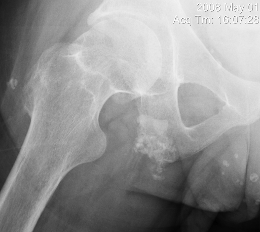
A plain radiograph was taken of the right hip and ischium. The study revealed a small amount of heterotopic bone formation under the right ischium (Figure 1).
Figure 1.
Radiograph of the right hip revealing evidence of heterotopic ossification near the ischial tuberosity.
With documented informed consent and with the patient lying in the left lateral decubitus position, approximately 3 mL of 1% lidocaine was injected in a peppered fashion into the region of the right ischial tuberosity. After approximately 5 minutes, pressure was reapplied to the region. This postinjection pressure did not produce the facial and arm sweating. The patient was then placed back in his wheelchair in the position that usually would trigger his hyperhidrosis, and the patient was observed for 10 minutes. The excessive sweating did not recur.
The patient was prescribed lidocaine transdermal patches (Lidoderm) to apply to the right ischium for 12 hours daily while sitting. His response to this treatment was monitored by telephone contact and subsequent clinic visits. The sweating did not recur with the use of the patches, although at times he found that the Lidoderm patches caused some inflammation of the skin below the ischium.
The patient subsequently participated in a wheelchair seating clinic at which he underwent pressure mapping both while sitting in his existing gel cushion as well as on a high-profile multiple-cell air-filled cushion (ROHO). This analysis showed increase pressure in the region of the right ischial tuberosity while sitting on his gel cushion. When placed on the air-filled cushion, he did not have the same pressure profile. The patient was subsequently switched to the air-filled cushion, and even without the lidocaine patch, he was able to sit for extended periods of time without experiencing hyperhidrosis.
DISCUSSION
Sweat glands are innervated by the sympathetic nervous system under control of the hypothalamus. Cervical and high thoracic spinal cord injury interrupts the descending fibers that form the sympathetic chain, and therefore prevents this normal supraspinal control (5). Reflex sweating occurs in cervical or high thoracic SCI when there is some type of irritative afferent stimulation from the body below the level of the lesion. In this case, the irritation was that of nocioception from pressure over the contralateral ischial tuberosity. Although there was no obvious pressure ulceration, there evidently was enough irritated tissue at the site of the ischium to cause afferent activation with pressure. This could have been made worse by the heterotopic bone, which might have included fracture fragments from the ischial tuberosity. It is interesting to note that the simulation came from the side of the body contralateral to the side that experienced the hyperhidrosis, suggesting a spinothalamic afferent pathway with decussation of fibers within the distal spinal cord. It is likely that the afferent impulse from around the site of injury triggered a sympathetic response in the cervical cord.
Traditionally, management of hyperhidrosis does involve removal of noxious afferent stimuli but does not involve directed blockage of the afferent pathway. Symptomatic treatments for this syndrome such as anticholinergic medications and sympathetic block traditionally focus on the efferent pathway of the reflex arc. In this case, however, by using a local lidocaine block through either injection or topical application, modulation of the afferent (nocioceptive) leg of the reflex pathway was achieved. Lidocaine works locally through the blockage of voltage gated sodium channels in the peripheral nerve, thereby preventing in this case the generation of afferent action potentials (6). This then prevented the reflex stimulation of the efferent sympathetic pathways innervating the sweat glands. As suggested by this case report, the ultimate, effective treatment was protection of the offending pressure point.
CONCLUSION
A case of hyperhidrosis secondary to a contralateral site of noxious stimulus in an individual with longstanding high cervical complete tetraplegia proved difficult to manage. Thorough physical examination revealed the source of noxious stimuli that precipitated the symptom. The site was confirmed by local anesthesia and ultimately treated by measures that reduced pressure on the damaged ischial area in the sitting position.
Acknowledgments
The author thanks Dr S. Sabin for his assistance and Dr M. Gonzalez-Fernandez for the radiograph. This case was initially presented in abstract form at the 2009 ASIA Meeting in Dallas, Texas.
References
- Krassioukov A, Warburton DE, Teasell R, Eng JJ. A systematic review of the management of autonomic dysreflexia after spinal cord injury. Arch Phys Med Rehabil. 2009;90(4):682–695. doi: 10.1016/j.apmr.2008.10.017. [DOI] [PMC free article] [PubMed] [Google Scholar]
- Andersen LS, Biering-Sorensen F, Muller PG, Jensen IL, Aggerbeck B. The prevalence of hyperhidrosis in patients with spinal cord injuries and an evaluation of the effect of dextropropoxyphene hydrochloride in therapy. Paraplegia. 1992;30(3):184–191. doi: 10.1038/sc.1992.53. [DOI] [PubMed] [Google Scholar]
- Glasauer FE, Czyrny JJ. Hyperhidrosis as the presenting symptom in post-traumatic syringomyelia. Paraplegia. 1994;32(6):423–429. doi: 10.1038/sc.1994.69. [DOI] [PubMed] [Google Scholar]
- Haas U, Geng V. Sensation of defecation in patients with spinal cord injury. Spinal Cord. 2008;46(2):107–112. doi: 10.1038/sj.sc.3102067. [DOI] [PubMed] [Google Scholar]
- Fast A. Reflex sweating in patients with spinal cord injury: a review. Arch Phys Med Rehabil. 1977;58(10):435–437. [PubMed] [Google Scholar]
- Cummins TR. Setting up for the block: the mechanism underlying lidocaine's use-dependent inhibition of sodium channels. J Physiol. 2007;582(Pt 1):11. doi: 10.1113/jphysiol.2007.136671. [DOI] [PMC free article] [PubMed] [Google Scholar]


