A Variety of new immunosuppressive drugs with distinct and diverse modes of action are currently being evaluated for their potential to control allograft and xenograft rejection. FK 506 is a macrolide lactone that, like cyclosporine A (CyA), inhibits selectively very early events in CD4+ T-lymphocyte activation and proliferation. In contrast, bredinin (BR; mizoribine), mycophenolic acid (MPA, the active moiety of RS61443), and brequinar sodium (BQR) inhibit later events in the cell cycle by interfering with purine (BR and MPA) or pyrimidine (BQR) biosynthesis. These antiproliferative drugs (BR, MPA, and BQR) are candidates to replace azathiopnne in combination therapy with CyA or FK 506 and steroids. Because there is little published information concerning the influence, if any, of BR, MPA, and BQR on production of lymphokine message, we have examined mRNA transcription for the immunoregulatory cytokines IL-2 and IL-10 in activated murine spleen cells.
MATERIALS AND METHODS
Immunosuppressive Agents
FK 506 (Fujisawa Pharmaceutical Co, Osaka, Japan) and MPA (Sigma Chemical Co, St Louis, Mo) were dissolved in absolute ethanol. BR (Asahi Chemical Co, Ltd, Tokyo, Japan), and BQR (Dupont Merck Pharmaceutical Co, Wilmington, Del) were dissolved in water before further dilution in cell culture medium.
Mouse T-Cell Activation and RNA Isolation
Spleens were removed asepticaily from male C57BL10 mice (8 to 12 weeks of age) and nucleated cell suspensions prepared and washed after red cell lysis in Tris-buffered NH4Cl, pH 7.2. The spleen ceils were resuspended finally in RPMI-1640 (Gibco, Grand Island, NY) with 10% (vol/vol) fetal bovine serum (Gibco). They were stimulated with concanavalin A (Con A) (5 μg/mL; Sigma) at a final cell concentration of 2.5 × 10⁶/mL in tissue culture tubes (Falcon, Becton Dickinson, Oxnard, Calif), for 24 hours in 5% CO2 in air at 37°C. FK 506, MPA, BR, or BQR were added at the beginning of the culture. Twenty-four hours later, cells were harvested and washed twice with RPMI-1640 before total RNA was isolated.
Cell Proliferation
Cultures of Con A–stimulated mouse spleen cells were established as described above. 3HTdR was added at 48 hours and cells harvested using a multiple cell harvester at 72 hours. DNA synthesis was expressed as counts per minute and results as percent inhibition relative to untreated Con A–stimuiated controls.
Detection of Cytokine Gene Expression by Polymerase Chain Reaction (PCR)
Cells were harvested at 24 hours after Con A stimulation and total RNA extracted by the guanidinium-isothiocyanate method (RNAzol, Cinna Biotecx, Friendswoods, Tex). A 1-μg RNA sample was reverse-transcribed into cDNA using oligo-dT primer and MMLV reverse transcriptase (Gibco BRL). This template cDNA was used for 30 or 40 cycles of PCR amplification using specific IL-2 and IL-10 oligonucleotide primers synthesized at the University of Pittsburgh DNA synthesis facility. The PCR amplification was conducted using Taq polymerase (Perkin-Elmer, Norwalk, Conn) and a model 480 DNA Thermal Cycler (Perkin-Elmer). The products of amplification, along with molecular weight markers for sizing, were separated on 2% agarose gels stained with ethidium bromide, and photographed in ultraviolet light. Semiquantitative analysis was achieved by comparing visibility of bands for serial 10-fold dilutions of input template DNA. β-Actin primers were used in a separate PCR reaction to control for amount of template cDNA.
RESULTS
Proliferation
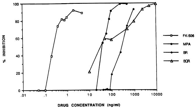
Figure 1 shows the inhibitory effects of the various drugs on cell proliferation. Each drug, when added with the stimulus at the initiation of culture, showed a dose–response curve with a maximum effect of >90% inhibition of proliferation. The 50% inhibitory concentration (IC50) for FK 506 was approximately 0.2 ng/mL. The IC50 for the other drugs was considerably higher: 30 ng/mL for BQR, 35 ng/mL for MPA, and 250 ng/mL for BR. The drug concentrations chosen for the gene expression experiments were FK 506,1 ng/mL; MPA, 500 ng/mL; BR, 1000 ng/mL; and BQR, 5000 ng/mL. At these concentrations, inhibition of DNA synthesis was >85% for each drug.
Fig 1.
Con A–stimulated mouse spleen cells were cultured with immunosuppressive drugs at the concentrations indicated. Drugs were added at the same time as the stimulus at the initiation of culture. Percent inhibition was calculated from the cpm value of 3HTdR incorporation compared with Con A–stimulated cells in media alone.
Cytokine Gene Expression
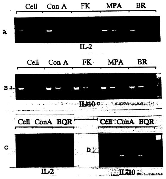
The reverse transcription PCR was performed using a standardized input amount of total RNA (1 μg), which was then reverse-transcribed into cDNA. In Fig 2 it can be seen that FK 506 strongly inhibited IL-2 production. However, MPA- and BR-treated cells showed the same level of IL-2 message as Con A–stimulated cells with no drug. As for IL-10, there was a high basal level expressed in cells alone. With Con A stimulation, this level increased. With FK 506, MPA, or BR, the IL-10 message was still present, at greater than basal levels. Recently, IL-10 has been shown to be present in other cell types (macrophages, B cells, and mast cells), and further experiments may be needed to confirm the cell of origin. It is clear that IL-2 mRNA was decreased by FK 506, but not IL-10 message. MPA and BR showed little effect on either of the cytokines studied.
Fig 2.
Effects of FK 506 (FK), MPA, BR, and BQR on expression of mRNA for IL-2 and IL-10. cDNA was amplified by PCR. IL-2: serial cDNA dilutions of 5, 0.5, and 0.05 ng (A) or 5 and 0.5 ng (C). IL-10: serial cDNA dilutions of 50, 5, and 0.5 ng (B) or 5 and 0.5 ng (D).
Figure 2 also shows the results for similar experiments using BQR. BQR did not selectively inhibit cytokine gene expression for IL-2 or IL-10. We observed a more general effect of BQR in reducing all mRNA levels in the cell, including constitutive genes β-actin and GAPDH (results not shown). This is probably due to its effect on pyrimidine biosynthesis pathways, and inhibition of uridine and cytidine production.
DISCUSSION
We have shown that, for several different immunosuppressive drugs at concentrations that markedly inhibit cell proliferation, cytokine gene expression is selectively inhibited only by FK 506, and not by MPA, BR, or BQR. This is not surprising, because FK 506 has been shown to act at an early stage to inhibit cytokine (such as IL-2) production,1 whereas MPA, BR, and BQR act at a step further along in the pathway from cell activation to proliferation. Our findings for BR are in agreement with those of Turka et al,2 who showed that BR failed to inhibit early events in T-cell activation, including expression of mRNA for IL-2. It was also noted that although FK 506 markedly reduced IL-2, IL-10 expression was spared. A differential effect of FK 506 on IL-4 and IL-10 gene expression has been published previously with regard to a mouse TH2 cell line,3 and may be linked to the immunosuppressive effect of the drug, because IL-10 has immunosuppressive properties.4
In conclusion, it is important to look at the effects of new immunosuppressive drugs at the molecular level, because these agents have the potential to be used in combination to act at various points in the cell activation/proliferation pathway. FK 506 acts on early events to block cytokine production, whereas MPA, BR, and BQR act on later events that prevent cell proliferation.
Acknowledgments
Supported by Grant DK29961-09 from the National Institutes of Health, Bethesda, Md.
REFERENCES
- 1.Tocci MJ, Matkovich DA, Collier KA, et al. J Immunol. 1989;143:718. [PubMed] [Google Scholar]
- 2.Turka L, Dayton J, Sinclair G, et al. J Clin Invest. 1991;87:940. doi: 10.1172/JCI115101. [DOI] [PMC free article] [PubMed] [Google Scholar]
- 3.Wang SC, Zeevi A, Jordan ML, et al. Transplant Proc. 1991;23:2920. [PubMed] [Google Scholar]
- 4.Taga K, Tosato G. J Immunol. 1992;148:1143. [PubMed] [Google Scholar]


