
Figure 1.
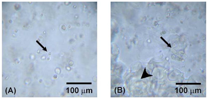
Light microscopy of OPF hydrogel composites containing rabbit MSCs. Small arrows indicate encapsulated rabbit MSCs, and big arrows indicate encapsulated microparticles. Figures (A) and (B) represent OPF hydrogel composites with only rabbit MSCs and with rabbit MSCs and TGF-β1-loaded microparticles at day 7, respectively.