Microclimatic edge effects and morpho-physiological characteristics are shaping lichen functional group distribution. Thus, lichen functional groups may be used as indicator for forest disturbance
Abstract
Background and aims
Forest edges created by fragmentation strongly affect the abiotic and biotic environment. A rarely studied consequence is the resulting impact on non-vascular plants such as poikilohydric lichens, known to be highly sensitive to changes in the microenvironment. We evaluated the impact of forest edge and forest interior on the distribution of two groups of crustose lichens characterized by the presence or absence of a cortex and sought explanations of the outcome in terms of photosynthetic response and water relations.
Methodology
Microclimate, distribution patterns and physiology of cortical and non-cortical lichens were compared at the edge and in the interior of an Atlantic rainforest fragment in Alagoas, Brazil. Ecophysiological aspects of photosynthesis and water relations were studied using chlorophyll a fluorescence analysis, and hydration and rehydration characteristics.
Principal results
Cortical and non-cortical functional groups showed a clear preference for interior and edge habitats, respectively. The cortical lichens retained liquid water more efficiently and tolerated low light. This explains their predominance in the forest interior, where total area cover on host tree trunks reached ca. 40 % (versus ca. 5 % for non-cortical lichens). Although non-cortical lichens exchanged water vapour efficiently, they required high light intensities. Consequently, they were able to exploit well-lit edge conditions where they achieved an area cover of ca. 19 % (versus ca. 7 % for cortical lichens). We provide some of the first data for lichens giving the relative quantity of incident light absorbed by the photosystem (absorptivity). The cortical group achieved higher absorptivity and quantum efficiencies, but at the expense of physiological plasticity; non-cortical lichens showed much decreased values of Fv/Fm and electron transport rates in the forest interior.
Conclusions
Morphological and physiological features largely determine the ecophysiological interaction of lichen functional groups with their abiotic environment and, as a consequence, determine their habitat preference across forest habitats. In view of the distinctiveness of their distribution patterns and ecophysiological strategies, the occurrence of cortical versus non-cortical lichens can be a useful indicator of undisturbed forest interiors in tropical forest fragments.
Introduction
In the light of progressive fragmentation of tropical rainforests, the creation of forest margins represents an increasing pressure on the remaining biota of forest remnants (Bierregaard et al., 2001; Laurance et al., 2002). Forest margins are commonly exposed to various impacts generally referred to as edge effects. These can be abiotic, biotic, direct or indirect (Murcia, 1995). On the biotic level, edge effects may, among others, directly influence functional diversity (de Melo et al., 2006; Tabarelli et al., 2008; Lopes et al., 2009) and species composition (Silva and Tabarelli, 2000) or indirectly influence plant–herbivore interaction (Wirth et al., 2008). These biotic effects are always based upon changes in abiotic conditions which are variable over space and time, and therefore difficult to quantify (Matlack, 1993; Camargo and Kapos, 1995; Newmark, 2001). It is generally held that edges are characterized by higher temperature, light intensity and wind disturbance, and lower relative humidity (RH) compared with forest interiors (Raynor, 1971; Chen et al., 1993; Matlack, 1993; Young and Mitchell, 1994; Newmark, 2001). In the tropics, however, very few studies have addressed microclimatic edge effects (Kapos, 1989; Camargo and Kapos, 1995; Newmark, 2001) and even less is understood regarding their impact on non-vascular plants.
As poikilohydric non-vascular plants are well known to interact quickly with their abiotic environment (Hartard et al., 2008), it is recognized that growth form and morphology play a major role in their successful establishment under varying environmental conditions (Larson and Kershaw, 1976; Larson, 1981; Sancho and Kappen, 1989; Bates, 1998; Lakatos et al., 2006). Across several biomes, microclimatic edge effects have been shown to change the biomass and distribution of numerous species (Sillett, 1994; Esseen and Renhorn, 1998; Hilmo and Holien, 2002; Rheault et al., 2003; Esseen, 2006; Boudreault et al., 2008), and diversity (Kivisto and Kuusinen, 2000). For example, Sundberg et al. (1997) attributed strongly decreased growth rates of two foliose lichens, Lobaria pulmonaria and Platismatia glauca, at the forest edge to physiological limitations which prevented the adjustment of photosynthetic acclimation to higher irradiances. As a consequence, lichens are frequently used as sensitive indicators of environmental change. Further, it has been shown that placing poikilohydric organisms into functional groups rather than just considering species composition can help elucidate microclimatic conditions as well as environmental impacts (Ellis and Coppins, 2006; Cornelissen et al., 2007) and land use change (Bergamini et al., 2005; Stofer et al., 2006). In the tropical understory, Lakatos et al. (2006) identified the following four functional groups of microlichens: (i) squamulous, (ii) filamentous umbrella-like, (iii) crustose with cortex and (iv) crustose without cortex. The latter two constitute the majority of lichens present, whereas macrolichens (e.g. foliose or fruticose forms) are restricted to montane forests (Sipman, 1989a,b; van Leerdam et al., 1990) or sporadic occurrences in the canopy of lowland forests (Cornelissen and Steege, 1989; Komposch and Hafellner, 2000) due to climatic limitations (Zotz 1999; Lakatos et al., 2006).
Crustose lichen groups are easily distinguished by the presence or absence of an upper cortex layer. This layer is formed by very dense glutinated fungal hyphae, creating a very firm, smooth and shiny upper surface, as exemplified in Dimerella, Lecidea, Myriotrema, Thallotrema, Thelotrema and Porina. In contrast, the upper surface of non-cortical lichens is composed of loosely interwoven hyphae, resulting in a byssoid-like structure. The resulting thalli are susceptible to mechanical damage and are of a non-shiny, somewhat whitish colour, as represented in the genera Chiodecton, Cryptothecia, Dichosporidium and Herpothallon (for more details on group characterization, see Lakatos et al., 2006). Thallus morphology as well as upper surface characteristics have a strong impact on physiological processes such as water uptake and evaporation (Sancho and Kappen, 1989; Valladares, 1994; de Vries and Watling, 2008), and hence largely determine photosynthetic activity. However, there has been little research into the physiology of tropical lichens. Although there are some studies on the ecophysiology of macrolichens in tropical montane forests (Lange et al., 1994, 2000; Zotz et al., 1998), studies in tropical lowland forests are extremely rare (Zotz and Winter, 1994; Zotz and Schleicher, 2003; Zotz et al., 2003). Only one study has addressed the ecophysiology of crustose microlichens to date (Lakatos et al., 2006).
In the present work, we expected to confirm the presence of microclimatic edge effects, which define two distinct microenvironments (forest edge and interior), on the performance and distribution of lichen. We hypothesized that cortical and non-cortical crustose lichens interact differently with their abiotic environment, and that this affects their different relative distributions across forest edge and interior. To test this hypothesis, we investigated microclimate, functional group composition and physiological mechanisms linked to morphology. We also studied light adaptation, desiccation and reactivation with water vapour, via chlorophyll a (Chl a) fluorescence, to provide possible explanations for differences in adaptation to edge and interior of the tropical forest understory.
Materials and methods
Study site
The study was conducted in the Atlantic forest north-east of Brazil at Usina Serra Grande (9°S, 35°52′W, 500–600 m above sea level), a private property located in the state of Alagoas. The region has a tropical climate with a 3-month dry season (<60 mm rain month−1). The annual rainfall is about 2000 mm with the wettest period occurring between April and August. The vegetation is characterized as a lower montane rainforest. This occurs at 100–600 m above sea level for Brazilian Atlantic rainforests (de Melo et al., 2006). Within this fragmented area, the study was carried out at Coimbra, comprising an area of 3500 ha. The fragment, created ∼60 years ago, is surrounded on all sides by sugarcane plantations, but currently shows no large-scale human disturbances. On a small scale, however, the forest is used by locals for firewood and hunting. Nevertheless, Coimbra is considered to be one of the largest preserved fragments in this region. Within the northern sector of the fragment, four plots (25 m × 25 m) were established at the forest edge and in the interior, respectively. Edge plot sites were 2–3 m from the forest border line. Undisturbed old-growth forest areas more than 150 m from the forest border were defined as interior sites.
Microclimate
The microclimate was recorded intermittently during field trips from 14 January 2007 to 19 April 2007. Temperature and RH at the forest edge and interior plots were measured 1 m above ground level at 1-min intervals using two-channel loggers with internal sensors (HOBO pro series; Onset Computer Corporation, NH, USA). One sensor operated as a reference outside the forest, being installed fully exposed over pasture to resemble matrix conditions closely. The period of investigation included a shift from dry (ending in February) to wet season. Hence, days of observation were grouped into three categories (dry, overcast and wet) according to the minimal daily RHrefmin as measured by the reference sensor. Categories reflect sunny dry days (RHrefmin < 50 %), overcast days where a very slight rain event might occur (RHrefmin = 50–60 %) and wet days with at least one heavy rain event (RHrefmin = 60–80 %).
Relative light intensity for trunk environments was measured using a 2π-quantum sensor and datalogger (190-A, LI 1400; LI-COR, Lincoln, NE, USA) at dusk and dawn to avoid direct radiation. Measurements were taken vertically in each direction (north, east, south and west) along the trunk 1.30 m above the ground. Values per direction (n = 3) and all directions per tree were averaged to obtain a representative value for the lichen light environment per tree.
Distribution patterns
Parameters of epiphytic lichen vegetation were assessed using a functional group approach according to the theoretical perspective adopted in this study. For this, all lichen thalli were classified into the two functional groups (with and without cortex) without further taxonomic identification. In view of the high species diversity of corticolous crustose lichen communities in the Atlantic forest of north-eastern Brazil (Cáceres et al., 2007), the functional group approach is expected to reduce the impact of species-specific characters and to increase the explanatory power of the variables studied. Examples from post hoc identifications of sampled thalli included species from the genera Letrouitia, Myriotrema, Porina and Thelotrema for the lichen functional group with cortex, whereas the group without cortex comprised genera such as Herpothallon and Cryptothecia. For information on the total species composition of corticolous crustose lichen communities in the region, see Cáceres et al. (2007).
For the assessment of functional group abundance, the relative area covered by lichens and the number of thalli were recorded on target trees in edge and interior plots. Only trees with a relatively smooth bark and a diameter at breast height (dbh) of 12–32 cm were sampled. A sample grid of 20 cm × 20 cm was placed on each selected tree using transparent plastic sheets fixed to the tree trunk 1.3 m above the ground. Five trees were sampled per plot on their north- and south-facing sides. In total, 40 records (20 trees) were sampled for each interior and edge habitat. A three-way analysis of variance (ANOVA) tested whether site, type of lichen or exposure (reflecting highest and lowest potential light impact at the microsite) affected lichen cover. Relative lichen cover (%) was square root transformed to meet the assumptions of ANOVA. Bonferroni's least significant difference (LSD) and Tukey's honestly significant difference (HSD) were applied as post hoc tests for multiple comparisons to determine significant differences between group means. Counts of thallus numbers were square root transformed and treated as ordinal data (Weißberg–Bingham test, WB = 0.994, P = 0.2). All statistical analysis, modelling and graphical diagnostics to check for compliance with the arithmetic assumptions of the tests and transformations were performed using the R Program (R 2.3.1; R Foundation for Statistical Computing, Vienna, Austria).
Ecophysiology
All physiological characteristics were determined via Chl a fluorescence measurements using pulse amplitude modulated equipment (IMAGING-PAM; H. Walz, Effeltrich, Germany). Parameters of maximum quantum yield of photosystem II (PSII) (Fv/Fm), quantum yield of PSII (ФPSII = (F′m − Ft)/F′m), non-photochemical quenching (NPQ = (Fm − F′m)/F′m) and electron transport rate (ETR = ФPSII × PFD × A × 0.5, where PFD is the photon flux density and A the absorptivity) were derived according to Genty et al. (1989) and Bilger et al. (1995). Absorptivity measures the relative quantity of incident light that is actually absorbed by the photosystem. Whereas for higher plants a general value of 0.84 is representatively used for the absorptivity (A) (Gabrielsen, 1948; Björkman and Demmig, 1987), absorptivity values for lichens have not been quantified. The chosen chlorophyll fluorometer provided the opportunity to actually measure the absorptivity, which was recorded for each sample and used to calculate accurate ETRs (see above).
Photosynthesis
Rapid light response curves (RLCs) were performed in situ to assess light adaptation in the respective habitats (edge/interior). We applied seven increasing light intensities (2, 5, 9, 22, 46, 71 and 104 µmol quanta m−2 s−1), each for a duration of 20 s. Although steady-state conditions may not be reached at each light intensity during RLCs, the method provides a good measure of the relative photosynthetic performance of the organism (Schreiber et al., 1997) and has been widely applied in studies of algae and higher plants in marine, aquatic and terrestrial ecosystems (Schreiber et al., 1997; White and Critchley, 1999; Rascher et al., 2000; Longstaff et al., 2002; Karim et al., 2003; Ralph and Gademann, 2005). Further, it has been shown by Lakatos et al. (2006) that short durations during light response curves are needed for tropical crustose lichens to prevent dehydration and rapid desiccation. A pair of lichen samples (cortical and non-cortical) per tree was darkened by a piece of black cloth the evening before measurements were made. Rapid light response curves were performed in situ on the trunk at a height of 0.4–1.40 m during early morning hours. Adequate hydration was secured by spraying with water prior to measurement. Ambient light was excluded by black cloth tied around the trunk as light intensities used in RLCs were fairly low. Twenty individuals each of cortical and non-cortical lichens at forest edge and interior, respectively, were measured, resulting in 80 RLCs. Fluorescence parameters were averaged for 10 areas spread randomly within the recorded fluorescence image. The relationship between ETR and photosynthetically active radiation was modelled using a non-rectangular hyperbola (Prioul and Chartier, 1977; Leverenz et al., 1990; Ogren and Evans, 1993):
| 1 |
and
| 2 |
where ϕ represents the initial slope of the curve and reflects the quantum efficiency of PSII; I is the photosynthetic PFD; P and Pmax are the actual and maximal rate of photosynthesis, respectively, represented by the ETR; Pmax estimates the plateau of the curve; while θ reflects the transition of the initial slope and the plateau. Light saturation is defined at 90 % of the maximal ETR fitted by the model and was derived by solving equation (1). Photophysiological parameters measured or derived from the light curve model were compared between lichen groups and habitat using Tukey's HSD post hoc test.
Desiccation
The desiccation and reactivation behaviour exposed to water vapour was investigated ex situ under controlled conditions in the laboratory of the University of Recife. Three trees with relatively smooth bark were chosen for sample collection, each providing three individuals of cortical and non-cortical lichen thalli of ∼30 mm in diameter (n = 9 for both experiments). Bark pieces holding one sample each were carefully removed from the tree and samples were immediately transported to Recife under moist conditions. In the laboratory, samples were kept under humid conditions with low light (∼20 µmol m−2 s−1). Experiments were conducted within a week of collection. Moist samples were fully hydrated by standardized spraying with liquid water. After 10 min, samples were carefully blotted and the maximum quantum yield was determined to ensure physiological fitness and sufficient hydration. Samples were exposed to desiccating conditions ranging within 30–33 °C and 55–65 % RH at low light conditions of 8 µmol m−2 s−1 (which were well below saturation). A pair of lichens (cortical and non-cortical) was measured consecutively to ensure homogeneous ambient conditions for one set of samples. The quantum yield of PSII was recorded at 1-min intervals until lichens were desiccated. Lichens were regarded as physiologically desiccated when the quantum yield was below 0.1 over the entire thallus. Differences in desiccation time between the groups were tested using paired t-tests.
Reactivation upon desiccation
Samples were placed in a box over silica and Fv/Fm was measured at 1-h intervals until lichens were desiccated (yield <0.1). Dry lichens were exposed to RH of 80–85 % at 27.5 ± 1.5 °C. The entire experiment was conducted in darkness and the maximal quantum yield was taken every minute for half an hour to explore the recovery of PSII by rehydration of the thallus with water vapour. Samples were subsequently fully hydrated as described above and the maximum quantum yield was taken.
Results
Microclimate at forest edge and interior
Differences in microclimate between forest edge and interior could be detected with respect to temperature and incident light. Diurnal ambient temperature ranges were always 1–3 °C higher at the edge: from 27 to 32 °C on sunny days, declining on overcast days to 25–28 °C and dropping to 22.5–25.2 °C on wet days. Average daily maximum temperature was significantly higher at the edge (26.9 ± 0.4 °C) than in the interior (25.9 ± 0.3 °C) on overcast days, but there were no differences regarding daily minimum temperature (Table 1). The diurnal temperature amplitude was larger at the forest edge: ∼6 °C compared with 4 °C in the interior on overcast days. At the forest edge, 1.5 % (±0.4 %) of the incident light reached the understory. In the intact forest interior, light intensity was significantly lower with only 0.7 % (±0.3 %) of the incident insolation reaching the understory (Table 1).
Table 1.
Table of means (±SD) for daily microclimatic parameters at the forest edge and interior, with Tmin, minimal temperature (°C); Tmax, maximum temperature; Tamp, temperature amplitude; RHmin, minimal RH (%); and rel. light, relative light intensity (%). Means were compared via Student's t-test. Days of observation were grouped into different weather categories with reference to the minimal daily RH outside the forest, with <50 % for sunny, <60 % for overcast and >60 % for wet days.
| Edge | Interior | t(6–12) | P-value | |
|---|---|---|---|---|
| Sunny days | ||||
| Tmin | 20.6 ± 1.3 | 21.5 ± 1.5 | 1.02 | n.s. |
| Tmax | 29.0 ± 1.6 | 27.9 ± 0.4 | −1.70 | 0.11 |
| Tamp | 8.6 ± 2.2 | 6.4 ± 1.4 | −1.88 | 0.09 |
| RHmin | 56.5 ± 6.6 | 60.9 ± 4.7 | 1.49 | 0.16 |
| Overcast days | ||||
| Tmin | 21.2 ± 0.9 | 21.6 ± 0.5 | 0.99 | n.s. |
| Tmax | 26.9 ± 0.4 | 25.9 ± 0.3 | −4.98 | <0.001 |
| Tamp | 5.9 ± 0.9 | 4.3 ± 0.3 | −3.81 | <0.01 |
| RHmin | 70.9 ± 4.3 | 72.9 ± 2.9 | 0.89 | n.s. |
| Wet days | ||||
| Tmin | 21.5 ± 0.9 | 21.6 ± 0.8 | 0.14 | n.s. |
| Tmax | 24.6 ± 0.4 | 23.8 ± 0.6 | −2.58 | <0.05 |
| Tamp | 3.4 ± 1.1 | 2.3 ± 0.7 | −1.58 | 0.16 |
| RHmin | 87.2 ± 7.8 | 90.1 ± 4.3 | 0.72 | n.s. |
| Rel. lightA | 1.46 ± 0.39 | 0.68 ± 0.28 | 6.99 | <0.001 |
A, log-transformed for statistical testing, means are back transformed in this table.
Abundance of cortical versus non-cortical lichens
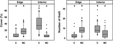
Data on lichen abundance were pooled (Fig. 1) since no effect could be detected between northern and southern exposures on the trunk. However, there was a significant habitat effect on lichen cover interacting with the lichen group (F1,152 = 8.24, P < 0.01), indicating the preferences of non-cortical lichens for the edge and corticals for the interior habitat. Mean lichen cover for lichens with a cortex (7 ± 2.5 %) was lower compared with non-cortical lichens (19.1 ± 8.9 %) at the forest edge. In contrast, this pattern reversed at the forest interior, where lichens with cortex showed an area cover of 39.6 ± 9.9 %, whereas non-cortical lichens only reached 4.6 ± 4.8 % (F1,152 = 17.3, P < 0.001). A similar pattern was observed regarding the number of individual lichen thalli. At the edge, the thallus number of cortical and non-cortical lichens averaged 10.1 ± 3.1 per 400 cm2 and 13.7 ± 5.6 per 400 cm2, respectively. Again, in the forest interior, lichens with cortex were more abundant, reaching 23.3 ± 5.4 thalli compared with 7.4 ± 4.9 per 400 cm2 for non-cortical lichens (F1,152 = 80.2, P < 0.001).
Fig. 1.
Lichen cover and number of thalli for cortical (C) and non-cortical (NC) lichens at the forest edge and interior (n = 40). Lower case letters indicate significant differences according to Tukey's HSD. If lower case letters are the same, groups cannot be distinguished at the P = 0.05 significance level.
Physiological light adaptation in situ
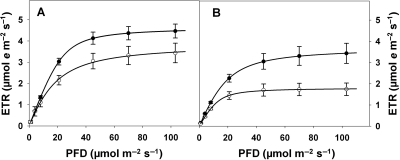
The physiological performance of lichens was related to effects of both habitat and functional group. Although the cortical group showed no physiological difference in performance in edge and interior habitats (Table 2), the non-cortical group exhibited greater physiological plasticity in photosynthetic performance. Both lichen functional groups show similar photosynthetic performance in general (Fig. 2), although two fundamental photophysiological parameters differed significantly between them: absorptivity and quantum efficiency of the ETR (Table 2). The cortical group achieved higher values of absorptivity of around 0.83, compared with 0.71 for non-cortical lichens. Moreover, the cortical group reached higher quantum efficiencies of 0.17 and 0.16, compared with 0.12 for the non-cortical group. The physiological plasticity of the non-cortical group is reflected by decreased values of Fv/Fm and ETRs in the forest interior, reaching only half the rates of the other functional group (Table 2). Moreover, non-corticals showed differences in absorptivities (Fisher's LSD) and light saturation PFDsat (Table 2). There were no differences between groups or habitats for NPQ at a PFD of 70 µmol m−2 s−1 as variation was quite large.
Table 2.
Cardinal points of light–response curves for lichens with and without a cortex. Absorptivity, proportion of photosynthetically active radiation that is absorbed; Fv/Fm, maximum quantum yield in the dark-adapted state; NPQ, non-photochemical quenching at a PFD of 70 µmol m−2s−1; Φ, quantum efficiency; ETRmax, maximum electron transport rate; PFDsat, photon flux density at 0.9 ETRmax; θ, convexity value. The latter four are derived by the fitted model. Means are shown with 1 SD (n = 14–20). If superscripts are the same, means cannot be distinguished significantly by Tukey's HSD or Fisher's LSD (in parentheses) at the P = 0.05 significance level. Numbers indicate transformation used for statistical testing: 1, log; 2, arcsinus transformation. Means are back transformed in this table.
| Edge |
Interior |
|||
|---|---|---|---|---|
| Cortex | No cortex | Cortex | No cortex | |
| Absorptivity | 0.84 ± 0.05a | 0.69 ± 0.05b | 0.83 ± 0.03a | 0.73 ± 0.06b(c) |
| Fv/Fm | 0.58 ± 0.06a | 0.58 ± 0.06ab(a) | 0.58 ± 0.04a | 0.53 ± 0.07b |
| NPQ1 | 0.43 ± 0.28a | 0.41 ± 0.21a | 0.34 ± 0.26a | 0.43 ± 0.16a |
| Φ | 0.173 ± 0.04a | 0.119 ± 0.03b | 0.158 ± 0.04a | 0.118 ± 0.03b |
| ETRmax1 | 4.6 ± 1.5a | 3.6 ± 2.1a | 3.6 ± 2.4a | 1.8 ± 1.1b |
| PFDsat1 | 45.7 ± 21.4a | 49.7 ± 31.1a | 46.8 ± 35.6a | 20.8 ± 7.9b |
| θ2 | 0.89 ± 0.09a | 0.90 ± 0.09a | 0.84 ± 0.19a | 0.91 ± 0.08a |
Fig. 2.
Response of ETR to increasing light intensity (PFD) for cortical (closed circles) and non-cortical (open circles) lichens at the forest edge (A) and interior (B). Curves were fitted by a non-rectangular hyperbola (Prioul and Chartier, 1977). Means of each individual fit are shown with 1 SEM (n = 14–20).
Desiccation and reactivation experiments
The duration required for desiccation until no photochemical processes could be detected via Chl a fluorescence was significantly different for the two lichen functional groups (Table 3). While, under controlled conditions, non-cortical lichens desiccated on average after ∼25 min, lichens with cortex retained photosynthetic activity for twice the time until the entire thallus was desiccated (∼50 min).
Table 3.
Results of desiccation and reactivation experiments. Samples were desiccating at 30–33 °C, 55–65 % RH and 8 µmol m−2s−1 PFD (n = 9). Samples were considered desiccated when the quantum yield of PSII was below 0.1 over the entire thallus. Desiccated samples were exposed to an average 27.5 °C and 80–85 % RH in darkness for 30 min (n = 5). Fv/Fmrehy, Fv/Fm (maximum quantum yield) measured upon the experiment when rehydrated with liquid water.
| Cortex | No cortex | t4–8 | P-value | |
|---|---|---|---|---|
| Dehydration | ||||
| Fv/Fm prior to experiment | 0.62 ± 0.02 | 0.56 ± 0.04 | −3.76 | <0.01 |
| Time till desiccation (min) | 26.1 ± 10.9 | 50.7 ± 14.2 | 6.35 | 0.000 |
| Reactivation with water vapour | ||||
| ΦPSII after 30 min | 0.33 ± 0.07 | 0.26 ± 0.03 | 1.57 | 0.16 |
| Fv/Fmrehy | 0.55 ± 0.04 | 0.44 ± 0.06 | 1.57 | 0.16 |
| % reached | 57.7 ± 10.7 | 59.6 ± 6.7 | −0.32 | n.s. |
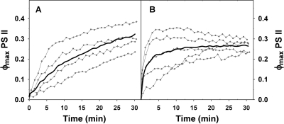
A distinct difference in behaviour was also observed during reactivation of photosynthesis by rehydration. Non-cortical lichens exhibited fast reactivation when exposed to high humidity at 80–86 % and 25–31 °C. After 5 min, the mean ΦPSII of lichens without a cortex was more than twice the ΦPSII for lichens possessing a cortex (Fig. 3). In contrast to non-cortical lichens, those with a cortex did not reach steady-state conditions within the 30 min period of the experiment. Values of Fv/Fm after rehydration were lower compared with those observed for lichens in situ (Table 3). Both types of lichen reached 58 % (with cortex) and 60 % (without cortex) of their maximum quantum yield prior to the experiment when subsequently allowed to rehydrate with liquid water, indicating some vulnerability to drought in both types.
Fig. 3.
Quantum yield of PSII (ϕPSII) of (A) cortical and (B) non-cortical lichens during reactivation with water vapour in the dark. The RH of the air was 80–85% at 27.5 ± 1.5 °C. Bold lines represent mean values, whereas grey lines show individual samples (n = 4–5).
Discussion
Edge-affected environmental conditions
The response of organisms after environmental change reflects their adaptive capacity in terms of morphological and physiological plasticity. This is particularly relevant for non-mobile and slow-growing organisms such as lichens inhabiting the understory of tropical humid rainforests (Zotz, 1999; Lakatos et al., 2006). As poikilohydric non-vascular plants, they are sensitive to environmental changes because their photosynthetic activity is largely dictated by the availability of light and water. Several studies have characterized forest edges by increased light levels, temperature and wind penetration, in association with lower relative humidities, compared with forest interiors (Raynor, 1971; Kapos, 1989; Chen et al., 1993; Matlack, 1993; Young and Mitchell, 1994; Camargo and Kapos, 1995; Newmark, 2001). Temperature, light and humidity measurements presented in this study confirm that the edge habitat is warmer, brighter and dryer than the forest interior (Table 1), suggesting that edge creation confronts lichens with a greater risk of desiccation. In contrast, the interior is characterized by more stable conditions that provide ready access to moisture at low light levels.
Distribution patterns at edge and interior environments
According to the microclimatic heterogeneity across forest habitats and based on distinct lichen functional groups, we expected different physiological performance to translate into group-specific habitat preferences. Our findings indeed revealed a pronounced shift in the distribution pattern of two lichen functional groups from the forest edge to its interior. Lichens without a cortex were significantly more abundant at the forest edge compared with the interior, where they were almost entirely non-existent (Fig. 1). In the interior, the cortical group was highly dominant, reaching a mean area cover of 40 %, while in certain instances covering up to 90 % of the bark surface. This distinct pattern is surprising since the criterion for classifying functional groups was only based on the simple morphological distinction of the presence versus absence of an upper cortex, while lichen species composition was random. In consideration of both the enormous tree species diversity and the known lack of host specificity of crustose tropical lichens (Cornelissen and ter Steege, 1989; Montfoort and Ek, 1989; Sipman, 1989b), it seems unlikely that the observed distribution pattern is strongly affected by tree species composition. Moreover, community formation is only weakly correlated with bark characteristics and microclimate in north-eastern Brazil (Sipman, 1989b; Cáceres et al., 2007). We only considered trees with a relatively smooth bark in order to avoid biases caused by substrate heterogeneity and to provide comparable conditions. Thus, there are strong indications that the observed lichen functional group distribution is a consequence of microclimatic edge effects.
Physiological explanation for distribution patterns
In accordance with our hypothesis, we found that the presence of a cortex strongly correlates with the physiological reactions of lichen functional groups influencing water exchange and light absorption. We found group-specific as well as habitat-specific differences in photosynthetic performance by lichens of the two functional groups. General group-specific differences in photophysiological parameters were uncovered irrespective of habitat for the absorptivity of incident light and the quantum efficiency of the ETR. Thus, while in cortical lichens the relative quantity of incident light that was absorbed by the photosystem (absorptivity) was estimated to be 0.83, a value similar to that of higher plants (Gabrielsen, 1948; Björkman and Demmig, 1987), lichens with no cortex showed markedly lower values of about 0.7 (Table 2). These are, to the best of our knowledge, the first published absorptivities for lichens. The values have implications for the calculation of absolute ETRs in lichens, which in the past have wrongly been assumed to be similar to those of higher plants (Bartak et al., 2000; Hajek et al., 2001). The reason for low light absorptivity in non-cortical lichens might be the byssoid thallus surface which scatters light to a much larger degree compared with a smooth cortex. Its white colour will also reflect more light. Therefore, the lower absorptivities of non-cortical lichens will contribute to their significantly lower quantum efficiency of photosynthesis. Non-cortical lichens require 8 quanta of incident light for the transport of one electron, compared with 6 quanta required by the cortical group (Table 2). This low light efficiency may well be crucial under the limiting light regimes present in the forest interior. Success in the interior environment requires effective exploitation of the limited amounts of light. This is achieved in cortical lichens by means of high absorptivities, high quantum efficiencies, maximal ETRs and maximum quantum yields (Fv/Fm). In these respects, the non-cortical lichens were inferior, indicating an inherent limitation to their photosynthetic performance in interior conditions. Moreover, their low Fv/Fm of 0.53 further suggests a decreased photosynthetic capacity of the non-cortical group when in the interior habitat. An overall average Fv/Fm of 0.58 was representative for tropical crustose lichens in this study. This value is similar to those for tropical crustose lichens studied previously (0.57–0.61; Lakatos et al., 2006), but lower than those of crustose or foliose lichens of temperate and boreal regions (Jensen, 1994; Schroeter, 1994; Gauslaa and Solhaug, 1996; Jensen et al., 1997; Renhorn et al., 1997).
At the forest edge, in contrast, photosynthetic performance by the non-cortical group was superior to that observed in the interior, and physiologically competitive with the cortical group in edge conditions where both groups revealed similar photosynthetic capacities (Fig. 2). Despite these similar photosynthetic capacities, non-cortical lichens exhibited higher abundances at the forest edge. This implies some advantage in this microhabitat. One possibility is that their low absorptivities may be advantageous in the context of photoprotection, particularly with regard to more frequent sunflecks (Lakatos et al., 2006). Another reason for the superior overall success is a basic difference in water retention characteristics. We found two contrasting strategies for regulating water relations in the cortical and non-cortical lichens: (i) rapid exchange of water vapour and (ii) efficient water retention. Because a cortex constitutes a diffusion barrier for water vapour, reactivation of photosynthesis with water vapour is slower than for non-cortical lichens. Exploitation of vapour is photosynthetically important since lichens already gain 60 % of their maximum photosynthetic capacity when in equilibrium with an RH of 80 % if water vapour is the exclusive source of water during rehydration. The same photosynthetic capacities were confirmed by Lakatos (2002) when rehydrating tropical crustose lichens at 70 % RH. However, the low resistances to water vapour diffusion created by the absence of a cortex will also favour fast desiccation. This, in turn, might serve as protection against the excessive light expected at the edge. In the forest interior, in contrast, greater water retention by cortical lichens enables this group to maintain an extended photophysiological activity, which is pivotal to exploiting the low light and infrequent sunflecks of this habitat (see also Lakatos et al., 2006).
Ecological applications
In tropical forest fragments, the identification of undisturbed interior conditions is often pivotal for management plans and conservation practices, especially in small fragments. However, assessment of the penetration depth of edge effects is difficult and laborious (Ewers and Didham, 2006). Because the distribution pattern of crustose lichens can be understood as a response of lichen functional traits to the microclimate regime, the two lichen functional groups could serve as useful bio-indicators of the presence of undisturbed forest interiors. In European boreal forests, crustose lichen species have been successfully identified as indicators of forest continuity (Tibell, 1992). However, species identification of tropical crustose lichens is often impossible for non-experts and requires sexually reproductive units, which are rare. Moreover, it is estimated that 50 % of the tropical lichens remain unidentified (Aptroot and Sipman, 1997). For tropical forests, Rivas Plata et al. (2008) proposed the use of species diversity of understory crustose lichen of the family Thelotremataceae as an index of ecological continuity. They found that Thelotremataceae diversity, all species of which possess a cortex, negatively correlates with the degree of disturbance and light exposure. To introduce this method to a broader spectrum of potential users, they suggested using a classification of 24 morphotypes based on apothecial and thallus morphology. Here, we suggest an even more straightforward classification to assess microclimatic conditions via classification into two easily identified groups. The proposed groups are easily recognized even by non-lichenologists due to the dichotomous distinction of the presence or absence of an upper cortex. This allows for rapid assessment of undisturbed forest interior and abiotic edge conditions. The low growth rates of tropical crustose lichens range from the almost immeasurably slow in the forest interior (Lakatos, 2002) to 4 ± 2 mm year−1 in a foliose lichen at the forest edge (Zotz and Schleicher, 2003). This should allow the detection of disturbances that persist for several years. Unpublished results (M. Lakatos) suggest that lichen functional group composition follows microclimatic gradients and may even thus be used to assess small-scale environmental heterogeneity.
Conclusions and forward look
At the well-lit forest edge, where non-cortical lichens dominate over those possessing a cortex, their inherent low light absorptivity and rapid desiccation rates of non-cortical lichens appear as adaptive photoprotective strategies. Furthermore, their capacity for especially rapid PSII reactivation when rehydrated at high relative humidities, and photosynthetic characteristics of high quantum efficiency and physiological plasticity, as indicated by decreased values of Fv/Fm and ETRs in the forest interior allow for an efficient response to fluctuating conditions. We therefore suggest that the non-cortical lichens can be characterized ecologically as light-demanding plants with physiological traits that appear unfavourable for the exploitation of low light conditions of the forest interior. On the other hand, lichens that possess a cortex benefit from enhanced interception of light and long water-holding capacities, and can be classified as shade tolerant. The substantial differences in photophysiological characteristics and hydration capacities between the cortical and the non-cortical groups clearly indicate a difference in thallus morphology (i.e. the presence and absence of a cortex in the thallus) as a crucial factor for the interaction with the abiotic environment. This, in turn, determines the distribution pattern of functional groups of crustose lichens across forest edge and interior habitats. Therefore, the relative distribution of lichen functional groups, based on the easily recognized presence or absence of a cortex, can be used as a rapid and dependable bio-indicator of undisturbed interiors and forest continuity in tropical forest fragments.
Sources of funding
The study was financed by the A.F.W. Schimper-Foundation for ecological research, the German Science Foundation (DFG, LA 1426/3-1) and the German Academic Exchange Service (DAAD, D/06/46401).
Contributions by the authors
M.L. was the principal investigator and supervisor of B.H. and A.P. The three authors were engaged in fund raising, field work, design of methods and data analyses. The project was a main part of the MSc thesis of A.P.
Conflict of interest statement
None declared.
Acknowledgements
We are very grateful to Inara Leal, Marcello Tabarelli, Mauro G. Santos and Sebastian Meyer for their hospitality and provision of facilities. Robert Lücking is thanked for species determination, Rainer Wirth and Gerhard Zotz for helpful comments on the manuscript, and Wanessa R. Almeida for committed assistance with field work.
References
- Aptroot A, Sipman HJM. Diversity of lichenized fungi in the tropics. In: Hyde KD, editor. Biodiversity of tropical microfungi. Hong Kong: University Press; 1997. [Google Scholar]
- Bartak M, Hajek J, Gloser J. Heterogeneity of chlorophyll fluorescence over thalli of several foliose macrolichens exposed to adverse environmental factors: interspecific differences as related to thallus hydration and high irradiance. Photosynthetica. 2000;38:531–537. doi:10.1023/A:1012405306648. [Google Scholar]
- Bates JW. Is ‘life-form’ a useful concept in bryophyte ecology? Oikos. 1998;82:223–237. doi:10.2307/3546962. [Google Scholar]
- Bergamini A, Scheidegger C, Stofer S, Carvalho P, Davey S, Dietrich M, Dubs F, Farkas E, Groner URS, Karkkainen K, Keller C, Lokos L, Lommi S, Máguas C, Mitchell R, Pinho P, Rico VJ, Aragon G, Truscott A-M, Wolseley PAT, Watt A. Performance of macrolichens and lichen genera as indicators of lichen species richness and composition. Conservation Biology. 2005;19:1051–1062. doi:10.1111/j.1523-1739.2005.00086.x. [Google Scholar]
- Bierregaard RO, Laurance WF, Gascon C, Benitez-Malvido J, Fearnside PM, Fonseca CR, Ganade G, Malcom JR, Martins MB, Mori S, Oliveira M, Merona JR-D, Scariot A, Spironello W, Williamson B. Principles of forest fragmentation and conservation in the Amazon. In: Bierregaard RO, Gascon C, Lovejoy TE, Mesquita RCG, editors. Lessons from Amazonia: the ecology and conservation of a fragmented forest. New Haven, USA: Yale University Press; 2001. pp. 371–385. [Google Scholar]
- Bilger W, Schreiber U, Bock M. Determination of the quantum efficiency of photosystem II and of nonphotochemical quenching of chlorophyll fluorescence in the field. Oecologia. 1995;102:425–432. doi: 10.1007/BF00341354. doi:10.1007/BF00341354. [DOI] [PubMed] [Google Scholar]
- Björkman O, Demmig B. Photon yield of O2 evolution and chlorophyll fluorescence characteristics at 77 K among vascular plants of diverse origins. Planta. 1987;170:489–504. doi: 10.1007/BF00402983. doi:10.1007/BF00402983. [DOI] [PubMed] [Google Scholar]
- Boudreault C, Bergeron Y, Drapeau P, Loopez LM. Edge effects on epiphytic lichens in remnant stands of managed landscapes in the eastern boreal forest of Canada. Forest Ecology and Management. 2008;255:1461–1471. doi:10.1016/j.foreco.2007.11.002. [Google Scholar]
- Cáceres M, Lücking R, Rambold G. Phorophyte specificity and environmental parameters versus stochasticity as determinants for species composition of corticolous crustose lichen communities in the Atlantic rain forest of northeastern Brazil. Mycological Progress. 2007;6:117–136. doi:10.1007/s11557-007-0532-2. [Google Scholar]
- Camargo JLC, Kapos V. Complex edge effects on soil moisture and microclimate in central Amazonian forest. Journal of Tropical Ecology. 1995;11:205–221. doi:10.1017/S026646740000866X. [Google Scholar]
- Chen J, Franklin JF, Spies TA. Contrasting microclimates among clearcut. Agricultural & Forest Meteorology. 1993;63:219–237. doi:10.1016/0168-1923(93)90061-L. [Google Scholar]
- Cornelissen JHC, ter Steege H. Distribution and ecology of epiphytic bryophytes and lichens in dry evergreen forest of Guyana. Journal of Tropical Ecology. 1989;5:131–150. doi:10.1017/S0266467400003400. [Google Scholar]
- Cornelissen JHC, Lang SI, Soudzilovskaia NA, During HJ. Comparative cryptogam ecology: a review of bryophyte and lichen traits that drive biogeochemistry. Annals of Botany. 2007;99:987–1001. doi: 10.1093/aob/mcm030. doi:10.1093/aob/mcm030. [DOI] [PMC free article] [PubMed] [Google Scholar]
- de Melo FPL, Dirzo R, Tabarelli M. Biased seed rain in forest. Biological Conservation. 2006;132:50–60. doi:10.1016/j.biocon.2006.03.015. [Google Scholar]
- de Vries MC, Watling JR. Differences in the utilization of water vapour and free water in two co-occurring foliose lichens from semi-arid southern Australia. Australian Ecology. 2008;33:975–985. [Google Scholar]
- Ellis CJ, Coppins BJ. Contrasting functional traits maintain lichen epiphyte diversity in response to climate and autogenic succession. Journal of Biogeography. 2006;33:1643–1656. doi:10.1111/j.1365-2699.2006.01522.x. [Google Scholar]
- Esseen PA. Edge influence on the old-growth forest indicator lichen Alectoria sarmentosa in natural ecotones. Journal of Vegetation Science. 2006;17:185–194. [Google Scholar]
- Esseen PA, Renhorn KE. Edge effects on an epiphytic lichen in fragmented forests. Conservation Biology. 1998;12:1307–1317. doi:10.1046/j.1523-1739.1998.97346.x. [Google Scholar]
- Ewers RM, Didham RK. Continuous response functions for quantifying the strength of edge effects. Journal of Applied Ecology. 2006;43:527–536. doi:10.1111/j.1365-2664.2006.01151.x. [Google Scholar]
- Gabrielsen EK. Effects of different chlorophyll concentrations on photosynthesis in foliage leaves. Physiologia Plantarum. 1948;1:5–37. doi:10.1111/j.1399-3054.1948.tb07108.x. [Google Scholar]
- Gauslaa Y, Solhaug KA. Differences in the susceptibility to light stress between epiphytic lichens of ancient and young boreal forest. Functional Ecology. 1996;10:344–354. doi:10.2307/2390282. [Google Scholar]
- Genty B, Briantais JM, Baker NR. The relationship between the quantum yield of photosynthetic electron transport and quenching of chlorophyll fluorescence. Biochimica et Biophysica Acta. 1989;990:87–92. [Google Scholar]
- Hajek J, Bartak M, Gloser J. Effects of thallus temperature and hydration on photosynthetic parameters of Cetraria islandica from contrasting habitats. Photosynthetica. 2001;39:427–435. doi:10.1023/A:1015194713480. [Google Scholar]
- Hartard B, Maguas C, Lakatos M. δ18O characteristics of lichens and their effects on evaporative processes of the subjacent soil. Isotopes in Environmental and Health Studies. 2008;44:111–125. doi: 10.1080/10256010801887521. doi:10.1080/10256010801887521. [DOI] [PubMed] [Google Scholar]
- Hilmo O, Holien H. Epiphytic lichen response to the edge environment in a boreal Picea abies forest in central Norway. Bryologist. 2002;105:48–56. doi:10.1639/0007-2745(2002)105[0048:ELRTTE]2.0.CO;2. [Google Scholar]
- Jensen M. Assessment of lichen vitality by the chlorophyll fluorescence parameter Fv/Fm. Cryptogamic Botany. 1994;4:187–192. [Google Scholar]
- Jensen M, Feige GB, Kuffer M. The effect of short-time heating on wet Lobaria pulmonaria: a chlorophyll fluorescence study. Bibliotheca Lichenologica. 1997;67:247–254. [Google Scholar]
- Kapos V. Effects of isolation on the water status of forest patches in the Brazilian Amazon. Journal of Tropical Ecology. 1989;5:173–185. doi:10.1017/S0266467400003448. [Google Scholar]
- Karim A, Fukamachi H, Hidaka T. Photosynthetic performance of Vigna radiata L. leaves developed at different temperature and irradiance levels. Plant Science. 2003;164:451–458. doi:10.1016/S0168-9452(02)00423-5. [Google Scholar]
- Kivisto L, Kuusinen M. Edge effects on the epiphytic lichen flora of Picea abies in middle boreal Finland. Lichenologist. 2000;32:387–398. doi:10.1006/lich.2000.0282. [Google Scholar]
- Komposch H, Hafellner J. Diversity and vertical distribution of lichens in a Venezuelan tropical lowland rain forest. Selbyana. 2000;21:11–24. [Google Scholar]
- Lakatos M. Ökologische Untersuchung wuchsformbedingter Verbreitungsmuster von Flechten im tropischen Regenwald. Germany: University of Kaiserslautern; 2002. PhD Thesis. [Google Scholar]
- Lakatos M, Rascher U, Büdel B. Functional characteristics of corticolous lichens in the understory of a tropical lowland rain forest. New Phytologist. 2006;172:679–695. doi: 10.1111/j.1469-8137.2006.01871.x. doi:10.1111/j.1469-8137.2006.01871.x. [DOI] [PubMed] [Google Scholar]
- Lange OL, Büdel N, Zellner H, Zotz G, Meyer A. Field measurements of water relation and CO2 exchange of the tropical, cyanobacterial basidiolichen Dictyonema glabratum in a Panamanian rainforest. Botanica Acta. 1994;107:279–290. [Google Scholar]
- Lange OL, Büdel B, Meyer A, Zellner H, Zotz G. Lichen carbon gain under tropical conditions: water relations and CO2 exchange of three Leptogium species of a lower montane rainforest in Panama. Flora. 2000;195:172–190. [Google Scholar]
- Larson DW. Differential wetting in some lichens and mosses: the role of morphology. The Bryologist. 1981;84:1–15. doi:10.2307/3242973. [Google Scholar]
- Larson DW, Kershaw KA. Studies on lichen-dominated systems. XVIII. Morphological control of evaporation in lichens. Canadian Journal of Botany. 1976;54:2061–2073. doi:10.1139/b76-220. [Google Scholar]
- Laurance WF, Lovejoy TE, Vasconcelos HL, Bruna EM, Didham RK, Stouffer PC, Gascon C, Bierregaard RO, Laurance SG, Sampaio E. Ecosystem decay of Amazonian forest fragments: a 22-year investigation. Conservation Biology. 2002;16:605–618. doi:10.1046/j.1523-1739.2002.01025.x. [Google Scholar]
- Leverenz JW, Falk S, Pilstrom CM, Samuelsson G. The effects of photoinhibition on the photosynthetic light-response curve of green plant cells Chlamydomonas reinhardtii. Planta. 1990;182:161–168. doi: 10.1007/BF00197105. doi:10.1007/BF00197105. [DOI] [PubMed] [Google Scholar]
- Longstaff BJ, Kildea T, Runcie JW, Cheshire A, Dennison WC, Hurd C, Kana T, Raven JA, Larkum AWD. An in situ study of photosynthetic oxygen exchange and electron transport rate in the marine macroalga Ulva lactuca (Chlorophyta) Photosynthesis Research. 2002;74:281–293. doi: 10.1023/A:1021279627409. doi:10.1023/A:1021279627409. [DOI] [PubMed] [Google Scholar]
- Lopes AV, Girao LC, Santos BA, Peres CA, Tabarelli M. Long-term erosion of tree reproductive trait diversity in edge-dominated Atlantic forest fragments. Biological Conservation. 2009;142:1154–1165. doi:10.1016/j.biocon.2009.01.007. [Google Scholar]
- Matlack GR. Microenvironment variation within and among forest edge sites in the eastern United States. Biological Conservation. 1993;66:185–194. doi:10.1016/0006-3207(93)90004-K. [Google Scholar]
- Montfoort D, Ek R. Vertical distribution and ecology of epiphytic bryophytes and lichens in a lowland rain forest in French Guyana. Utrecht, The Netherlands: 1990. MSc Thesis. [Google Scholar]
- Murcia C. Edge effects in fragmented forests: implications for conservation. Trends in Ecology and Evolution. 1995;10:58–62. doi: 10.1016/S0169-5347(00)88977-6. doi:10.1016/S0169-5347(00)88977-6. [DOI] [PubMed] [Google Scholar]
- Newmark WD. Tanzanian forest edge microclimatic gradients: dynamic patterns. Biotropica. 2001;33:2–11. [Google Scholar]
- Ogren E, Evans JR. Photosynthetic light-response curves: I. The influence of carbon dioxide partial pressure and leaf inversion. Planta. 1993;189:182–190. [Google Scholar]
- Prioul JL, Chartier P. Partitioning of transfer and carboxylation components of intracellular resistance to photosynthetic carbon dioxide fixation: a critical analysis of the methods used. Annals of Botany. 1977;41:789–800. [Google Scholar]
- Ralph PJ, Gademann R. Rapid light curves: a powerful tool to assess photosynthetic activity. Aquatic Botany. 2005;82:222–237. doi:10.1016/j.aquabot.2005.02.006. [Google Scholar]
- Rascher U, Liebig M, Lüttge U. Evaluation of instant light-response curves of chlorophyll fluorescence parameters obtained with a portable chlorophyll fluorometer on site in the field. Plant, Cell and Environment. 2000;23:1397–1405. doi:10.1046/j.1365-3040.2000.00650.x. [Google Scholar]
- Raynor GS. Wind and temperature structure in a coniferous forest and a contiguous field. Forest Science. 1971;17:351–363. [Google Scholar]
- Renhorn KE, Esseen PA, Palmqvist K, Sundberg B. Growth and vitality of epiphytic lichens. 1. Responses to microclimate along a forest edge-interior gradient. Oecologia. 1997;109:1–9. doi: 10.1007/s004420050051. doi:10.1007/s004420050051. [DOI] [PubMed] [Google Scholar]
- Rheault H, Drapeau P, Bergeron Y, Esseen PA. Edge effects on epiphytic lichens in managed black spruce forests of eastern North America. Canadian Journal of Forest Research. 2003;33:23–32. doi:10.1139/x02-152. [Google Scholar]
- Rivas Plata E, Lücking R, Lumbsch HT. When family matters: an analysis of Thelotremataceae (Lichenized Ascomycota : Ostropales) as bioindicators of ecological continuity in tropical forests. Biodiversity and Conservation. 2008;17:1319–1351. doi:10.1007/s10531-007-9289-9. [Google Scholar]
- Sancho LG, Kappen L. Photosynthesis and water relations and the role of anatomy in Umbilicariaceae (lichens) from Central Spain. Oecologia. 1989;81:473–480. doi: 10.1007/BF00378954. doi:10.1007/BF00378954. [DOI] [PubMed] [Google Scholar]
- Schreiber U, Gademann R, Ralph PJ, Larkum AWD. Assessment of photosynthetic performance of Prochloron in Lissoclinum patella in hospite by chlorophyll fluorescence measurements. Plant and Cell Physiology. 1997;38:945–951. [Google Scholar]
- Schroeter B. In situ photosynthetic differentiation of the green algal and the cyanobacterial photobiont in the crustose lichen Placopsis contortuplicata. Oecologia. 1994;98:212–220. doi: 10.1007/BF00341474. doi:10.1007/BF00341474. [DOI] [PubMed] [Google Scholar]
- Sillett SC. Growth rates of two epiphytic cyanolichen species at the edge and in the interior of a 700-year-old Douglas fir forest in the western Cascades of Oregon. Bryologist. 1994;97:321–324. doi:10.2307/3243465. [Google Scholar]
- Silva JMC, Tabarelli M. Tree species impoverishment and the future flora of the Atlantic forest of northeastern Brazil. Nature. 2000;404:72–74. doi: 10.1038/35003563. doi:10.1038/35003563. [DOI] [PubMed] [Google Scholar]
- Sipman HJM. Lichen zonation in the Parque los Nevados transect. Studies on Tropical Andean Ecosystems. 1989a;3:461–483. [Google Scholar]
- Sipman HJM. Lichens. In: Lieth H, Werger MJA, editors. Tropical rain forest ecosystems. Amsterdam, The Netherlands: Elsevier Science; 1989b. [Google Scholar]
- Stofer S, Bergamini A, Aragon G, Carvalho P, Coppins BJ, Davey S, Dietrich M, Farkas E, Karkkainen K, Keller C, Lokos L, Lommi S, Máguas C, Mitchell R, Pinho P, Rico VJ, Truscott AM, Wolseley PA, Watt A, Scheidegger C. Species richness of lichen functional groups in relation to land use intensity. Lichenologist. 2006;38:331–353. doi:10.1017/S0024282906006207. [Google Scholar]
- Sundberg B, Palmqvist K, Esseen PA, Renhorn K-E. Growth and vitality of epiphytic lichens. II. Modelling of carbon gain using field and laboratory data. Oecologia. 1997;109:10–18. doi: 10.1007/s004420050052. doi:10.1007/s004420050052. [DOI] [PubMed] [Google Scholar]
- Tabarelli M, Lopes AV, Peres CA. Edge effects drive tropical forest fragments towards an early-successional system. Biotropica. 2008;40:657–661. doi:10.1111/j.1744-7429.2008.00454.x. [Google Scholar]
- Tibell L. Crustose lichens as indicators of forest continuity in boreal coniferous forests. Nordic Journal of Botany. 1992;12:427–450. doi:10.1111/j.1756-1051.1992.tb01325.x. [Google Scholar]
- Valladares F. Texture and hygroscopic features of the upper surface of the thallus in the lichen family Umbilicariaceae. Annals of Botany. 1994;73:493–500. doi:10.1006/anbo.1994.1061. [Google Scholar]
- van Leerdam A, Zagt RJ, Veneklaas EJ. The distribution of epiphyte growth-forms in the canopy of a Columbian cloud-forest. Vegetatio. 1990;87:59–71. doi:10.1007/BF00045656. [Google Scholar]
- White AJ, Critchley C. Rapid light curves: a new fluorescence method to assess the state of the photosynthetic apparatus. Photosynthesis Research. 1999;59:63–72. doi:10.1023/A:1006188004189. [Google Scholar]
- Wirth R, Meyer ST, Leal IR, Tabarelli M. Plant herbivore interactions at the forest edge. Progress in Botany. 2008;69:423–448. doi:10.1007/978-3-540-72954-9_17. [Google Scholar]
- Young A, Mitchell N. Microclimate and vegetation edge effects in a fragmented podocarp-broadleaf forest in New Zealand. Biological Conservation. 1994;67:63–72. doi:10.1016/0006-3207(94)90010-8. [Google Scholar]
- Zotz G. Altitudinal changes in diversity and abundance of non-vascular epiphytes in the tropics: an ecophysiological explanation. Selbyana. 1999;20:256–260. [Google Scholar]
- Zotz G, Schleicher T. Growth and survival of the foliose lichen Parmotrema endosulphureum in the lowland tropics of Panama. Ecotropica. 2003;9:39–44. [Google Scholar]
- Zotz G, Winter K. Photosynthesis and carbon gain of the lichen Leptogium azureum in a lowland tropical forest. Flora. 1994;189:179–186. [Google Scholar]
- Zotz G, Büdel B, Mayer A, Zellner H, Lange OL. In situ studies of water relations and CO2 exchange of the tropical macrolichen Sticta tomentosa. New Phytologist. 1998;139:525–535. doi:10.1046/j.1469-8137.1998.00215.x. [Google Scholar]
- Zotz G, Schultz S, Rottenberger S. Are tropical lowlands a marginal habitat for macrolichens? Evidence from a field study with Parmotrema endosulphureum in Panama. Flora. 2003;198:71–77. [Google Scholar]


