Abstract
In addition to providing a semi permeable barrier that protects a cell from harmful stimuli, lipid membranes occupy a central role in hosting a variety of biological processes, including cellular communications and membrane protein functions. Most importantly, protein-membrane interactions are implicated in a variety of diseases and therefore many analytical techniques were developed to study the basis of these interactions and their influences on the molecular architecture of the cell membrane. In this study, sum frequency generation (SFG) vibrational spectroscopy is used to investigate the spontaneous membrane insertion process of Cytochrome b5 and its mutants. Experimental results show a significant difference in the membrane insertion and orientation properties of these proteins, which can be correlated with their functional differences. In particular, our results correlate the non-functional property of a mutant cytochrome b5 with its inability to insert into the lipid bilayer. The approach reported in this study could be used as a potential rapid screening tool in measuring the topology of membrane proteins as well as interactions of biomolecules with lipid bilayers in-situ.
Integral membrane proteins constitute a third of all proteins in nature and are responsible for a host of biological processes such as ion transport, cellular communications and metabolism of compounds.1–3 Normally membrane proteins are directed, in a co-translational manner, to the plasma membrane via a specific signal sequence located near the N-terminus of the polypeptide chains.4,5 Interestingly, for tail-anchored membrane proteins, this specific signal sequence is absent. Instead, a hydrophobic segment located near the C-terminus serves to anchor the proteins to the bilayer in a post-translation manner.4,5 Members belonging to this class of proteins, in particular Cytochrome b5 (Cyt-b5), exhibit unusual membrane insertion property that remains unclear.4–6 One of the major problems in interrogating interactions between proteins and membranes is the lack of an analytical technique with adequate sensitivity and temporal resolution that allows for the studies to be conducted at physiologically relevant protein concentrations. Recently, sum frequency generation (SFG) vibrational spectroscopy has been shown to be able to overcome this limitation. SFG is a surface sensitive second order nonlinear optical technique,7–17 which has been applied to investigate interfacial structures of peptides and proteins.18–34 SFG is capable of detecting adsorption of peptides/proteins onto a model membrane surface in a sub-µM concentration.35 Although SFG is successful in interrogating interactions of small peptides with lipid bilayers, which serves as models for cell membranes, its application to study membrane protein has not been well explored.36 In this study, membrane-bound cytochrome b5 (Cyt-b5) and its inactive mutants are used to demonstrate the efficiency of SFG for high-throughput studies of membrane proteins. Cyt-b5 is a 16 kDa tail-anchored membrane protein whose interaction with Cytochrome P450 is crucial in drug metabolism.4–6 Cyt-b5 comprises of three distinct domains with vastly different dynamics: a heme-containing soluble domain, a membrane-spanning anchor, and a linker region connecting the former two.4–6 (The amino acid sequences of the wild-type Cyt-b5 and its mutants are given in Fig. S1 of the Supporting Information.) The spontaneous insertion of Cyt-b5 into membrane is of particular interest as this property seems to be an exception rather than the norm for most tail-anchored membrane proteins.4–6 More importantly, the function of Cyt-b5 is related to its ability to anchor into the ER (endoplasmic reticulum) membrane as functional assays have demonstrated that when the transmembrane helix is removed, the protein becomes inactive.4–6 Since the membrane anchor of Cyt-b5 lies near the C terminus, it is unable to insert into the membrane via a co-translational manner,4–6 suggesting the existence of a post-translational mechanism that facilitates the spontaneous membrane insertion of Cyt-b5 both in vitro and in vivo.4–6 However, such a mechanism received little attention thus far and remains poorly understood.
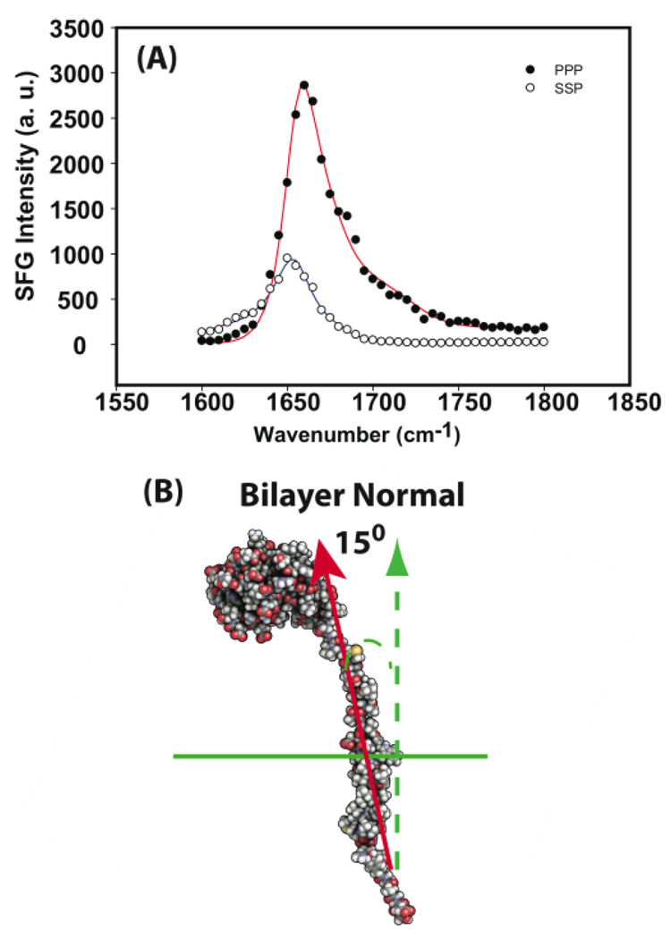
In this study, a series of SFG experiments were used to elucidate the spontaneous membrane insertion property of Cyt-b5 into lipid bilayers. In an SFG experiment, a single substrate supported lipid bilayer was used as a model cell membrane. (Details about the SFG experiments can be found in the Supporting Information.) SFG spectra in the amide I frequency region were collected from wild-type Cyt-b5 in a supported deuterated dimyristoylphosphatidylcholine (dDMPC/dDMPC) bilayer at 25 °C using ssp (s-polarized SFG signal, s-polarized input visible, and p-polarized input IR beam) and ppp (p-polarized SFG signal, p-polarized input visible, and p-polarized input IR beam) polarization combinations of the input and output beams shown in Figure 1A. A peak centered at 1655 cm−1, arising from an α-helix, dominates the SFG spectra.33 Since Cyt-b5 contains α-helical structures in both soluble and transmembrane domains4–6, a software package, namely NLOPredict,34 was used to determine the contribution of SFG signals from the soluble domain. From the NLOPredict program, no substantial SFG signal was generated from helices in the soluble domain as their dipole moments point in opposite directions, which lead to the cancellation of their SFG signals (Figs. 2S and 3S in the Supporting Information). Therefore, the SFG signals mainly originate from the α-helical transmembrane domain and the orientation of the helix was determined from the best-fitting ppp and ssp signal strength ratio of the peak at 1655 cm−1 as shown in Figure 1A.33 Based on our analysis, the Cyt-b5 membrane-anchoring helix inserts into the dDMPC/dDMPC bilayer with a 15° tilt angle relative to the bilayer normal as depicted in Figure 1B. This angle agrees with previous solid-state NMR result of 17°, which was measured from magnetically-aligned DMPC:DHPC bicelles.37–39 This excellent agreement between the SFG and solid-state NMR results validates the SFG method in the determination of topology and helical tilt angles for Cyt-b5. Also, SFG has recently been combined with NMR in studying interfacial peptides, which demonstrates the effectiveness of combining these techniques for the studies of surface bound peptides.40
Figure 1.
(A) ssp and ppp polarized SFG amide I signals of Cyt-b5 in a dDMPC/dDMPC lipid bilayer at 25°C. The dependence of the ppp/ssp ratio with respect to the helical tilt angle is shown in Supporting information. Thus, from the experimentally measured ppp/ssp ratio, it is possible to calculate the tilt angle of an alpha helix from an SFG experiment. (B) A proposed model of Cyt-b5 describing its orientation and topology in lipid bilayers.
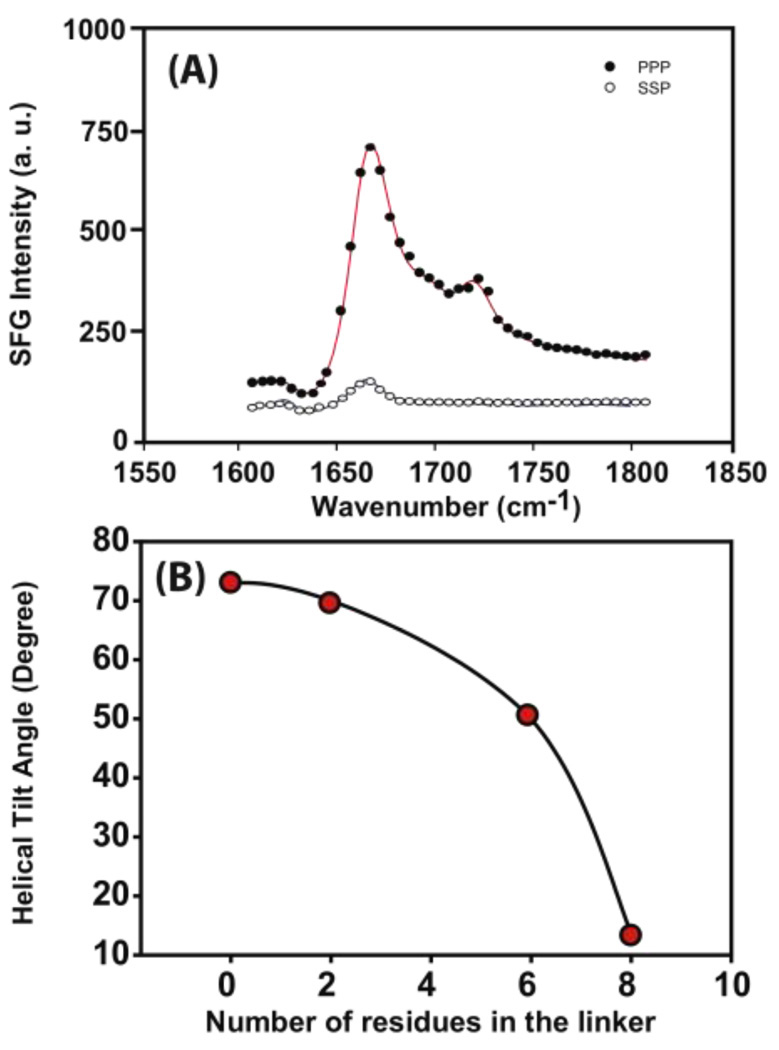
In addition to wild-type Cyt-b5, an inactive mutant Cyt-b5 (m-Cyt-b5) that lacks eight amino acids in the linker region was used to investigate the role and the synergy of the various domains played in the membrane insertion process of Cyt-b5. 41 Surprisingly, the SFG amide I signal from the m-Cyt-b5 detected in a dDMPC/dDMPC bilayer at 25 °C is weaker compared to that of its wild-type counterpart as shown in Figure 2A. Assuming similar membrane coverage, the tilt angle of the m-Cyt-b5 (m: mutant) helix is determined to be 70° with respect to the bilayer normal while using the intensity difference in the ppp SFG spectra between Cyt-b5 and m-Cyt-b5. This result was confirmed by an independent SFG measurement using the signal strength ratio of the ppp and ssp spectra and the tilt angle was calculated to be 73°. Therefore, m-Cyt-b5 most likely tilts towards the membrane surface instead of inserting into the membrane, suggesting that the linker region can indeed influence the manner of membrane insertion of Cyt-b5. To further investigate the influence of the linker length on the membrane insertion property of Cyt-b5, several Cyt-b5 mutants that differ in their linker length were used. SFG results on different mutants in a dDMPC/dDMPC bilayer at 25 °C inferred that the length of the linker region can indeed influence its membrane insertion: as the length of the linker region increased, the tilt angle of the helical membrane anchor decreased, indicative of membrane insertion as shown in Figure 2B.
Figure 2.
(A) ssp and ppp polarized SFG amide I signals of a mutant-Cyt-b5 in a dDMPC/dDMPC lipid bilayer at 25 °C. (B) The dependence of the experimentally measured tilt angle of the transmembrane helix on the number of residues in the linker region of the protein.
SFG experiments were also carried out to measure the effect of lipid acyl chain length on the membrane insertion property of Cyt-b5 and its mutants. The results are summarized in Table 1. Interestingly, the wild-type Cyt-b5 inserts readily as long as the bilayer temperature is above the gel-to-liquid crystalline phase transition temperature (Tm) of the lipid. On the other hand, the insertion of m-Cyt-b5 requires a higher temperature and is partially dependent on the lipid phase. For instance, the gel-to-liquid crystalline phase transition temperature of dilauroylphosphatidylcholine (DLPC) is 4 °C, but m-Cyt-b5 fails to insert into the DLPC bilayer even at 30 °C, which indicates an additional thermal energy is required for membrane insertion. Furthermore, the thickness of the lipid bilayer influences the membrane orientation of Cyt-b5. This is a consequence of the hydrophobic mismatch between the length of the hydrophobic segment of the transmembrane helix and the hydrophobic thickness of the lipid bilayer.41,42 Therefore, to minimize the exposure of the hydrophobic residues in the transmembrane helical region to the aqueous environment, the helix needs to orient such that the length of its hydrophobic segment matches with the hydrophobic bilayer thickness.41 Since a cell membrane is often composed of a mixture of lipids with different chain lengths, membrane proteins adjust their orientation to match the hydrophobic thickness of the bilayers. Therefore, our results demonstrate that the orientation of a membrane protein is dynamic and is a reflection of the nature of the bilayer.
Table 1.
The membrane orientation of the wild-type Cyt-b5 and a mutant Cyt-b5 (with a deletion of eight amino acids in the linker region) in various phospholipid bilayers as a function of temperature.
| Lipid | Tm (°C) | Temperature (°C) | Helical tilt angle | |
|---|---|---|---|---|
| wild-type | mutant | |||
| dDLPC | 4 | 30 | 20° | 75° |
| 45 | - | 26° | ||
| dDMPC | 23 | 25 | 14° | 73° |
| 30 | - | 76° | ||
| 45 | - | 14° | ||
| dDPPC | 40 | 30 | N/A | N/A |
| 45 | 10° | N/A | ||
N/A refers to no detectable SFG amide I signal from the protein and Tm is the gel-to-liquid-crystalline phase transition temperature of a lipid.
dDLPC: deuterated dilauroylphosphatidylcholine; dDPPC: deuterated dipalmitoylphosphatidylcholine; dDMPC: dimyristoylphosphatidylcholine. Since the wild-type Cyt b5 can insert into the lipid bilayer at room temperature, we did not perform the measurements at higher temperatures (indicated by dashes).
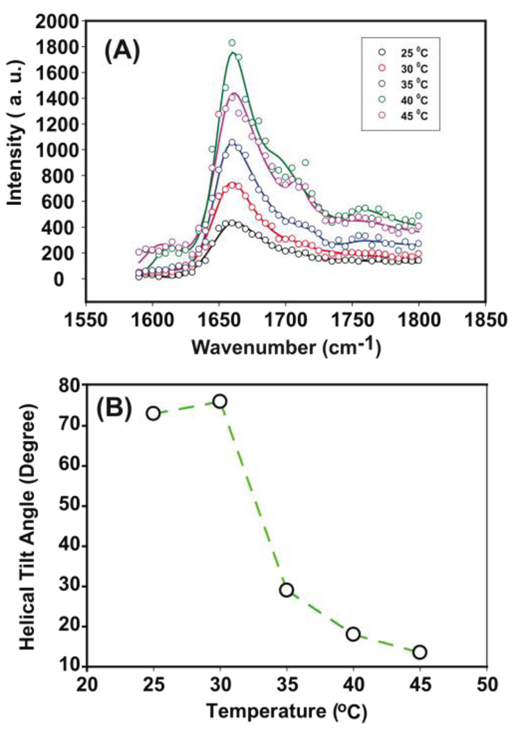
While the m-Cyt-b5 (with a deletion of eight amino acids in the linker region) fails to insert into the lipid bilayer at 25°C, it remains associated with the membrane surface. This raises a question of whether the surface bound 8-deletion m-Cyt-b5 can insert into the membrane if experimental condition changes. To address this question, temperature-dependent SFG experiments were conducted on the dDMPC/dDMPC bilayer surface bound m-Cyt-b5 and the results are given in Figure 3. Since the excess m-Cyt-b5 in the aqueous phase were removed after flushing the system several times with water, the changes in the observed SFG signals will be solely due to the reorientation of the surface bound 8-deletion m-Cyt-b5. Interestingly, the SFG signal intensity increases as a function of temperature (Fig. 3A), suggesting a reorientation of m-Cyt-b5 into the lipid bilayers. The angles deduced from the ppp/ssp signal stretch ratios detected at different temperatures (Fig. 3B) confirm the dependence of the helical anchor orientation on temperature. Therefore, a kinetic barrier seems to prevent m-Cyt-b5 from penetrating into the hydrophobic region of the bilayer at 25°C. This barrier is likely related to protein dynamics. In order for insertion to occur, a range of molecular motions is required that permits reorientation, permeation and translocation of the m-Cyt-b5 helical anchor into the membrane. Importantly, the presence of the linker region can increase the mobility of the protein; in fact, it is the length of the linker that influences the membrane insertion property of Cyt-b5 as shown in our experimental data as well as the functional properties of the mutant proteins.42 Therefore, the synergy between the various domains holds the key in the spontaneous membrane insertion of Cyt-b5.
Figure 3.
(A) ppp polarized SFG amide I band of a 8-deletion mutant-Cyt-b5 in a dDMPC/dDMPC lipid bilayer as a function of temperature. The increase in the intensity of ppp polarized SFG amide I band indicates a reorientation of the protein. The intensity of ppp polarized SFG amide I band at 45° is lower compared to that of 40°, which can be attributed to the desorption of protein from the lipid bilayer surface. (B) Tilt angle of a 8-deletion mutant-Cyt-b5 as a function of temperature determined using SFG ppp/ssp signal strength ratio.
In conclusion, we have demonstrated that it is feasible to probe, in real time, the interaction between a membrane protein and lipid bilayers using SFG experiments with unprecedented sensitivity as demonstrated for Cyt-b5. The significant difference observed in the membrane insertion properties of the wild-type and mutant Cyt-b5 suggests that the length of the linker region can mediate the dynamics of the protein as well as its function, which is in excellent agreement with the functional studies reported in the literature.43 Therefore, the approach reported in this study could be used as a potential rapid screening tool in determining the topology of membrane proteins as well as interactions of biomolecules with lipid bilayers in-situ, which in combination with solid-state NMR could be a solution to the present problems in the structural studies of membrane proteins in their native environment.
Supplementary Material
ACKNOWLEDGMENT
This research is supported by the National Institute of Health (1R01GM081655-01A2 to ZC, GM084018 and RR023597 to AR, and GM035533 to LW), CRIF-NSF, VA Merit Review Grant to LW, and the Office of Naval Research (N00014-08-1-1211 for ZC). The authors thank Dr. Thennarasu for help with fluorescence measurements to determine the membrane binding affinity of cytochrome b5.
Footnotes
SUPPORTING INFORMATION AVALIABLE: List of abbreviations, amino acid sequences of Cyt-b5 and its mutants, NLOPredict simulations, methods, and SFG theory are available free of charge via the internet at http://pubs.acs.org
REFERENCES
- 1.White SH. Nature. 2009;459:344–346. doi: 10.1038/nature08142. [DOI] [PubMed] [Google Scholar]
- 2.Hessa T, White SH, von Heije G. Science. 2005;307:1427. doi: 10.1126/science.1109176. [DOI] [PubMed] [Google Scholar]
- 3.Ahuja S, Smith SO. Trends Pharmacol. Sci. 2009;9:494–502. doi: 10.1016/j.tips.2009.06.003. [DOI] [PubMed] [Google Scholar]
- 4.Renthal R. Cell Mol. Life Sci. 2010;67:1077–1088. doi: 10.1007/s00018-009-0234-9. [DOI] [PMC free article] [PubMed] [Google Scholar]
- 5.Colombo SF, Longhi R, Borgese N. J. Cell Sci. 2009;122:2383–2392. doi: 10.1242/jcs.049460. [DOI] [PubMed] [Google Scholar]
- 6.Dürr UHN, Ramamoorthy A, Waskell L. Biochim. Biophys. Acta. 2007;1768:3235–3259. doi: 10.1016/j.bbamem.2007.08.007. [DOI] [PubMed] [Google Scholar]
- 7.Shen YR. The principles of nonlinear optics. New York: John Wiley& Sons; 1984. [Google Scholar]
- 8.Eisenthal KB. Chem. Rev. 1996;96:1343–1360. doi: 10.1021/cr9502211. [DOI] [PubMed] [Google Scholar]
- 9.Richmond GL. Chem. Rev. 2002;102:693–2724. doi: 10.1021/cr0006876. [DOI] [PubMed] [Google Scholar]
- 10.Perry A, Neipert C, Space B, Moore PB. Chem. Rev. 2006;106:1234–1258. doi: 10.1021/cr040379y. [DOI] [PubMed] [Google Scholar]
- 11.Gopalakrishnan S, Liu DF, Allen HC, Kuo M, Shultz MJ. Chem. Rev. 2006;106:1155–1175. doi: 10.1021/cr040361n. [DOI] [PubMed] [Google Scholar]
- 12.Chen Z, Shen YR, Somorjai GA. Ann. Rev. Phys. Chem. 2002;53:437–465. doi: 10.1146/annurev.physchem.53.091801.115126. [DOI] [PubMed] [Google Scholar]
- 13.Geiger FM. Ann. Rev. Phys. Chem. 2009;60:61–83. doi: 10.1146/annurev.physchem.59.032607.093651. [DOI] [PubMed] [Google Scholar]
- 14.Baldelli S. Acc. Chem. Res. 2008;41:421. doi: 10.1021/ar700185h. [DOI] [PubMed] [Google Scholar]
- 15.Ye HK, Abu-Akeel A, Huang J, Katz HE, Gracias DH. J. Am. Chem. Soc. 2006;128:6528. doi: 10.1021/ja060442w. [DOI] [PubMed] [Google Scholar]
- 16.Li QF, Hua R, Cheah IJ, Chou KC. J. Phys. Chem. B. 2008;112:694. doi: 10.1021/jp072147j. [DOI] [PubMed] [Google Scholar]
- 17.Carter JA, Wang ZH, Dlott DD. Acc. Chem. Res. 2009;42:1343–1351. doi: 10.1021/ar9000197. [DOI] [PubMed] [Google Scholar]
- 18.Koffas TS, Kim J, Lawrence CC, Somorjai GA. Langmuir. 2003;19:3563–3566. [Google Scholar]
- 19.Mermut O, Phillips DC, York RL, McCrea KR, Ward RS, Somorjai GA. J. Am. Chem. Soc. 2006;128:3598–3607. doi: 10.1021/ja056031h. [DOI] [PubMed] [Google Scholar]
- 20.Phillips DC, York RL, Mermut O, McCrea KR, Ward RS, Somorjai GA. J. Phys. Chem. C. 2007;111:255–261. [Google Scholar]
- 21.Chen X, Sagle LB, Cremer PS. J. Am. Chem. Soc. 2007;129:15104–15105. doi: 10.1021/ja075034m. [DOI] [PMC free article] [PubMed] [Google Scholar]
- 22.Jung SY, Lim SM, Albertorio F, Kim G, Gurau MC, Yang RD, Holden MA, Cremer PS. J. Am. Chem. Soc. 2003;125:12782–12786. doi: 10.1021/ja037263o. [DOI] [PubMed] [Google Scholar]
- 23.Kim G, Gurau MC, Lim SM, Cremer PS. J. Phys. Chem. B. 2003;107:1403–1409. [Google Scholar]
- 24.Dreesen L, Sartenaer Y, Humbert C, Mani AA, Méthivier C, Pradier CM, Thiry PA, Peremans A. ChemPhysChem. 2004;5:1719–1725. doi: 10.1002/cphc.200400213. [DOI] [PubMed] [Google Scholar]
- 25.Evans-Nguyen KM, Fuierer RR, Fitchett BD, Tolles LR, Conboy JC, Schoenfisch MH. Langmuir. 2006;22:5115–5121. doi: 10.1021/la053070y. [DOI] [PubMed] [Google Scholar]
- 26.Doyle AW, Fick J, Himmelhaus M, Eck W, Graziani I, Prudovsky I, Grunze M, Maciag T, Neivandt DJ. Langmuir. 2004;20:8961–8965. doi: 10.1021/la0484220. [DOI] [PubMed] [Google Scholar]
- 27.Weidner T, Apte JS, Gamble LJ, Castner DG. Langmuir. 2009;26:3433–3440. doi: 10.1021/la903267x. [DOI] [PMC free article] [PubMed] [Google Scholar]
- 28.Weidner T, Samuel NT, McCrea K, Gamble LJ, Ward RS, Castner DG. Biointerphases. 2010;5:9–16. doi: 10.1116/1.3317116. [DOI] [PMC free article] [PubMed] [Google Scholar]
- 29.Li F, Gang M, Elsa C, Yan Y. J. Am. Chem. Soc. 2010;132:5405–5412. doi: 10.1021/ja909546b. [DOI] [PubMed] [Google Scholar]
- 30.Chen X, Wang J, Boughton AP, Kristalyn CB, Chen Z. J. Am. Chem. Soc. 2007;129:1420–1427. doi: 10.1021/ja067446l. [DOI] [PubMed] [Google Scholar]
- 31.Chen X, Wang J, Paszti Z, Wang F, Schrauben JN, Tarabara VV, Schmaier AH, Chen Z. Anal. Bioanal. Chem. 2007;388:65–72. doi: 10.1007/s00216-006-0999-8. [DOI] [PubMed] [Google Scholar]
- 32.Chen X, Wang J, Sniadecki JJ, Even MA, Chen Z. Langmuir. 2005;21:2662–2664. doi: 10.1021/la050048w. [DOI] [PubMed] [Google Scholar]
- 33.Tan NguyenK, Le Clair SV, Ye S, Chen Z. J. Phys. Chem B. 2009;113:12169–12180. doi: 10.1021/jp904153z. [DOI] [PMC free article] [PubMed] [Google Scholar]
- 34.Moad A, Moad C, Perry J, Wampler R, Goeken GS, Begue N, Shen T, Heiland R, Simpson G. Comput. Chem. 2007;28:1996–2002. doi: 10.1002/jcc.20706. [DOI] [PubMed] [Google Scholar]
- 35.Ye S, Nguyen KT, Le Clair SV, Chen Z. J. Struct. Biol. 2009;168:61–77. doi: 10.1016/j.jsb.2009.03.006. [DOI] [PMC free article] [PubMed] [Google Scholar]
- 36.Chen X, Boughton AP, Tesmer JJG, Chen Z. J. Am. Chem. Soc. 2007;129:12658–12659. doi: 10.1021/ja075542w. [DOI] [PubMed] [Google Scholar]
- 37.Xu J, Dürr UHN, Im SC, Gan Z, Waskell L, Ramamoorthy A. Angew Chem Int Ed Engl. 2008;47:7864–7867. doi: 10.1002/anie.200801338. [DOI] [PMC free article] [PubMed] [Google Scholar]
- 38.Dürr UHN, Yamamoto K, Im SC, Waskell L, Ramamoorthy A. J. Am. Chem. Soc. 2007;129:6670–6671. doi: 10.1021/ja069028m. [DOI] [PMC free article] [PubMed] [Google Scholar]
- 39.Soong R, Smith PES, Yamamoto K, Im SC, Waskell L, Ramamoorthy A. J. Am. Chem. Soc. 2010;132:5779–5788. doi: 10.1021/ja910807e. [DOI] [PMC free article] [PubMed] [Google Scholar]
- 40.Weidner T, Breen NF, Li K, Drohny GP, Castner DG. Proc. Natl. Acad. Sci. U. S. A. 2010;107:13288–13293. doi: 10.1073/pnas.1003832107. [DOI] [PMC free article] [PubMed] [Google Scholar]
- 41.Ramamoorthy A, Kandasamy SK, Lee DK, Kidambi S, Larson RG. Biochemistry. 2007;46:965–975. doi: 10.1021/bi061895g. [DOI] [PMC free article] [PubMed] [Google Scholar]
- 42.Kandasamy SK, Lee DK, Nanga RPR, Xu J, Santos JS, Larson R, Ramamoorthy A. Biochim. Biophys. Acta. 2009;1788:686–695. doi: 10.1016/j.bbamem.2008.11.009. [DOI] [PubMed] [Google Scholar]
- 43.Clarke TA, Im SC, Bidwai A, Wakell L. J. Bio. Chem. 2004;279:36009–36818. doi: 10.1074/jbc.M406055200. [DOI] [PubMed] [Google Scholar]
Associated Data
This section collects any data citations, data availability statements, or supplementary materials included in this article.


