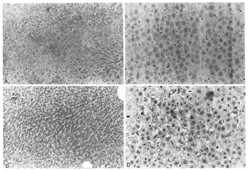
Fig. 7.

Histological appearance of representative livers obtained from animals studied. (A) Medium-power photomicrograph of hepatic lobule in group 1 animal. Wide swaths of coagulative necrosis are infiltrated by neutrophils. Residual preserved hepatocytes extend into the center of the field (H & E, original magnification × 100). (B) Residual hepatocytes in group 1 animal. Single mitosis is identified in the center of the photograph. Other animals in this group had higher mitotic activity (H & E, original magnification × 250). (C) Lobular field from group 3 animal. Even at this power, sublethal hepatocyte damage in the form of cytoplasmic vacuolization is observed. However, no overt necrosis is seen (H & E, original magnification × 100). (D) Brisk mitotic activity is seen in hepatocytes from this group 3 animal (H & E, original magnification × 250).
