Abstract
We report a case of pyoderma gangrenosum occurring at the site of a laparoscopic port insertion following laparoscopic inguinal hernia repair.
Keywords: Pyoderma gangrenosum, Laparoscopy, Port insertion, Inguinal hernia repair
Pyoderma gangrenosum is a rare condition characterised by non-infective, necrotising ulceration of the skin with an incidence of approximately 1 person per 100,000 per year. The aetiology of the condition is unknown; however, in 50% of cases, it is associated with underlying systemic disease particularly inflammatory bowel disease, arthropathy (rheumatoid and spondyloarthritis), multiple myeloma and other paraproteinaemias and acute leukaemia. In 30% of patients, the cutaneous ulcerations of pyoderma gangrenosum occur follow skin trauma.
Case report
A 73-year-old man was admitted as an emergency with a 4-day history of a painful lump in the right groin. A diagnosis of an incarcerated right inguinal hernia was made and the patient underwent a laparoscopy at which a right inguinal hernia was confirmed and repaired using mesh. Following hernia repair, the palpable groin lump was still present; therefore, a local exploration was performed revealing a cyst of the spermatic cord, which was excised. Following this, the patient was well and discharged home.
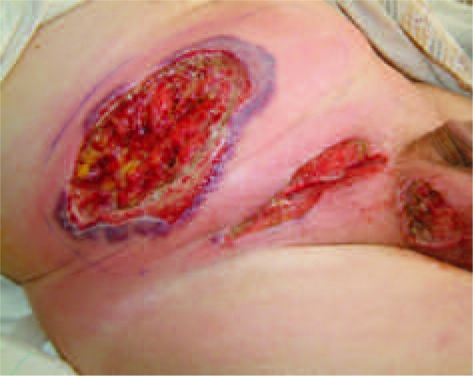
Two weeks later, the patient represented via the accident and emergency department. A superficial dehiscence of the groin wound had occurred secondary to a haematoma with minor discharge. Whilst this had initially responded to oral antibiotic therapy, he was now experiencing further problems not only in the groin but also at the site of the right 5-mm port-site. The area surrounding the port site was erythematous with purple discolouration of the skin and associated vesicle formation and discharge. This was associated with systemic symptoms of fever, lethargy and dizziness. Blood tests revealed a leukocytosis of 22.8 × 10⁹/l and a C-reactive protein level of 169 mg/l. The patient was admitted to hospital and commenced on intravenous erythromycin (penicillin allergy). Ultrasonography was performed and failed to reveal any subcutaneous abscess/collection. Over the following 24 h, the area of skin discolouration increased in size and ulceration was apparent. With the concern of a spreading necrotising infection, the patient was commenced on intravenous Meropenem and Linezolid and underwent surgical debridement. Microscopy of tissue revealed leukocytes but no organisms; cultures grew Proteus mirabilis sensitive to Meropenem. Histology revealed massive neutrophilic infiltration with florid, suppurative necrosis of the overlying epidermis extending into the subcutaneous fat. Neutrophils were identified around and within blood vessel walls, which was considered to be a secondary process. Following debridement, the wound appearance continued to deteriorate with further discolouration and ulceration associated with pyrexia and general malaise. Further debridement was performed; however, a similar wound response was observed (Fig. 1). At 72-h post admission, microbiological and histological results were available and it was felt that the clinical appearances were inconsistent with the microbial picture. The possibility of a pyoderma gangrenosum type reaction was suggested. Rheumatology advice was sought and the patient was commenced on high-dose intravenous methylprednisolone. Following the commencement of steroids, the wound appearance improved with no new areas of necrosis developing and sloughing of existing necrosis. The patient was converted to oral prednisolone and made a slow, but steady, recovery. All subsequent immunological screening and investigations proved normal.
Figure 1.
Following surgical debridement to clean and healthy tissue, ulceration and skin necrosis rapidly returned with a spreading surrounding cellulitis.
Discussion
This is the first reported case of pyoderma gangrenosum occurring in a port site following laparoscopic surgery. An additional unusual feature is the lack of pre-existing perceived risk factors for its development.
Pyoderma gangrenosum is a rare condition, which is recognised to occur following trauma or in response to surgical wounds. It more commonly affects patients who suffer from an associated systemic disease such as ulcerative colitis or rheumatoid arthritis; this is reflected in previous case reports of post-surgical pyoderma gangrenosum.1–3
The early appearances of pyoderma gangrenosum in our patient mimicked a postoperative wound infection with pain, erythema, slough and superficial dehiscence at the wound site together with fever and leukocytosis. In view of the clinical findings of necrosis, the patient was started on intravenous antibiotics and underwent debridement of the affected area. Subsequent histology confirmed a marked suppurative, necrotic process affecting the epidermis with neutrophils forming vesicles and large bullae. In this case, however, the microbiology culture results failed to reveal any organism commonly associated with spreading or necrotic infections and the cultured bacteria (Proteus spp.) had proven sensitivities to the antibiotics administered. Despite treatment, rapidly progressive skin changes were observed with no response to antibiotics or debridement. The continuing cutaneous deterioration taken in context with the microbiology results threw into question the initial diagnosis. The possibility of pyoderma gangrenosum was raised and the patient was commenced on high-dose intravenous steroids, with rapid improvement. Pyoderma gangrenosum exhibits the Koebner phenomenon, also called the ‘Koebner response’ or the ‘isomorphic response’, which refers to skin lesions appearing on lines of trauma. As in our case, surgical debridement often aggravates the condition causing further tissue damage.
The histological findings in our patient of a massive neutrophilic infiltration with necrosis of the overlying epidermis, neutrophils around and within blood vessel walls but without a full vasculitic picture, are typical of pyoderma gangrenosum;4 however, these histological features simulate cellulitis or abscess.
Treatment for pyoderma gangrenosum generally involves local or systemic corticosteroids or immunomodulating therapies together with the management of any associated underlying disease. However, no specific therapy is uniformly effective for patients with pyoderma gangrenosum.4 Our patient responded well to corticosteroids.
If patients present with necrotising, ulcerating or ‘breaking-down’ wounds and fail to respond to conventional treatment or have an unusual bacteriological picture, particularly in the presence of an associated systemic disease, alternative diagnoses should be considered. This case highlights the need for clinicians to be aware of pyoderma gangrenosum as a postoperative phenomenon in order to allow appropriate medical therapy to be instigated and to prevent unnecessary and extensive surgical debridement.
References
- 1.Lilford RJ, Tindall VR, Batchelar AG. Post-surgical pyoderma gangrenosum of the vaginal vault associated with ulcerative colitis: a case report. Eur J Obstet Gynecol Reprod Biol. 1989;31:93–4. doi: 10.1016/0028-2243(89)90030-0. [DOI] [PubMed] [Google Scholar]
- 2.Pishori T, Quoreshi AH. Post-colectomy peristomal pyoderma gangrenosum. J Coll Phys Surg Pak. 2005;15:121–2. [PubMed] [Google Scholar]
- 3.Long CC, Jessop J, Young M, Holt PJA. Minimising the risk of post-operative pyoderma gangrenosum. Br J Dermatol. 1992;127:45–8. doi: 10.1111/j.1365-2133.1992.tb14826.x. [DOI] [PubMed] [Google Scholar]
- 4.Jackson JM, Callen JP. Pyoderma gangrenosum. 2008. < http://emedicine.medscape.com/article/1123821-overview>.


